4587
2D k-space waves for silent EPI acquisitions1Donders Institute for Brain, Cognition and Behaviour, Radboud University Nijmegen, Nijmegen, Netherlands, 2Department of Radiology, University Medical Center Utrecht, Utrecht, Netherlands, 3MR Coils BV, Zaltbommel, Netherlands, 4Erwin L. Hahn Institute for Magnetic Resonance Imaging, University of Duisburg-Essen, Essen, Germany
Synopsis
High acoustic noise levels in fMRI-acquisitions are not only problematic in terms of undesired activation patterns in the brain, but also with respect to patient comfort. A 2D-EPI sequence is presented which is capable of acquiring fMRI-data in silent mode by using a head insert z-gradient coil. For silent data acquisition, a wave-like k-space trajectory is then required. The sequence is compared to a standard FLASH and EPI acquisition. The measured acoustic noise of the silent 2D-EPI is in the order of the idle mode of the scanner and arises from the sound of the continuously active helium pump.
Purpose
MRI acquisitions, in particular fMRI studies suffer from high acoustic noise levels. The acoustic noise caused by the gradients induces a BOLD signal in the auditory cortex which is particularly undesired in auditory experiments. Additionally, it can severely discomfort or even frighten patients during the examination. [1]
The main acoustic noise in an EPI sequence arises from the fast gradient switching of the intense readout. Several methods have been introduced to smooth the slew rates and therewith reduce the acoustic sound. [2]
Latest technical developments made it feasible to build a head z-gradient coil driven by a sinusoidal waveform operating at 20kHz which lies in the inaudible frequency range of humans. [3]
Here, we present a new sequence design benefiting from the head z-gradient coil specifications which is introduced as a fourth gradient axis to the sequence. The modified silent RO-segmented EPI sequence will be able to soundlessly acquire fMRI data by using a modified k-space acquisition scheme.
Methods
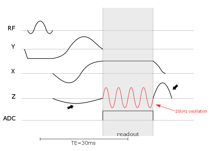
The sinusoidal waveform of the z-gradient insert will be externally controlled and inaudibly operational during the readout time of the ADC as shown in figure 1 (red line). The maximal gradient strength is 41 mT/m with a maximum slew rate of 5200 T/m/s.
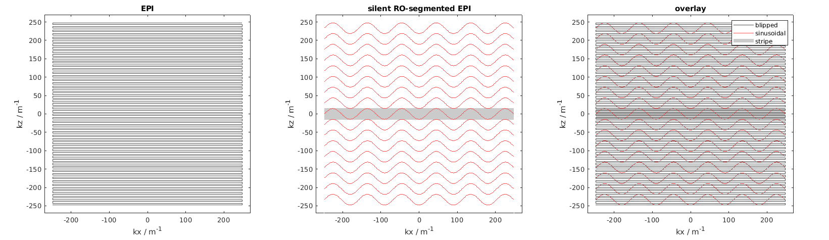
A standard 2D-EPI acquisition has thereupon been modified in the following way. To silently cover the desired k-space trajectory (see figure 2), a constant readout gradient in x-direction was implemented. Targeting an acquisition at 3T with a typical TE=30ms and TR=50ms, the deadtime between the excitation and readout can be used to smooth the residual gradients in the sequence and obtain a completely soundless acquisition. To achieve full k-space coverage, the acquisition is performed in segments. The combination of the 20kHz-oscillating gradient along z and a simultaneous constant RO-gradient in x results in k-space stripes which are moved along kz with standard phase-encoding blips for each segment. For a 2mm resolution, a maximum of 17 k-space stripes/segments is necessary each covering an area of up to 6 standard k-space lines per stripe depending on the constant RO-gradient strength.
Here, the sequence was tested without the z-gradient insert to evaluate the acoustic noise level.
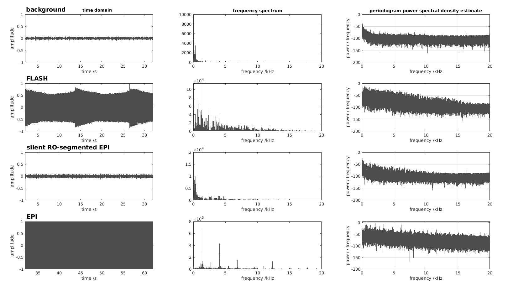
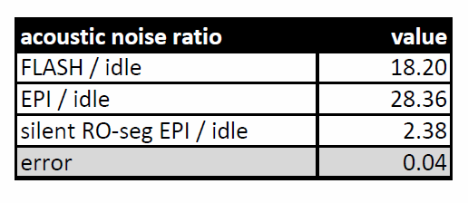
The modified silent RO-segmented EPI sequence was compared to a standard FLASH and EPI sequence. To measure the acoustic noise levels, a microphone was placed in the scanner room and the sound was recorded over 30s for each sequence individually. The acoustic noise levels were determined by the ratio of the sums of the Fourier transformed signal within the audible range window of [10Hz 20kHz] in arbitrary units. $$ \text{acoustic noise ratio} = \frac{\sum{P_{\text{event}}}}{\sum{P_{\text{idle}}}} \quad \text{with} \quad P_x = \left| FT(S_x) \times \text{rect}\left([10\text{Hz}, 20\text{kHz}]\right) \right|^2 $$
An error for the measurement was estimated from the background acquisition: $$\text{error} = \frac{P_{\text{idle3}}/P_{\text{idle1}}- P_{\text{idle2}}/P_{\text{idle1}}}{2}$$
Results
While the standard FLASH and EPI sequences generated typical acoustic noise levels, the scanner remains silent when playing out the modified silent RO-segmented EPI (figure 3). The recorded acoustic noise during the acquisition is only arising from the sound of the helium cold head in the background as confirmed by a background measurement in idle mode. The values of the acoustic noise measurements are shown in table 1 in arbitrary units on a logarithmic scale $$$10 \times \text{log}_{10}(\text{acoustic noise ratio})$$$.Discussion
Due to the fact that the gradient insert can only be applied in z-direction, acquisitions are limited to coronal and sagittal images.
To only test the sound level of the sequence, the silent RO-segmented EPI was run without the z-gradient insert which will currently not produce any image.
In fact, the RO-segmented silent EPI can also easily be extended to a silent 3D acquisition by adding according phase-encoding blips along y played out simultaneously with the slice-select rewinder.
Applications profiting most from a completely silent acquisition would be EPI (fMRI) and SWI sequences.
Conclusion
By using the combination of a fast and inaudible z-gradient coil and a constant readout gradient, a completely silent fMRI scan can be acquired using a 2D wave-like k-space trajectory.Acknowledgements
This work was funded by the European Union (EU) through the "MR Brain" European Regional Development Fund (ERDF), http://ec.europa.eu/regional_policy/en/funding/erdf/.References
1. Ravicz M. et al, Acoustic noise during functional magnetic resonance imaging, J Acoust Soc Am, 2000, 108(4):1683-1696
2. Schmitter S. et al, Silent echo planar imaging for auditory fMRI, Magn Reson Mater Phy, 2008, 21:317-325
3. “EP 3364205 A1 20180822 - Method and apparatus for ultrasonic gradients in magnetic resonance imaging.” https://data.epo.org/gpi/EP3364205A1-METHOD-AND-APPARATUS-FOR-ULTRASONIC-GRADIENTS-IN-MAGNETIC-RESONANCE-IMAGING.
Figures