4586
Supersonic imaging with a silent gradient axis driven at 20 kHz1University Medical Centre Utrecht, Utrecht, Netherlands, 2Spinoza Center for Neuroimaging, Amsterdam, Netherlands
Synopsis
Gradient inserts allow for faster switching and higher gradient strengths than conventional whole-body gradient coils. However, the higher gradient performance is accompanied by an increase in acoustic sound pressure. We present a gradient insert that switches at 20 kHz (above human hearing perception) and therefore allows for imaging with an inaudible gradient axis. Additionally, we introduce a readout scheme for imaging at 20 kHz, and show the first imaging results on a phantom and a healthy volunteer using an inaudible gradient axis.
Background
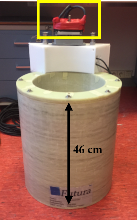
The gradient coils are the main source of acoustic noise during an MR-exam. This noise originates from the rapidly switching currents and their interaction with the main magnetic field through the Lorentz force, causing the gradient coil to vibrate. Gradient inserts provide higher gradient performance than whole-body gradient systems with only limited peripheral nerve stimulation (PNS). Acoustic sound pressure levels, however, do increase due to this increased gradient performance and close proximity to the subject 1,2. Previously, a lightweight single-axis (z-axis) gradient insert was presented that can produce high slew rates (1300 T/m/s) and gradient strengths (200 mT/m) 3 (Figure 1). This coil features a low-inductance and resistance, which makes it ideal for fast switching applications. In this work, we present a modified version of this gradient insert to allow for supersonic imaging by switching the coil at 20 kHz (above human hearing perception). In addition, a special readout scheme was implemented to allow for efficient signal acquisition and adequate k-space coverage.Methods
The gradient coil was paired with an 18 kW audio amplifier (Powersoft,Italy) to ensure a sufficient bandwidth for producing the 20 kHz driving current. A pair of capacitors was used to make the coil resonant at 20 kHz and matched to the audio amplifier, maximizing the current available to generate the gradient field. The silent gradient axis was controlled by an external waveform generator that was triggered by the MR-system (Philips Achieva 7T, Netherlands).
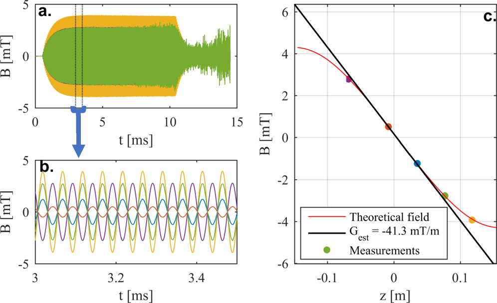
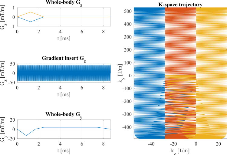
Imaging: The gradient amplitude was measured using a set of field probes (Skope, Switzerland) along the z-axis of the gradient insert to determine the spatiotemporal field behavior. Furthermore, we implemented a readout sequence that fills k-space in a number of sequential lanes (segmented readout) with a width proportional to the amplitude of the resonant gradient (Figure 2). The whole-body z-gradient was used to produce the gradient area needed to move these lanes in k-space to increase the resolution in the z-direction. Spatial encoding perpendicular to the z-direction was achieved using a constant gradient in the direction perpendicular to the slice orientation.
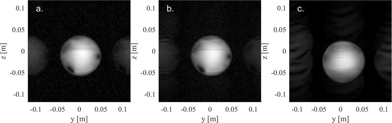
The aforementioned sequence was used to image a water-filled phantom and the head of a volunteer. Images of different resolution were obtained by varying the overlap of lanes (more overlap increases the k-space sampling density) and the gradient amplitude. Each of these acquisitions featured 61 lanes and a fixed resolution of 1.2 mm in the y-direction. The in-vivo acquisition was performed using a 32-channel receive coil (Nova Medical, USA). Reconstruction was performed offline and involved mapping the measured signal to the expected non-cartesian k-space trajectory. Interpolation was performed to grid the data to a cartesian grid.
Results and discussion
Figure 3 shows the gradient field measured with the field probes. Here, we measured a steady-state gradient amplitude of 41 mT/m (reached after 2.5 ms) when the coil was driven at 20 kHz, which corresponded to a peak slew rate of 5200 T/m/s. Despite the high slew rate, no peripheral nerve stimulation was experienced by the volunteer, which could be attributed to the short pulse duration (25 µs per ramp), and the short field extent of the gradient insert. Moreover, no sound was perceived by the volunteer when driving solely the insert gradient at 20 kHz and 41 mT/m in the 7T magnet.
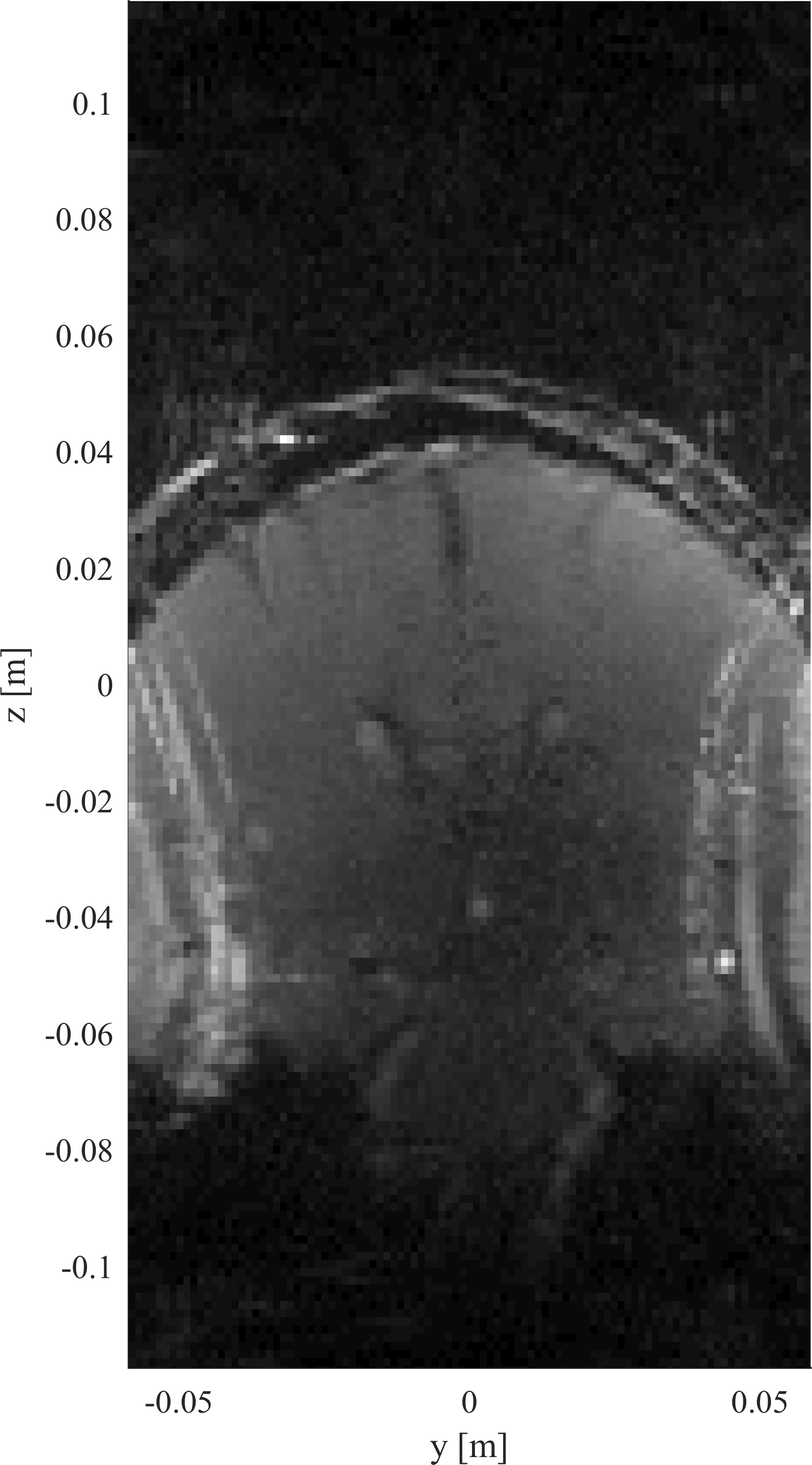
Figure 4 shows the images acquired on the water-filled phantom. Here, we successfully reconstructed images for the different degrees of overlap and gradient amplitudes. In all cases, the imaged sphere featured limited geometrical distortion, as no correction for gradient non-linearity was applied. Figure 5 shows the first in-vivo image acquired on a brain. Here, almost full brain coverage was observed in the z-direction. However, the tight field-of-view in the y-direction resulted in an aliasing artefact, which can be removed once SENSE is implemented. The ghosting visible in all images was caused by small deviations between the calculated and actual k-space trajectory. The incorporation of measured fields in the reconstruction pipeline may further improve the reconstructed images 4.
The current implementation of the sequence is not completely inaudible yet, as the pre-winder gradients still produce some audible sounds. The segmented readout can also be acquired in a single-shot or multi-shot EPI fashion.
Conclusion
We have presented a setup that can drive a gradient insert at 20 kHz with 41mT/m and 5200 T/m/s at 7T without PNS nor sound experience, and an acquisition scheme that utilizes four physical gradient axes (whole-body + z-axis gradient insert) to image at 20 kHz. The first imaging results show that it is feasible to image using a completely silent gradient axis.Acknowledgements
No acknowledgement found.References
1. Weiger M, Overweg J, Rösler MB, et al. A high-performance gradient insert for rapid and short-T 2 imaging at full duty cycle. Magn. Reson. Med. [Internet] 2017;00:1–11. doi: 10.1002/mrm.26954.
2. Winkler SA, Schmitt F, Landes H, DeBever J, Wade T, Alejski A, Rutt BK. Gradient and shim technologies for ultra high field MRI. Neuroimage [Internet] 2016:0–1. doi: 10.1016/j.neuroimage.2016.11.033.
3. Van der Velden TA, Van Leeuwen CC, Huijing ER, Borgo M, Luijten PR, Klomp DWJ, Siero JCW. (2017), A lightweight gradient insert coil for high resolution brain imaging ISMRM , #4329
4. Dietrich BE, Brunner DO, Wilm BJ, Barmet C, Gross S, Kasper L, Haeberlin M, Schmid T, Vannesjo SJ, Pruessmann KP. A field camera for MR sequence monitoring and system analysis. Magn. Reson. Med. 2016;75:1831–1840. doi: 10.1002/mrm.25770.
Figures