4585
Wave-CAIPI accelerated whole brain structure imaging using three-dimensional T1 weighted SPACE sequence1Lauterbur Research Center for Biomedical Imaging, Shenzhen Institutes of Advanced Technology, Chinese Academy of Sciences, Shenzhen, China, 2Research Center for Medical AI, Shenzhen Institutes of Advanced Technology, Chinese Academy of Sciences, Shenzhen, China
Synopsis
Three-dimensional (3D) SPACE (sampling perfection with application optimized contrast using different flip angle evolutions) sequences are the workhorse for volume imaging with isotropic spatial resolution. However, spatial resolution is often scarified to achieve clinically acceptable scan time. Conventional one- and two-dimensional parallel imaging techniques could help reducing the scan time but would lead to deteriorated signal-to-noise (SNR) performance at submillimeter spatial resolutions. In this study, three-dimensional parallel imaging technique-Wave-CAIPI is utilized to improve the SNR performance for whole brain SPACE imaging with isotropic 0.6 mm resolution. In vivo results demonstrated that Wave-CAIPI could improve the SNR at 5x acceleration.
Introduction
Three-dimensional SPACE sequence1 is commonly used for large volume imaging with isotropic submillimeter spatial resolutions. However, simultaneous large spatial coverage and high spatial resolution often lead to clinically impractical long scan time. The achievable acceleration factor using one-dimensional parallel imaging is often less than 4. Two-dimensional parallel imaging techniques such as 2D GRAPPA2 and CAIPIRINHA3 could achieve 4x or 6x acceleration but suffers from significant degradation of signal-to-noise ratio (SNR) especially for high resolution imaging. This work utilizes the newly proposed 3D parallel imaging technique, Wave-CAIPIRINHA4, to achieve better imaging SNR for whole brain T1 weighted SPACE imaging5 with high isotropic spatial resolution of 0.6 mm.Methods
Pulse Sequence: The sequence diagram of developed Wave-SPACE is shown in Figure 1. Wave gradients of sinusoidal waveform are played simultaneously along the phase (Gy) and partition (Gz) encoding dimensions during the readout. A π/2 phase shift is imposed between the two waveforms4. Large wave gradient amplitude is desired to achieve effective geometry factor reduction by Wave-CAIPI6. However, high-resolution SPACE sequence requires high bandwidth and small echo spacing for optimized T1 contrast and SNR1. To obtain the maximal wave gradient amplitude while not exceeding the gradient slew-rate limitation, a slight modification is made to the wave gradient on the partition encoding dimension (Gz). Specifically, 1/4 cycle in the head and 3/4 cycle at the tail (i.e. one cycle in total) are truncated in the sinusoidal Gz wave gradient. The wave gradient along Gz is then allowed to moderately ramp up from zero point and ramp down to zero point and leads to reduced slew rates.
In Vivo Experiments: The IRB approved study was performed on a 3T Siemens Tim Trio MRI system with a commercial 32-channel head coil. T1 weighted SPACE was used for whole brain structure imaging with an isotropic resolution of 0.6 mm. A 25 years-old healthy volunteer was prospectively recruited with informed consent being obtained. Three scans using different subsampling schemes were performed after localization: (1) 2x2 Uniform, (2) 2x2 Wave-CAIPI, and (3) 2x2 CAIPI. Common imaging parameters were: TE/TR = 8.5/850 ms, ETL = 42, matrix size = 304x304x256, FOV = 180x180x154 mm3, bandwidth = 567 Hz/pixel. Each scan took 4:51 minutes to be completed. Both uniform and CAIPI accelerations embedded a 24x24 calibration region in the k-space center for coil sensitivity estimation. Separate ACS data with k-space size of 304x24x24 was acquired immediately before Wave-CAIPI scan to estimate the coil sensitivity map. Four calibration scans taking single slice projection data were also acquired before imaging to characterize the Point Spread Function (PSF) for wave-encoding model. These PSF calibration scans used the same wave amplitude and cycle as the imaging scan, but used longer TR and shorter ETL to guarantee the SNR in estimated PSF.
Image Reconstruction: All datasets were reconstructed offline in MATLAB (Mathworks, Natick, MA, USA). ESPIRiT algorithm was firstly utilized for estimating the three-dimensional coil sensitivity map. Then, 3D SENSE reconstruction model was iteratively solved by LSQR algorithm to reconstruct Uniform and CAIPI accelerated datasets. For Wave-CAIPI accelerated dataset, PSF model characterizing the wave encoding was firstly estimated and integrated into the 3D SENSE model. Then the parallel imaging model was solved by the LSQR iteration. Finally, geometry factor maps showing the spatial distribution of noise amplification were derived using theoretical analysis.
Results
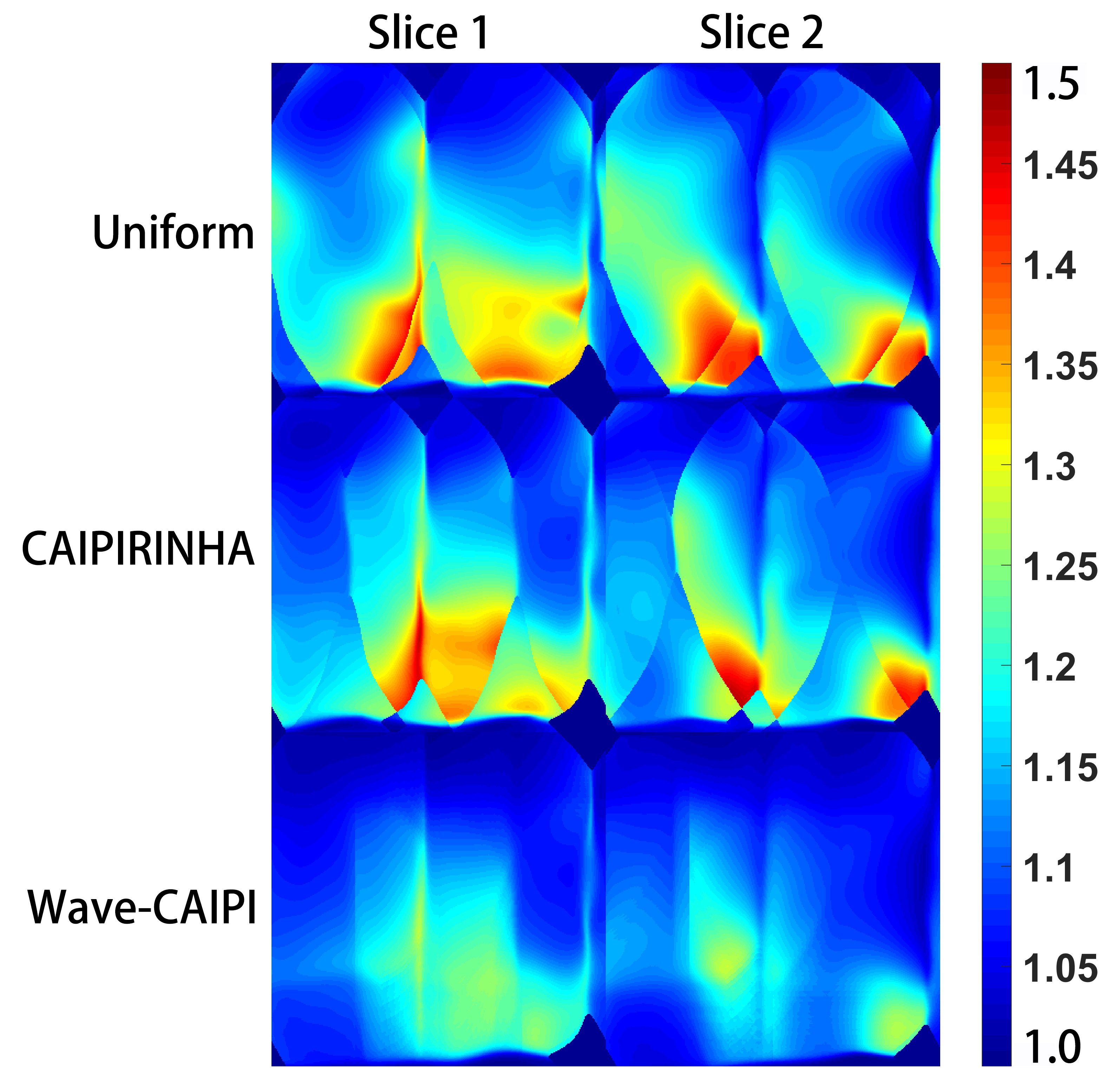
Figure 2 depicts the two sagittal slices of reconstructed 3D images for the three acquisition methods: uniform, 2D CAIPIRINHA and wave-CAIPI with R=2x2 fold acceleration. Wave-CAIPI yields to reconstruction with higher SNR and less artifact penalties than the other two subsampling schemes. The corresponding g-factor maps are shown in Figure 3. Decreased and more uniform g-factor map was obtained by using the wave-CAIPI technique.Discussion
In this study, 3D parallel imaging method Wave-CAIPI is successfully integrated into the SPACE sequence. The geometry factor at 5x acceleration is improved comparing with 2D parallel imaging methods. The SNR performance improvement of wave-CAIPI is not significant at present. One reason may be only small wave gradient amplitude was applied currently. The second reason is the challenging PSF estimation, which is susceptible to heavy noise at high-resolution imaging. Further work will focus on improving G-factor reduction via increasing the amplitude of wave gradient. The signal-to-noise of projection scan will also be improved to achieve accurate PSF calibration for wave-CAIPI accelerated high-resolution T1 SPACE.
Acknowledgements
This work was supported in part by the grant from the National Science Foundation of China (61871373, 61471350, 81729003), Guangdong Provincial Key Laboratory of Medical Image Processing (2017A050501026), and the National Science Foundation of Guangdong Province (2018A0303130132).References
[1] Mugler III JP. Optimized Three-Dimensional Fast-Spin-Echo MRI. J Magn Reson Imaging 2014; 39:745-767.
[2] Blaimer M, Breuer FA, Mueller M, et al. 2D-GRAPPA-Operator for Faster 3D Parallel MRI. Magn Reson Med 2006; 56:1359-1364.
[3] Breuer FA, Blaimer M, Mueller MF, et al. Controlled Aliasing in Volumetric Parallel Imaging (2D CAIPIRINHA). Magn Reson Med 2006; 55:549-556.
[4] Bilgic B, Gagoski BA, Cauley SF, et al. Wave-CAIPI for Highly Accelerated 3D Imaging. Magn Reson Med 2015; 73:2152-2162.
[5] Park J, Mugler III JP, Horger W, Kiefer B. Optimized T1-weighted contrast for single-slab 3D Turbo Spin-Echo Imaging with long echo trains: application to whole-brain imaging.
[6] Qiu Z, Wang JJ, Ying L, Liu X, Liang D. Parameter Optimization of Wave-CAIPI Based on Theoretical Analysis. In Proceedings of the 26th Annual Meeting of ISMRM, Paris, France, 2018. p. 3506.
Figures