4582
Simultaneous T2* and T2 weighted imaging based on ultrafast SPEN MRIQingjia Bao1 and Lucio Fydman2
1Weizmann Institute of Science, Rehovot, Israel, 2Weizmann Institute of Science, Rehovot, AB, Israel
Synopsis
This work presents new sequences to acquire multislice images with different contrasts –a T2* weighted one for enhancing BOLD and a T2 weighted one for faithful location– in a single shot. The sequences rely on SPatiotemporal ENcoding (SPEN), an ultrafast MRI method with immunity to artifacts, and they utilize a “full-refocusing” mode to obtain a T2 weighted image and a “
INTRODUCTION
Pulse sequences that acquire multiple images with different contrasts in a single shot exhibit advantages over single-contrast imaging, not only in terms of improving the acquisition time efficiency but, equally important, thanks to their immunity to an interscan variability that can compromise direct in vivo comparisons. This has proven particularly valuable in fMRI, where concurrent GE EPI and SE EPI scanning evidenced advantages vs separate-scan acquisitions1,2. This work introduces two variants based on SPatiotemporal Encoding (SPEN), which in the same shot provide “fully-refocused” images that are purely T2-weighted, and “non-fully-refocused” images subject to T2* weighting3,4. The variants differ in their order of these T2 and T2* weighted acquisitions; their reliability and robustness are tested in vivo and compared against EPI counterparts.METHODS
The pulse sequences are shown in Figure 1. They utilize a “full-refocusing” mode whereby each spin packet fully refocuses its T2*/shift effects at the instant of its signal emission to deliver pure T2 weighted images, and a “non full-refocusing” mode to obtain T2* weighted images. In Figure 1a, the first echo train is collected immediately after the chirp encoding pulse and hence is acquired under “non-fully-refocused” conditions; after a pulse and suitable delay, a second echo train will be “fully refocused”3 and deliver pure T2-weighted images. Conversely, in Figure 1b the first echo train is “fully-refocused” while the second (and subsequent) echo train(s) is(are) T2* weighted. To assess this strategy’s ability to deliver T2*/T2 weighted images data were collected on a 7T/120mm horizontal Agilent MRI scanner using a quadrature Millipede® coil; comparisons were run against EPI, including single-shot and interleaved versions. RESULTS & DISCUSSION
Figure 2 shows results for simultaneously collected T2*/T2-weighted images based on the pulse sequences shown in Figure 1. The upper two rows show multi-slice data acquired from Figure 1a, in which T2* contrast is obtained first and T2 contrast later. The lower two show the data acquired from Figure 1b, in which this order is reversed. Figure 3 shows results from in vivo brain experiments collected in mice, comparing single- and multi-shot (interleaved) SPEN acquisitions performed with the sequence in Fig. 1a, with results arising from scanner-supplied gradient- and spin-echo (single-contrast) EPI experiments. Besides the obvious reduction in acquisition time, it is interesting to notice (i) the weaker artifacts arising in the T2*-weighted SPEN images vis-à-vis GE EPI counterparts as a result of their bigger effective bandwidths, and (ii) the added image faithfulness arising in the T2-only weighted SPEN images vis-à-vis SE EPI counterparts, as a result of the full refocusing condition. Figure 4 shows an additional application, this time to in vivo scans of the abdominal region. This is a more challenging target in terms of susceptibility and motions than brain. Thus not only are the advantages mentioned earlier for the case of brain noticed in these experiments, but also a substantial reduction of motional artifacts in the SPEN-derived data.CONCLUSION
Simultaneous acquisition of T2* and T2 weighted images based on ultrafast SPEN opens new routes to obtain multi-parametric images with speed and robustness. This in turn can enable valuable routes in functional and real-time MRI, susceptibility weighted imaging, etc. These extensions are being explored both in preclinical and human MR settings.Acknowledgements
We are grateful to Ricardo Martinho and Zhiyong Zhang (WIS) for valuable discussions. Financial support came from the NIH human placenta project (R01HD086323), the Israel Science Foundation (grants 2508/17 and 965/18), the Kimmel Institute for Magnetic Resonance (Weizmann Institute) and the generosity of the Perlman Family Foundation.References
1. Schwarzbauer, C., Mildner, T., Heinke, W., Brett, M. & Deichmann, R. Dual echo EPI - The method of choice for fMRI in the presence of magnetic field inhomogeneities? Neuroimage 49, 316–326 (2010). 2. Glielmi, C. B., Xu, Q., Craddock, R. C. & Hu, X. Simultaneous acquisition of gradient echo/spin echo BOLD and perfusion with a separate labeling coil. Magn. Reson. Med. 64, 1827–1831 (2010). 3. Rita Schmidt; Lucio Frydman New Spatiotemporal Approaches for Fully Refocused, Multislice Ultrafast 2D MRI. 71, 711-722 (2014). 4. Bao, Q., Solomon, E., Liberman, G., Cousin, S. & Fydman, L. Single-scan multi-spin-echo {SPEN} for dynamic T2 mapping and for 3D T2 weighted anatomical imaging. Proc. Jt. Annu. Meet. ISMRM-ESMRMB, Paris, Fr. 380 (2018).Figures
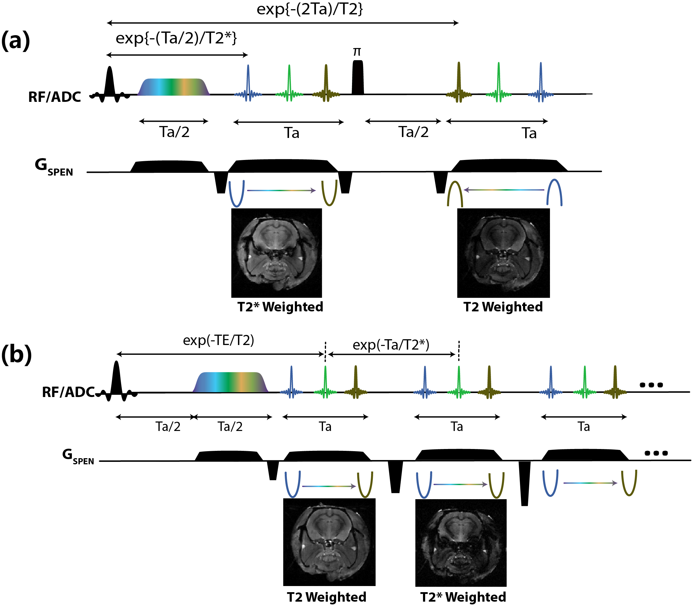
Figure 1 Variants for the simultaneous acquisition of T2*/T2-weighted images based on SPEN, with the readout and slice selection axes/gradients omitted for ease of understanding. A chirped pulse –illustrated with multiple colors, each one addressing spatially-separated spins. During Ta where a gradient progressively brings the spin-packets into focus. (a) the first echo is acquired under “non-fully-refocused” conditions; after a hard pulse and a suitable delay (Ta/2), a second echo train will be “fully refocused” and deliver a T2*-free, solely T2-weighted images. (b) the first echo is “fully-refocused”, and subsequent echoes are collected using gradient reversals and are hence subject to T2* weightings.
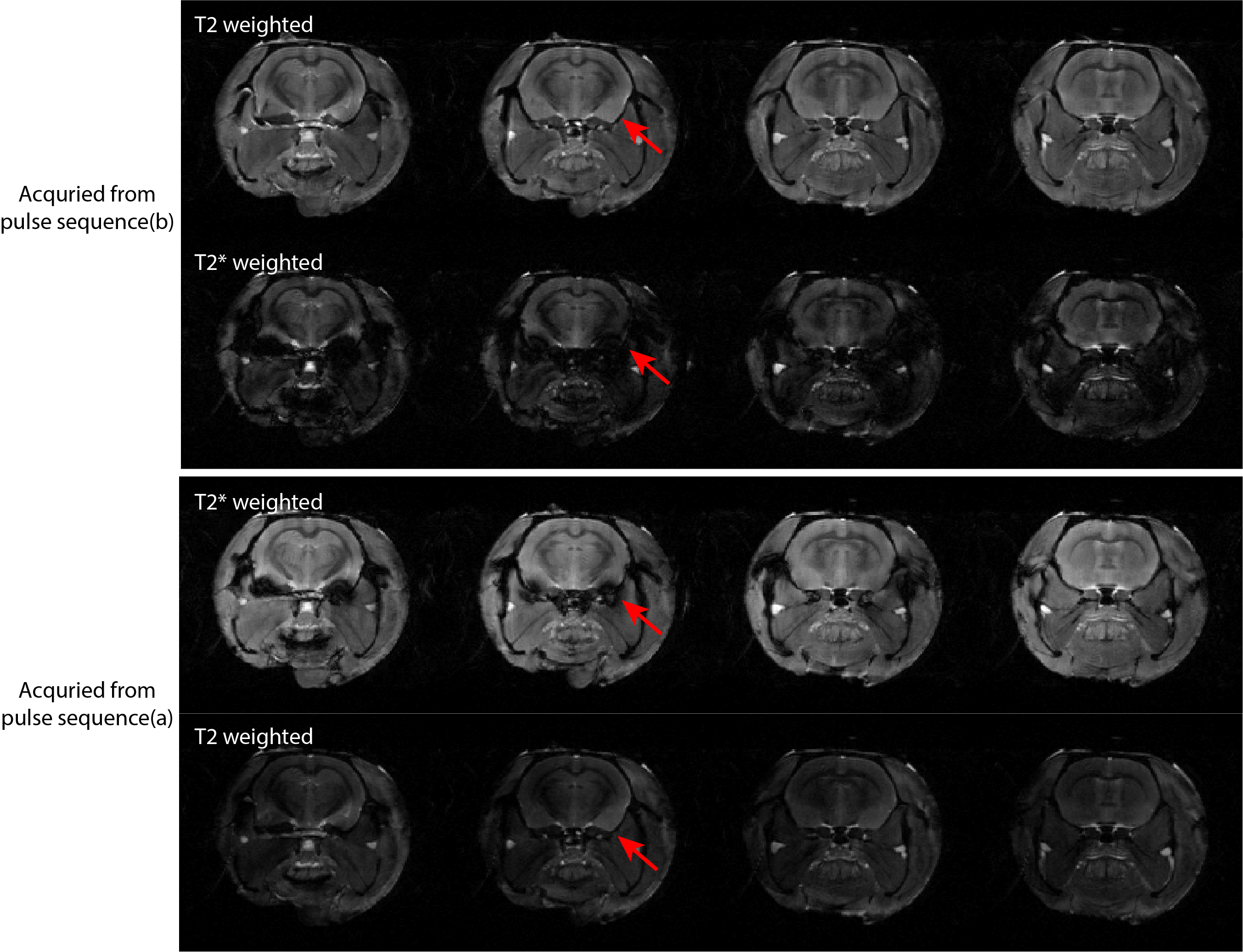
Figure 2. Multi-slice results
for simultaneously T2*/T2 contrast images based on the pulse sequences shown in
Figure 1, collected on an ex vivo rat
head. The different intensities of the T2/T2*-weighted images in each row
reflect their acquisition orders. Red arrows highlight some of the differences
noted with/without T2* refocusing.
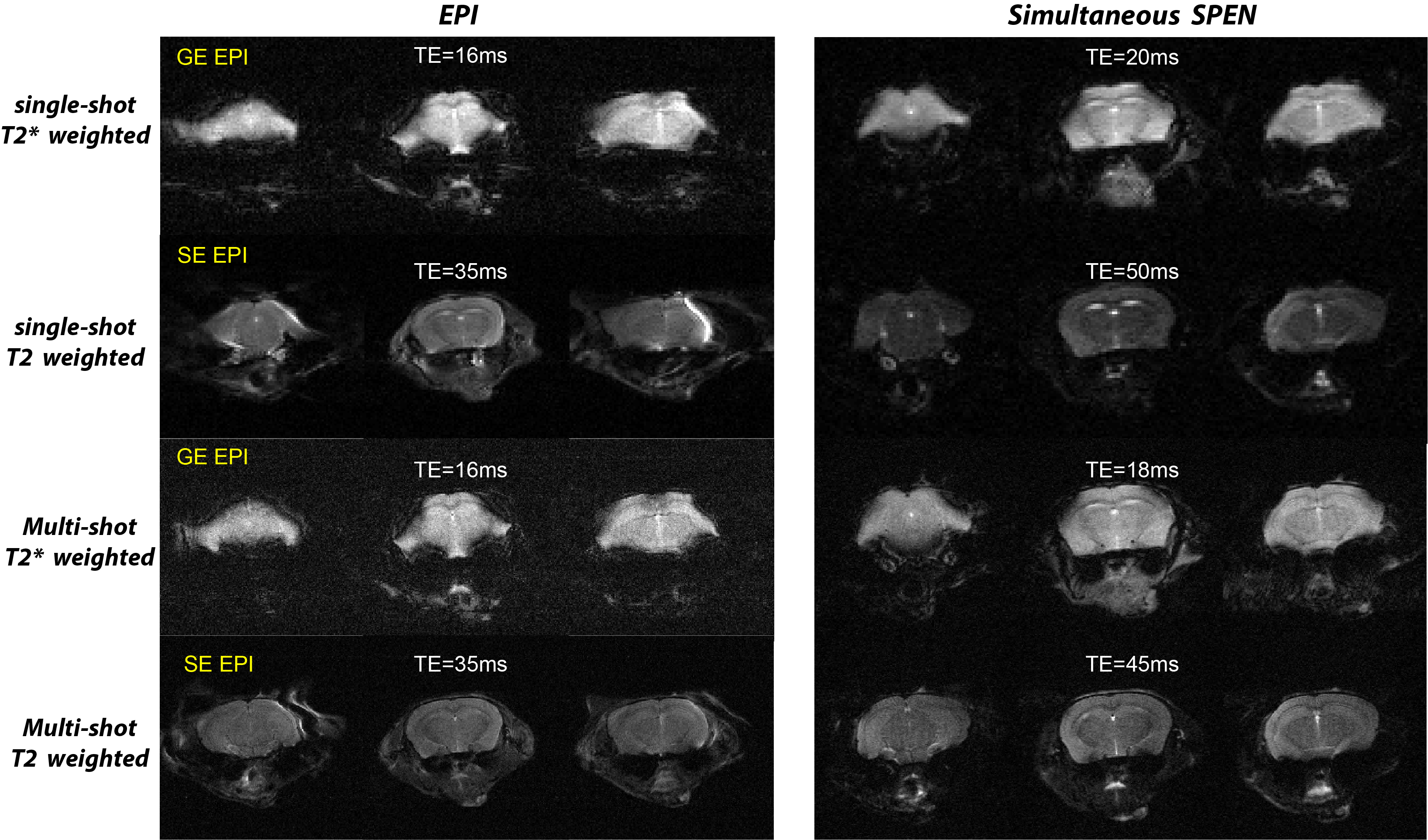
Figure 3. In vivo mouse brain results collected with single shot (top) and interleaved (bottom) GE EPI and SE EPI experiments (left-hand column), and with the simultaneously T2*/T2-weighted pulse sequence introduced in Figure 1a (right-hand column). Shown in each case are the TE times corresponding to the centers of the acquisitions; data interleaving involved the combination of five shots.
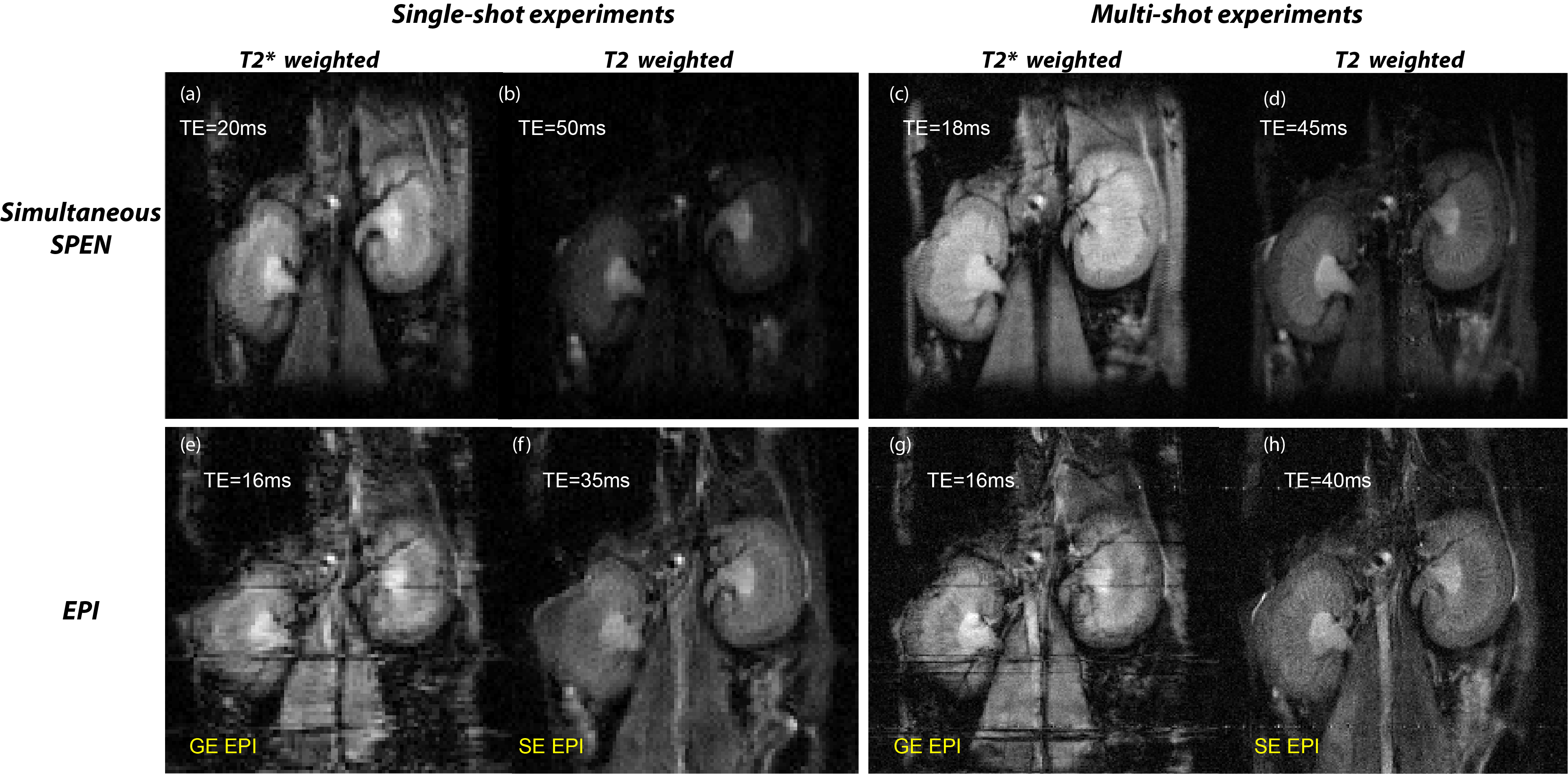
Figure 4. Similar as in Fig 3, but for in vivo mouse
kidney experiments. The horizontal lines in the EPI images reflect problems in
post-processing, arising from motions between the reference scan and the
collected data (SPEN is free from these as it does not require an independent
reference scan).