4581
Twisted radial echo planar trajectory (EPIstar) for 3D self-navigated golden angle structural and functional MRI1Medicine, University of Hawaii, Honolulu, HI, United States
Synopsis
A new 3D trajectory design for efficient, self-navigated golden angle high-resolution MRI acquisition is presented along with results in SWI and BOLD functional MRI.
Introduction
Radial sampling is having resurgence for segmented brain and body MRI due to its intrinsic self-navigation from oversampling the k-space center making it robust to motion without the need for special hardware or pulse sequence modifications.(1-4) Radial sampling has the additional advantage of producing benign “streaking” aliasing artifacts compared to Cartesian undersampling that allows for large accelerations and an efficient use of parallel imaging or Compressed Sensing.(5,6) For these reasons radial EPI sampling has been proposed for segmented volumetric fMRI (TURBINE) as well as other applications.(7-11) We propose a novel three-dimensional radial EPI trajectory (EPIstar) that consists of a twisted stack-of-spirals design for improved sampling and parallel imaging performance in self-navigated golden angle MRI. The EPIstar method is applied to both SWI and fMRI as example brain applications.Trajectory Design
The EPIstar trajectory is derived from the radial EPI design in which straight readout lines are replaced by in-out spirals in the x,y-plane and consecutive z-phase encodes are twisted along z using x,y-gradient blips. For the spiral design the whirl trajectory is used,(12) which is a hybrid between radial and spiral, and is further optimized to traverse rapidly through the k-space center with non-zero magnitude to shorten the readout length (as opposed to decreasing to zero). The resulting EPIstar trajectory is characterized by improved incoherent coverage of 3D k-space at the cost of prolonged readouts. For high-resolution imaging an interleaved segmentation scheme along kz is applied to avoid severe susceptibility induced image distortion and signal loss by extending the readout length beyond 50ms. Therefore, EPIstar gradients are designed that only cover a fraction (Rz) of the required kz-planes. In analogy to standard 3D imaging, each of the Rz shots uses an additional z-encoding blip to shift the k-space trajectory in kz and fill in the missing planes of the previous shots. The combination of all Rz shots then gives one fully-sampled EPIstar blade. A benefit of having multiple excitations is that a large twist between the k-space segments can be incorporated without further extending the length of the trajectory.
Methods
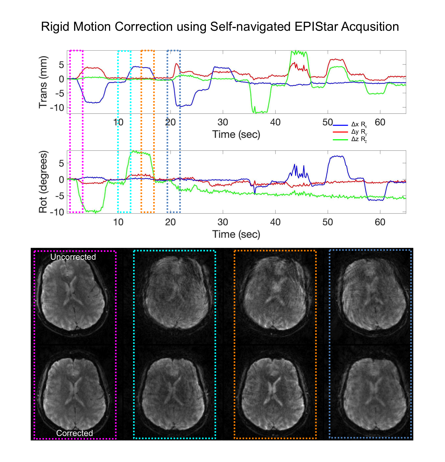
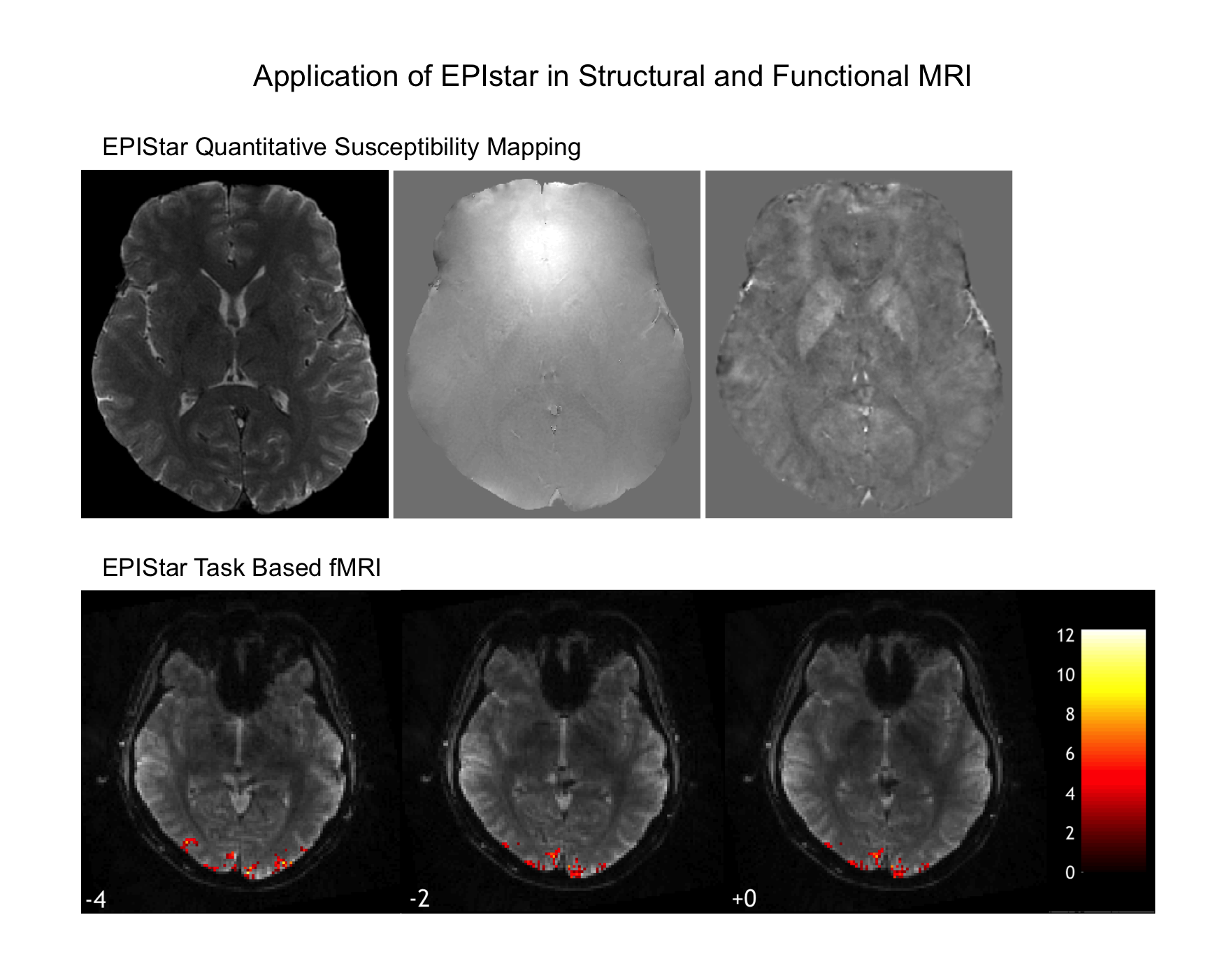
Acquisition: MRI data were acquired using a 3D gradient echo sequence (20° slab select excitation, TR=50-85ms and TE=17-35ms) and gradient spoiling. For the segmented acquisition all segments of a full blade were acquired prior to advancing to the next blade. In either case consecutive blades were rotated by the golden angle (≈111.25°). Self-navigation and Reconstruction: A generalized SENSE reconstruction method using the Michigan Image Reconstruction Toolbox (MIRT) was applied. The k-space trajectories, off-resonance and coil sensitivity maps and data were inserted directly into the signal equation and solved numerically using a time-segmented non-uniform fast Fourier transform (NUFFT) within a regularized conjugate gradient algorithm.(13) All reconstructions were performed offline using Matlab. For the purpose of self-navigation a series of images of reduced quality/resolution but high temporal resolution was obtained by reconstructing datasets from only 3 blades. Image were realigned volume-by-volume using MCFLIRT from the FSL software library (FMRIB, Oxford, UK). The rigid motion parameters (6 degrees of freedom) were used to adjust the k-space input, which was then used to reconstruct the final high-resolution images. SWI Maps: Four echo images (TR=85ms,TE=17,23,29,35ms) at a resolution of 0.8x0.8x2mm3 were obtained (TA=4x34s). The quantitative susceptibility maps were generated using the Morphology Enabled Dipole Inversion (MEDI) toolbox (http://pre.weill.cornell.edu/mri/pages/qsm.html). BOLD Maps: fMRI was performed using a visual paradigm consisting of six repetitions of 5s of flashing checkerboard at 8Hz and 15s rest. Images were reconstructed at a 1.7x1.7x3mm3 resolution after correcting for rigid motion using N=30 blades (TR=65ms/TE=25ms). The data were analyzed at a temporal resolution of 1.95s using a general linear model as implemented in SPM12 (FIL Lab, UCL).Results
The EPIstar design presents an efficient and versatile sampling strategy for fast 3D golden angle MRI acquisitions (Figure 1). Images show a remarkably benign aliasing pattern even for extreme degrees of undersampling (Figure 2) allowing it to be used for self-navigated motion correction at high temporal resolution (Figure 3). On the other hand high-quality T2*-weighted images are obtained when more blades are considered for each image. The results for high-resolution SWI and motion-corrected fMRI are shown in Figure 4.Discussion
The results using the EPIstar golden angle approach indicate the vast potential of this new trajectory design in motion-robust functional and structural MRI in view of increasingly popular techniques such as reordering and self-navigation. The addition of compressed sensing and other elaborate reconstruction techniques is expected to further improve the performance especially of largely undersampled data. A major technical challenge with EPIstar, however, is distortion and signal loss from magnetic susceptibility variations. Long readout times and echo times magnify these effects, which primarily manifest in the “slow” through-plane direction.Acknowledgements
This project was supported by the NIH grants R01DA019912, R21EB02076References
1. Benkert T, Feng L, Sodickson DK, Chandarana H, Block KT. Free-breathing volumetric fat/water separation by combining radial sampling, compressed sensing, and parallel imaging. Magn Reson Med 2017;78(2):565-576.
2. Chandarana H, Feng L, Ream J, Wang A, Babb JS, Block KT, Sodickson DK, Otazo R. Respiratory Motion-Resolved Compressed Sensing Reconstruction of Free-Breathing Radial Acquisition for Dynamic Liver Magnetic Resonance Imaging. Invest Radiol 2015;50(11):749-756.
3. Feng L, Axel L, Chandarana H, Block KT, Sodickson DK, Otazo R. XD-GRASP: Golden-angle radial MRI with reconstruction of extra motion-state dimensions using compressed sensing. Magn Reson Med 2016;75(2):775-788.
4. Lee GR, Griswold MA, Tkach JA. Rapid 3D radial multi-echo functional magnetic resonance imaging. NeuroImage 2010;52(4):1428-1443.
5. Breuer FA, Blaimer M, Heidemann RM, Mueller MF, Griswold MA, Jakob PM. Controlled aliasing in parallel imaging results in higher acceleration (CAIPIRINHA) for multi-slice imaging. Magnetic resonance in medicine : official journal of the Society of Magnetic Resonance in Medicine / Society of Magnetic Resonance in Medicine 2005;53(3):684-691.
6. Lustig M, Donoho D, Pauly JM. Sparse MRI: The application of compressed sensing for rapid MR imaging. Magn Reson Med 2007;58(6):1182-1195.
7. Graedel NN, McNab JA, Chiew M, Miller KL. Motion correction for functional MRI with three-dimensional hybrid radial-Cartesian EPI. Magn Reson Med 2017;78(2):527-540.
8. Chiew M, Graedel NN, McNab JA, Smith SM, Miller KL. Accelerating functional MRI using fixed-rank approximations and radial-cartesian sampling. Magn Reson Med 2016;76(6):1825-1836.
9. Silva AC, Barbier EL, Lowe IJ, Koretsky AP. Radial echo-planar imaging. J Magn Reson 1998;135(1):242-247.
10. McNab JA, Gallichan D, Miller KL. 3D steady-state diffusion-weighted imaging with trajectory using radially batched internal navigator echoes (TURBINE). Magn Reson Med 2010;63(1):235-242.
11. Jonathan SV, Vakil P, Jeong YI, Menon RG, Ansari SA, Carroll TJ. RAZER: a pulse sequence for whole-brain bolus tracking at high frame rates. Magn Reson Med 2014;71(6):2127-2138.
12. Pipe JG. An optimized center-out k-space trajectory for multishot MRI: comparison with spiral and projection reconstruction. Magn Reson Med 1999;42(4):714-720.
13. Sutton BP, Noll DC, Fessler JA. Fast, iterative image reconstruction for MRI in the presence of field inhomogeneities. IEEE Trans Med Imaging 2003;22(2):178-188.
Figures
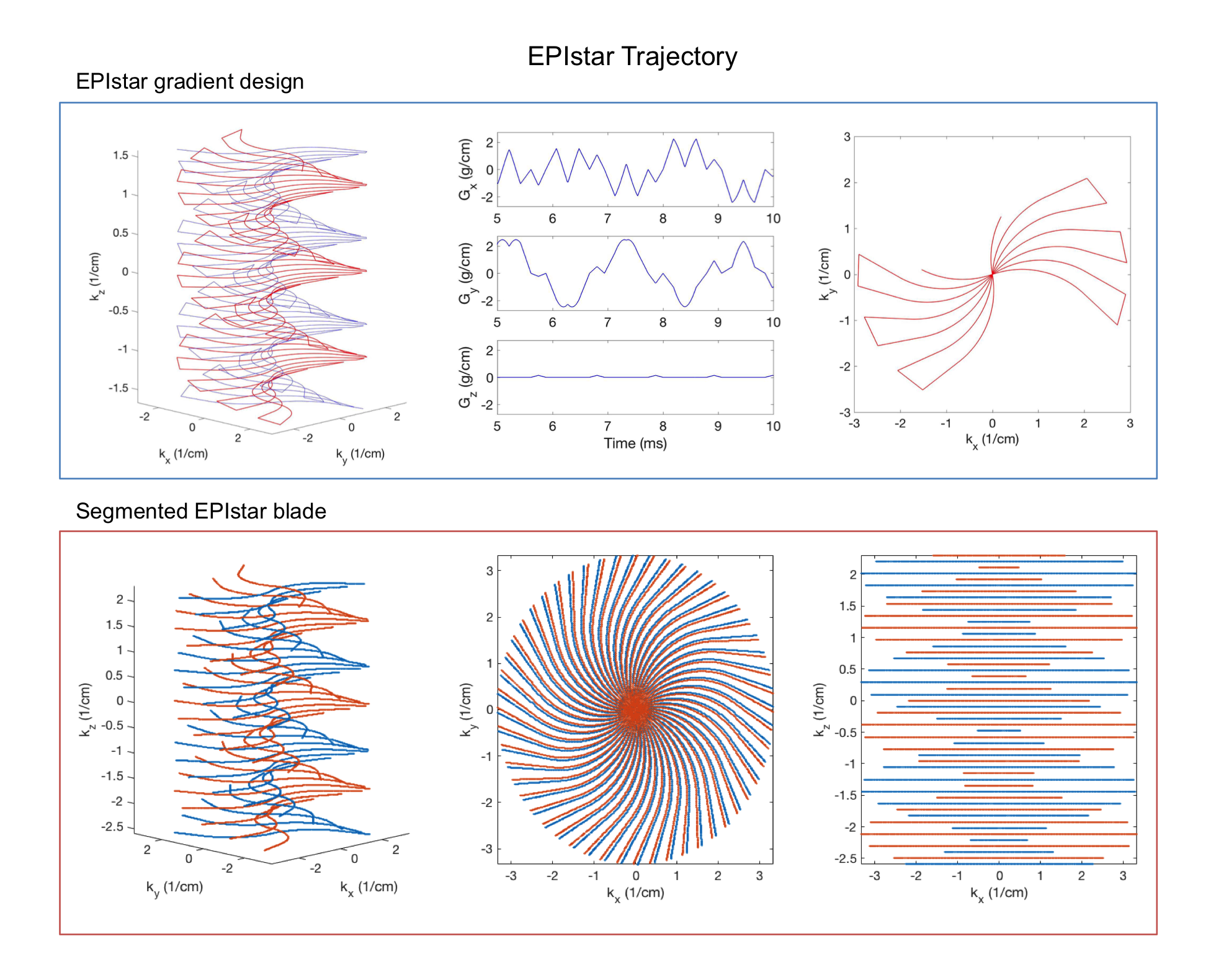
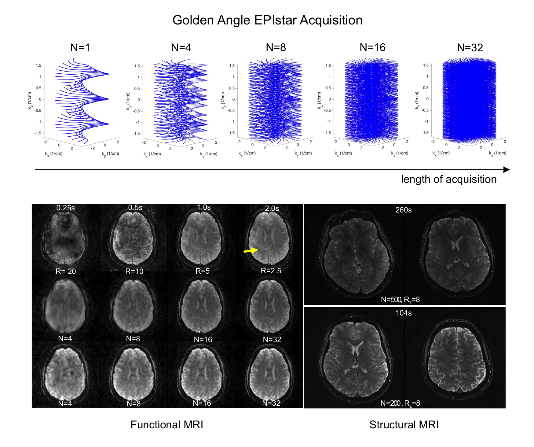
Figure 2. The increasing k-space coverage for an EPIStar acquisition is shown as it is rotated about the golden angle through time(top). The comparison of brain images (1.7x1.7x3mm3) for radial EPI,EPIstar and blipped-CAIPI EPI trajectories is shown below. Images were reconstructed considering increasing numbers of blades and consequently longer acquisition times which was matched by corresponding blipped-CAIPI EPI scans. The strong performance especially for low numbers of blades is demonstrated for EPIstar sampling which leads to a more benign aliasing pattern. High-quality T2*-weighted images are obtained in reasonable time using a segmented EPIstar readout(bottom-right). Structural brain images were obtained at a 0.5x0.5x1mm3(top) and 0.86x0.86x1mm3(bottom) resolution.