4579
Highly Accelerated 3D EPI using Compressed Sensing1University of Erlangen-Nuremberg, Erlangen, Germany, 2Siemens Healthcare GmbH, Erlangen, Germany, 3University of Glasgow, Glasgow, United Kingdom
Synopsis
In previous work, Echo-Planar Imaging (EPI) has been used in combination with a CAIPIRINHA undersampling scheme, as in SMS blipped CAIPI or 3D CAIPI EPI, for highly accelerated BOLD, perfusion and diffusion weighted imaging. In a separate development, Compressed Sensing (CS) was employed in combination with parallel imaging to significantly accelerate a range of non-EPI 3D imaging sequences. In general, this is achieved by using a variable-density randomized sampling scheme which gives aliasing artefacts a noise like appearance. This work explores the use of CS to accelerate 3D EPI acquisitions and demonstrates an improved performance compared to the CAIPIRINHA approach.
Introduction
3D Echo Planar Imaging (EPI) suffers from distortions and signal dropout due to the long echo-train readout (RO). To address these two effects, 3D EPI is often combined with interleaved acquisitions for anatomical T2* imaging1. However, this prolongs the measurement time. A number of acceleration methods exist aiming to reduce the acquisition time. For example, a 3D EPI sequence CAIPIRINHA2 undersampling can be employed3. 3D EPI scan time has also been reduced by using the WAVE-CAIPI4,5 technique, although the reduction in g-factor is limited by the slew rate of the gradient system. Another way to accelerate a 3D acquisition is Compressed Sensing (CS)6 with a randomized variable-density undersampling scheme. This work introduces a highly accelerated 3D EPI sequence that combines interleaving with CS to reduce signal drop out and distortions at short scan times.Methods
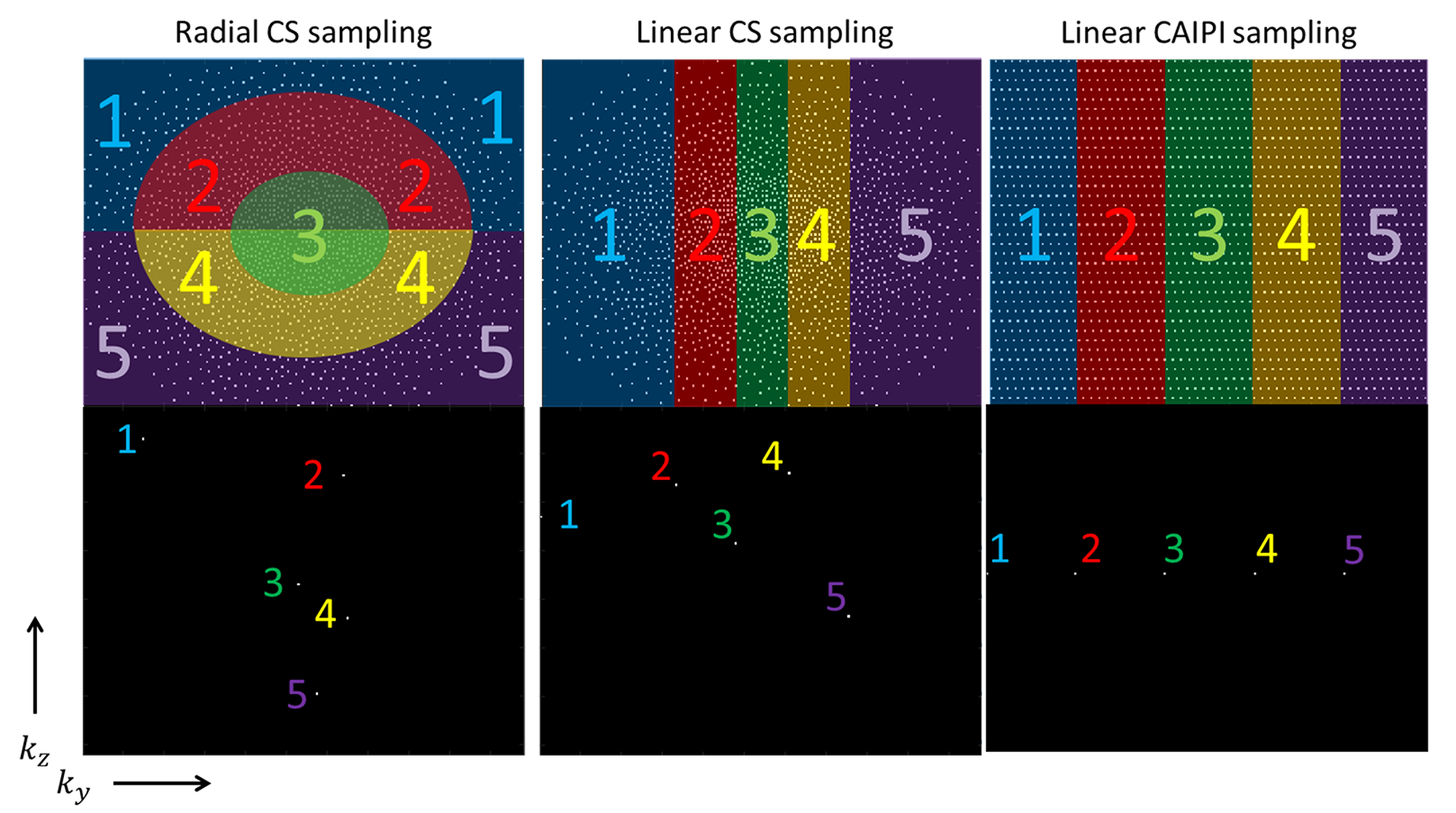
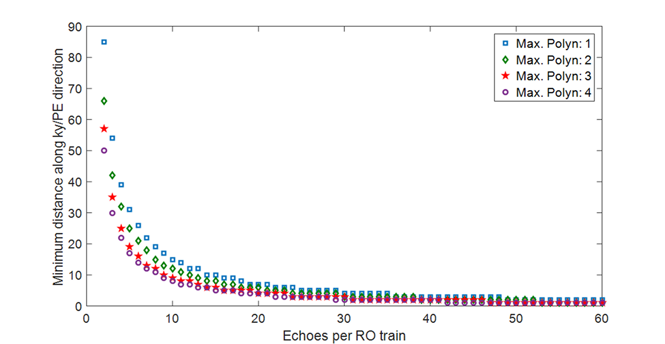
Two ky/kz sampling schemes were investigated: a linear and a radial scheme. In each case, the schemes were designed to provide a set of sample points that corresponded to a variable-density Poisson disk. The different sampling methods are shown in Fig. 1. The size of the blipped gradient pulse is variable, but kept within a certain range. Distortions are defined by the minimum blip size along a given direction. For the linear sampling scheme, the minimum blip size depends on the number of echoes and the density variation (compare Fig. 2). Echo time shifting is applied to all of these sampling schemes7.
The performances of the two sampling schemes were investigated by acquiring a test data set corresponding to fully sampled 3D multi-contrast, gradient-echo images with 1 mm isotropic resolution and 12 equidistant TE values ranging from 2 to 30 ms. Data were acquired on a 7T MAGNETOM Terra system (Siemens Healthcare GmbH, Erlangen, Germany) with a 1Tx32Rx head coil (Nova Medical, Wilmington, MA, USA). Because the test data set included all ky and kz sample points at each echo time, it was possible to test each EPI trajectory retrospectively by selecting, for each raw data point, the appropriate echo position in the EPI echo train.
Results and Discussion
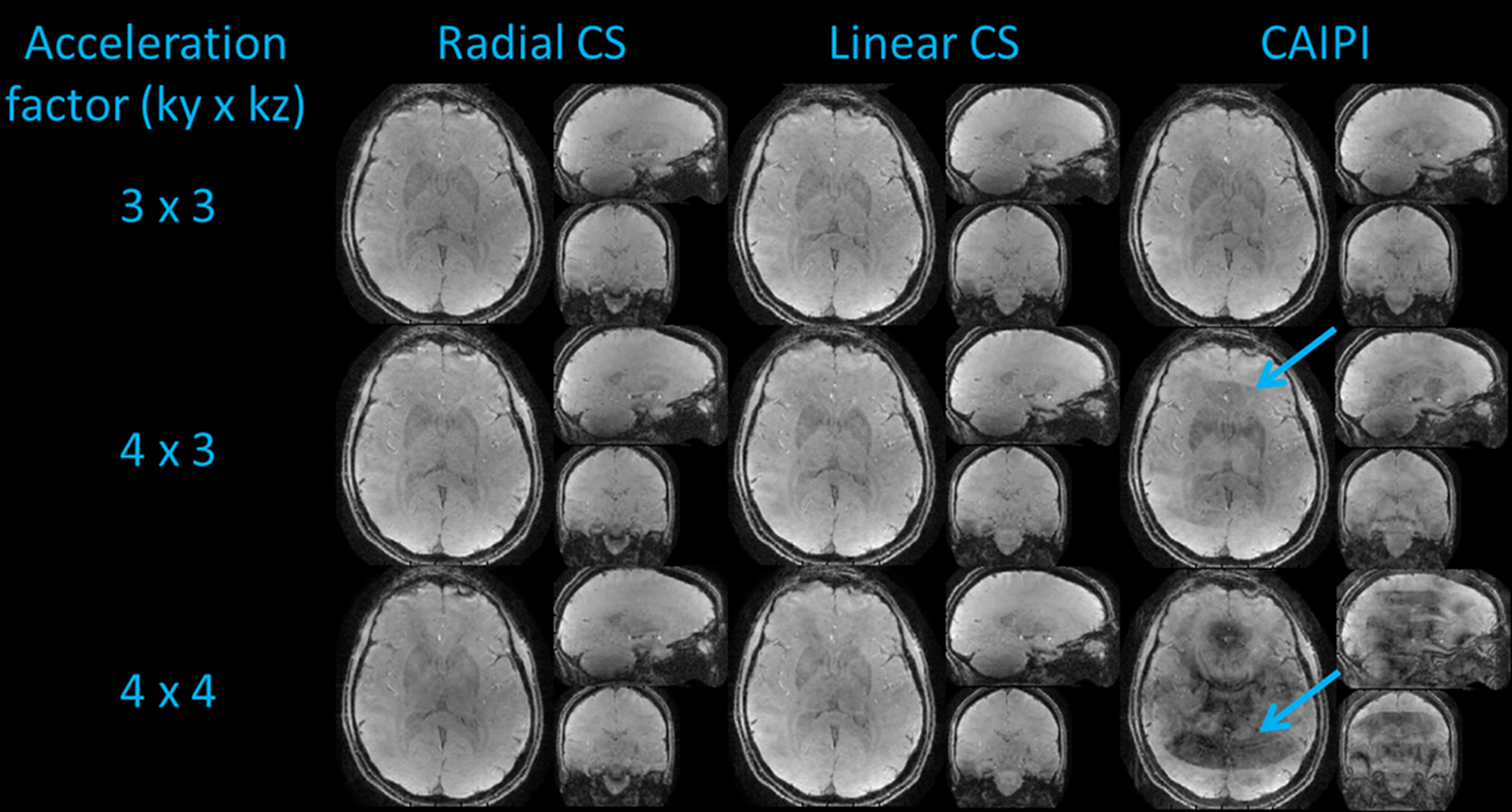
Figure 3 shows the reconstructed images in transverse, sagittal and coronal views. The reconstructed images with CS show fewer artefacts and a higher apparent SNR. The 16-fold accelerated images show minor artefacts with CS, whereas the corresponding CAIPIRINHA approach shows strong aliasing artefacts.
In the linear sampling scheme, distortions only appear along y direction, whereas for the radial scheme distortions appear along both y and z directions. The maximum achievable number of echoes depends on the resolution and the density variation. For linear and radial sampling, 13 echoes were acquired per shot, resulting in a minimum blip size of 9$$$\Delta$$$k (with 1$$$\Delta$$$k as the Nyquist sampling step size). The distortion level of this EPI acquisition corresponds to a single-shot scan with nine-fold acceleration in the y direction. For an echo-spacing of 1 ms the effective echo-spacing is 0.11 ms and consequently results in minimal distortions. The overall image quality between the 9-fold CAIPI undersampling and the CS 16-fold undersampled acquisitions are comparable. Hereby the time to acquire one volume can be reduced from 5 seconds (CAIPIRINHA 9-fold) to 2.8 seconds (CS 16-fold), which is equivalent to a reduction of 43 %.
Conclusion
CS-3D-EPI enables highly accelerated imaging with minimal distortions and short echo trains. It can be integrated on standard MR imaging systems without significant demands on the gradient slew rate. Compared to 3D EPI with a CAIPIRINHA undersampling scheme, CS provides a substantial reduction in the level of artefacts and provides access to higher acceleration factors. For the radial sampling scheme, an alternative kooshball trajectory8 could be investigated in future work. The CS 3D EPI method has a number of potential applications, including multi-echo fMRI, multi-echo SWI and T2*-weighted imaging for anatomical or dynamic perfusion studies. Due to the fact that all k-space points are aligned on a Cartesian grid, fast Cartesian CS algorithms can be employed, allowing reasonable reconstruction times on standard equipment.Acknowledgements
We would like to thank Roosie Woodward and Tracy Hopkins for helping with the volunteer measurements.References
1. Zwanenburg, J. J. M., Versluis, M. J., Luijten, P. R. & Petridou, N. Fast high resolution whole brain T2* weighted imaging using echo planar imaging at 7T. NeuroImage 56, 1902–1907 (2011).
2. Breuer, F. A. et al. Controlled aliasing in parallel imaging results in higher acceleration (CAIPIRINHA) for multi-slice imaging. Magn Reson Med 53, 684–691 (2005).
3. Narsude, M., Gallichan, D., van der Zwaag, W., Gruetter, R. & Marques, J. P. Three-dimensional echo planar imaging with controlled aliasing: A sequence for high temporal resolution functional MRI. Magn Reson Med 75, 2350–2361 (2016).
4. Bilgic, B. et al. Wave-CAIPI for highly accelerated 3D imaging. Magn Reson Med 73, 2152–2162 (2015).
5. Poser, B. A. et al. Echo-planar imaging with wave-CAIPI acquisition and reconstruction. in (2017).
6. Lustig, M., Donoho, D. & Pauly, J. M. Sparse MRI: The application of compressed sensing for rapid MR imaging. Magn Reson Med 58, 1182–1195 (2007).
7. Feinberg, D. A. & Oshio, K. Phase errors in multi-shot echo planar imaging. Magnetic Resonance in Medicine 32, 535–539 (1994).
8. Glover, G. H., Pauly, J. M. & Bradshaw, K. M. Boron-11 imaging with a three-dimensional reconstruction method. J Magn Reson Imaging 2, 47–52 (1992).
9. Uecker, M., Tamir, J., Ong, F., Holme, C. & Lustig, M. Bart: Version 0.4.01. (2017). doi:10.5281/zenodo.817472
Figures