4577
Variable Frequency Wave-encoded 3D Turbo Spin Echo Imaging1Philips Research North America, Cambridge, MA, United States, 2Vascular Imaging Lab, Department of Radiology, University of Washington, Seattle, WA, United States, 3Center for Biomedical Imaging Research, Department of Biomedical Engineering, Tsinghua University, Beijing, China, 4Philips Research Hamburg, Hamburg, Germany
Synopsis
Wave encoding is an emerging approach that can take better usage of the three-dimensional (3D) spatial encoding power of multi-channel coils employed in parallel imaging (PI). In this work, a variable frequency (VF) wave encoding approach is proposed to improve the aliasing propagation property and reduce the side lobe amplitude of the transformed point spread function. This VF approach can also induce amplitude modulated wave encoding gradients to reduce eddy currents and improve the slice selection profile. The preliminary results demonstrated its improved PI performance for 3D turbo spin echo imaging over Cartesian and constant frequency wave encoding schemes.
Introduction
Wave encoding techniques can improve the parallel imaging (PI) performance of three-dimensional (3D) Cartesian encoding techniques by propagating the in-plane subsampled aliasing further along the readout direction1, and have demonstrated their benefits for accelerated volumetric imaging2,3 and simultaneous multi-slice imaging4,5. The wave encoding k-space trajectory is typically designed with constant frequency (CF) sinusoidal waveform and the existing optimization strategy6 focuses mostly on tuning the amplitude and number of cycles, where the larger amplitude can contribute to more improved aliasing spreading effects. However, increasing the gradient amplitude may cause eddy current issue and degrade the slice/slab selection profile. In this study, a variable frequency (VF) wave encoding k-space trajectory is developed to provide an alternative waveform optimization strategy that can reduce the eddy current issue while better distributing the aliasing along the readout direction. The PI reconstruction performance of VF wave encoding scheme is evaluated for 3D turbo spin echo (TSE) imaging and compared with Cartesian and CF wave encoding schemes.Methods
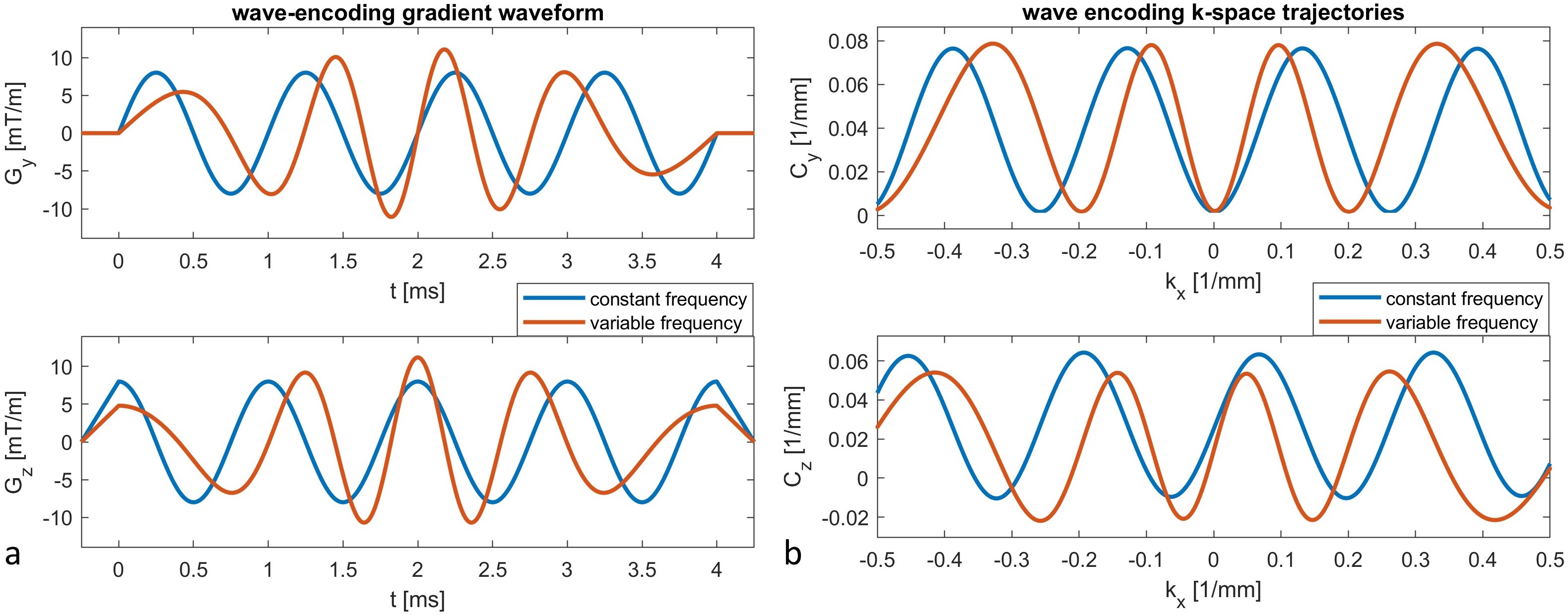
Variable frequency wave encoding gradient waveform design: The instantaneous frequency $$$f(t)$$$ is designed as a CF component modulated by a nonlinear symmetric VF component within the data acquisition time ($$$T_{acq}$$$) such that the corkscrew shaped readout trajectory rotates more frequently at the central k-space while behaving oppositely at the peripheral k-space. So the sampling spacing is varying along the readout direction which can mimic the variable density sampling to reduce the maximum side lobes of transformed point spread function (TPSF). In this work, $$$f(t)=f_c-2{\pi}{\beta}/T_{acq}cos(2{\pi}t/T_{acq}), t\in[0,T_{acq}]$$$ is used, where parameter $$$\beta$$$ can be further optimized the performance and adapted to the hardware constraints including maximum gradient strength (Gmax) and slew rate. The VF wave encoding k-space trajectories can be scaled by a pair of amplitude parameters ($$$A_y$$$ and $$$A_z$$$) and represented as $$$C_y(t)=A_y(1-cos(2{\pi}\int_0^tf(\tau)d\tau))$$$ and $$$C_z(t)=A_zsin(2{\pi}\int_0^tf(\tau)d\tau))$$$, which will also apply an additional amplitude modulation for the wave encoding gradient waveform (figure 1a) to reduce eddy currents and to achieve similar slice/slab selection profiles in comparison to Cartesian encoding scheme.
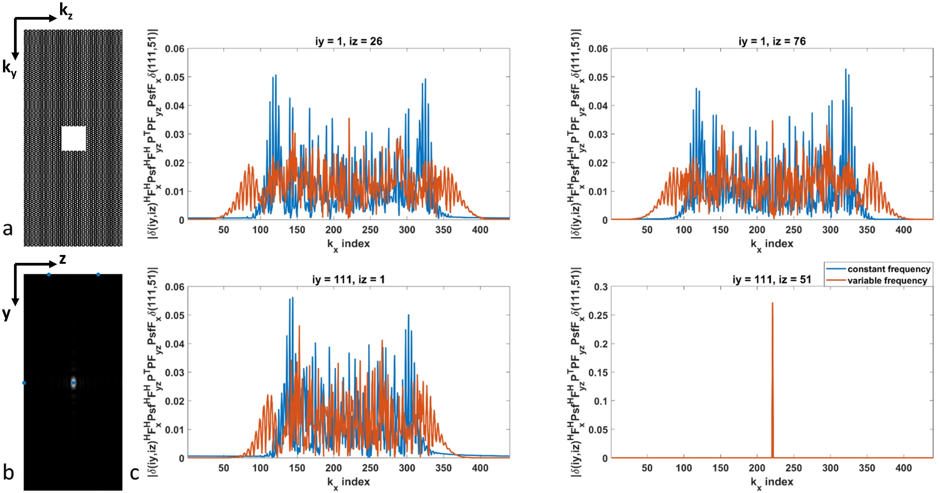
Wave encoding PI reconstruction and TPSF analysis: After CF and VF wave encoding MR scans, the wave point spread function ($$$Psf$$$) and coil sensitivities can be calibrated with the previously developed self-calibration method7 with different subspace models for k-space trajectory representation, and SPIRiT8 based PI method can be performed for image reconstruction. The TPSF between two voxels $$$r$$$ and $$$\rho$$$ defined by $$$TPSF(r,\rho)=\delta_r^HF_x^HPsf^HF_{yz}^HP^TPF_{yz}PsfF_x\delta_{\rho}$$$, where $$$P^TP$$$ stands for the undersampling mask, can be further analyzed and compared between CF and VF wave encoding schemes.
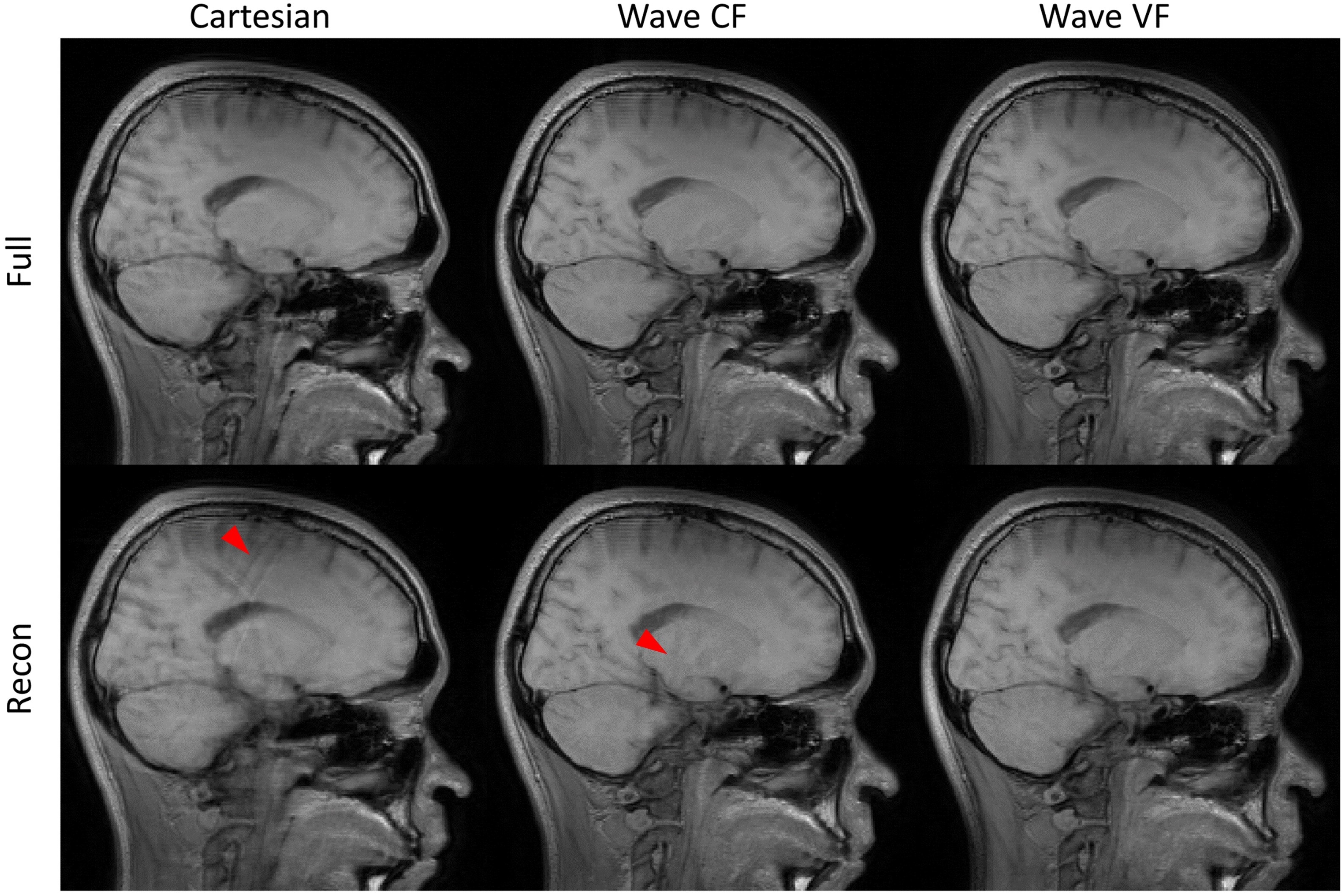
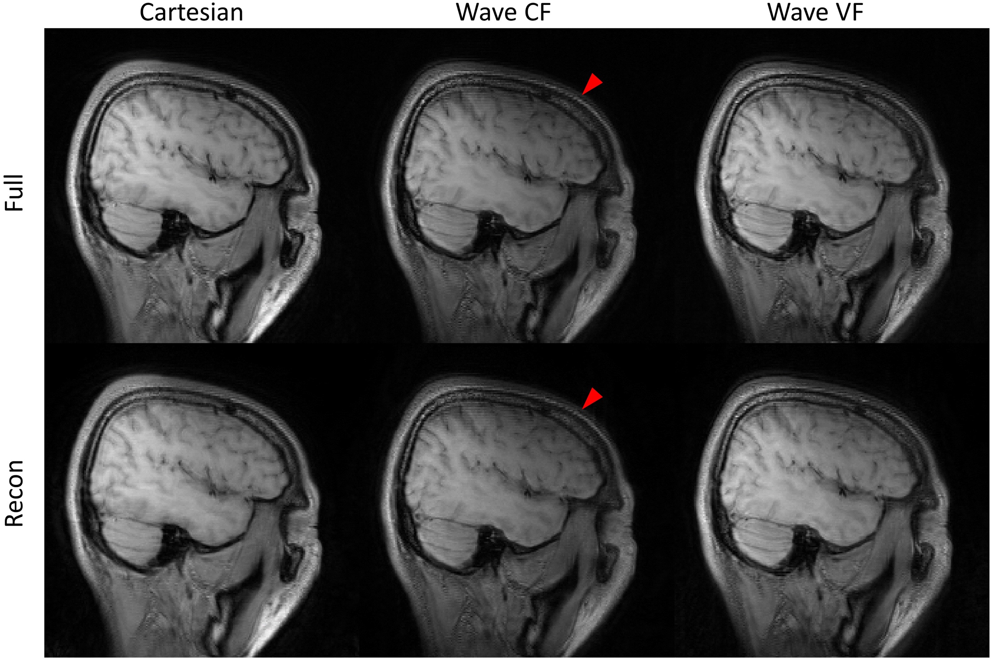
MR experiments: The fully sampled wave encoded (CF & VF) and Cartesian encoded whole brain datasets were acquired by a 15-channel head coil on Philips Ingenia 3.0T scanner using the 3D TSE sequence (FOV = 230x230x198mm3, resolution = 1x1x2mm3, TE/TR = 15ms/800ms, TSE factor = 30) with or without wave encoding gradients (CF: Gmax=8mT/m, 4 cycles; VF: Gmax=11mT/m, 4 cycles). The acquired datasets were retrospectively subsampled with a 25x25 central calibration area and CAIPI undersampling pattern9 in peripheral k-space.
Results
Comparison of TPSF between CF and VF wave encoding: Figure 1b illustrates the calibrated CF and VF wave encoding k-space trajectories and demonstrates the adaptive capacity of previously developed self-calibration method (7). With the calibrated wave encoding k-space trajectories, the simulation results in figure 2 demonstrate that VF wave encoding method can improve the aliasing propagation width along the readout direction and reduce the side lobe amplitudes of TPSF.
Comparison of PI reconstruction performance: For 3x2 CAIPI in vivo brain acceleration experiment, VF wave encoding can provide better aliasing suppression results in both central and lateral slices (figure 3 and 4). In addition, VF wave encoding can significantly reduce the signal loss due to eddy current induced slice profile degradation in lateral slice (figure 4).
Discussion and Conclusion
The VF wave encoding is able to further extend the subsampled aliasing propagation along the readout direction and reduce the side lobe amplitude of the TPSF. Also, the VF wave encoding gradients can be adapted to reduce eddy current effects and thus improve the slice selection profile for 3D TSE imaging by the additionally induced amplitude modulation. With the previously developed wave encoding self-calibration method, VF wave encoding PI reconstruction has demonstrated improved image quality in comparison to Cartesian and CF wave encoding methods.Acknowledgements
This study is partially supported by the grant from National Institutes of Health (5R01NS092207).References
1. Bilgic B, Gagoski BA, Cauley SF, Fan AP, Polimeni JR, Grant PE, Wald LL, Setsompop K. Wave-CAIPI for highly accelerated 3D imaging. Magn Reson Med. 2015;73(6):2152-62.
2. Polak D, Setsompop K, Cauley SF, Gagoski BA, Bhat H, Maier F, Bachert P, Wald LL, Bilgic B. Wave-CAIPI for highly accelerated MP-RAGE imaging. Magn Reson Med. 2018 Jan;79(1):401-406.
3. Chen F, Zhang T, Cheng JY, Shi X, Pauly JM, Vasanawala SS. Autocalibrating motion-corrected wave-encoding for highly accelerated free breathing abdominal MRI. Magn Reson Med. 2017;78(5):1757-1766.
4. Gagoski BA, Bilgic B, Eichner C, Bhat H, Grant PE, Wald LL, Setsompop K. RARE/turbo spin echo imaging with Simultaneous Multislice Wave-CAIPI. Magn Reson Med. 2015;73(3):929-38.
5. Poser BA, Bilgic B, Gagoski BA, Uludag K, Stenger VA, Wald LL, Setsompop K. Echo-Planar Imaging with Wave-CAIPI Acquisition and Reconstruction. ISMRM 2017. p1198.
6. Polak D, Cauley S, Huang S, Longo M, Bilgic B, Raithel E, Wald L, Setsompop K. Highly-accelerated volumetric brain protocol using optimized Wave-CAIPI encoding. ISMRM 2018. p0937.
7. Zhou Z, Yuan C, Börnert P. Self-calibrating wave-encoded 3D turbo spin echo imaging using subspace model based autofocus. ISMRM 2018. p0939.
8. Lustig M, Pauly JM. SPIRiT: Iterative self-consistent parallel imaging reconstruction from arbitrary k-space. Magn Reson Med. 2010;64(2):457-71.
9. Breuer FA, Blaimer M, Mueller MF, Seiberlich N, Heidemann RM, Griswold MA, Jakob PM. Controlled aliasing in volumetric parallel imaging (2D CAIPIRINHA). Magn Reson Med. 2006;55(3):549-56.
Figures