4576
Silent T2* Imaging on 7T using ZTE Combined with Gradient-Echo BURST1GE Healthcare, Munich, Germany, 2IRCCS Stella Maris Foundation and IMAGO7, Pisa, Italy
Synopsis
ZTE combined with gradient-echo BURST enables silent 3D radial T2* imaging. It was implemented on 7T and T2* weighted images were acquired with isotropic resolutions of 1-3mm. From the series with different echo times, both phase and T2* maps were extracted.
Introduction
Zero Echo-Time (ZTE) imaging is a silent encoding method to acquire predominantly proton-density weighted images [1,2,3]. Combining ZTE imaging with BURST [4] allows to recall gradient echoes exhibiting T2* decay [5]. Goal of this work was to implement this novel imaging sequence on 7T and explore potential applications.Methods
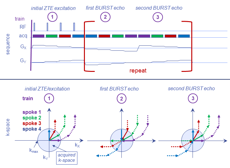
The ZTE-BURST sequence recalling gradient echoes is depicted in Fig. 1. After an initial ZTE train acquiring the FID, the gradients are reversed in both time and amplitude, while RF is switched off, hence recalling gradient-echoes. Another BURST echo can be acquired by playing out the same trajectory as during the initial ZTE/FID train. Further repetition of the two BURST trains recalls further echoes at later echo times.
Data was reconstructed by first applying a few corrections, mainly adjusting the gradient delay and a zero-order phase correction extracted from the centre of k-space from the BURST echoes of the 3D radial spokes as explained in [5]. This was followed by gridding the spokes onto a 3D Cartesian grid, and 3D Fourier transformation. Everything was implemented in Matlab.
Images were acquired in the brain of a healthy volunteer on a 7T whole-body MRI (GE Healthcare, Milwaukee, WI, USA) using a 32-channel head receive coil combined with a quadrature-mode birdcage transmit coil (Nova Medical, Wilmington, MA, USA).
Results and Discussion
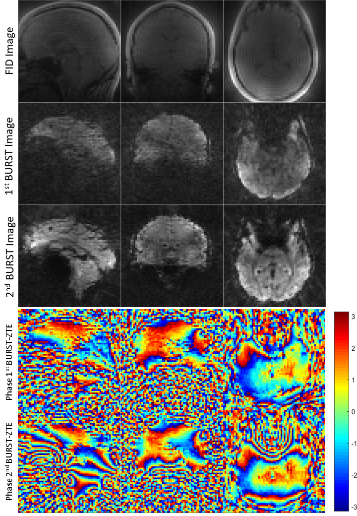
Figures 2-4 show T2* ZTE-BURST images acquired at 7T at different resolutions. Phase maps were extracted from the 3mm isotropic resolution images in Fig. 2. Background phase was removed by subtracting the phase of the ZTE-FID images from the phase of the BURST images, hence exhibiting a phase evolution purely due to magnetic field variations.
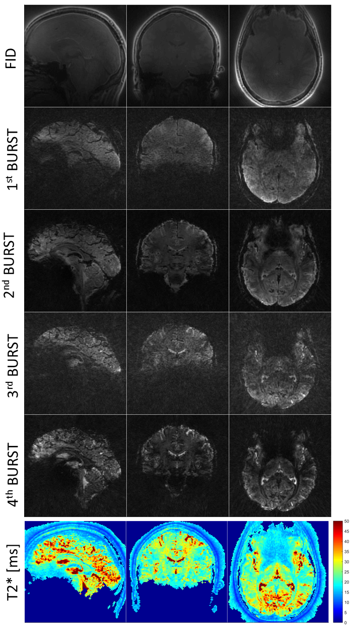
T2* maps were extracted from the 1.5mm isotropic resolution images in Fig. 3 by fitting the images from different echo times to an exponential decay. Even BURST echoes exhibit a good image quality, slightly better than the odd BURST echoes, which could be due to inconsistent echo times for the different spokes in the odd BURST echoes.
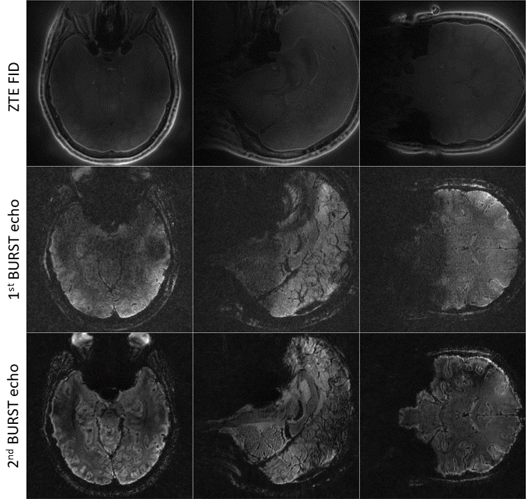
High resolution T2* weighted images with an isotropic resolution of 1mm are shown in Fig. 4. Image quality enables visualisation of microstructures unique to imaging at 7T.
Conclusion
Combining ZTE with gradient-echo BURST is a silent and elegant method to encode T2* contrast, yielding good image quality.Acknowledgements
Partially funded by EU Horizon 2020 grant #801075 NICI.References
[1] Fast imaging in liquids and solids with the back-projection low angle shot (BLAST) technique. Hafner S. Magn Reson Imaging 1994;12:1047-1051.
[2] Ultra-fast imaging using low flip angles and fids. Madio DP, Lowe IJ. Magn Reson Med 1995;34:525–529.
[3] Density of organic matrix of native mineralized bone measured by water- and fat-suppressed proton projection MRI. Wu Y, Ackerman JL, Chesler DA, Graham L, Wang Y, Glimcher MJ. Magn Reson Med. 2003;50:59-68.
[4] Burst imaging. Hennig J, Hodapp M. MAGMA. 1993; 1:39-48.
[5] Silent T2* and T2 Encoding using ZTE Combined with BURST. Schulte RF, Buonincontri G, Costagli M, Menini A, Wiesinger F, Solana AB. Magn Reson Med. In press.
Figures