4575
Spin-Echo ZTE-BURST for Quiet T2-Weighted Imaging1GE Healthcare, Munich, Germany
Synopsis
ZTE was combined with single spin-echo BURST encoding for acquiring T2 weighted images in a relatively quiet manner.
Introduction
ZTE is a silent acquisition method, yielding predominantly proton density weighted images [1,2,3]. Combining ZTE with BURST [4] can potentially yield images with the clinically more relevant T2 contrast [5]. In the initial spin-echo ZTE-BURST implementation, double spin-echoes were required to compensate for the unknown phase [5], resulting in relatively long echo times and overall acquisition durations. In this work, a single echo BURST acquisition and reconstruction led to significant reduction of scan time and clinically useful TEs.Methods
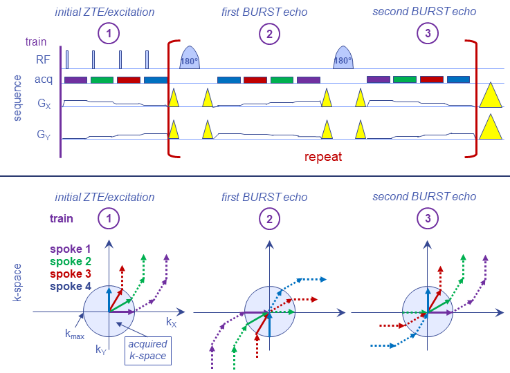
The spin-echo ZTE-BURST sequence is shown and explained in Fig. 1. Similar to the gradient-echo version [5], the magnetisation of the individual excitations is stored in k-space. Main difference is that gradients of odd BURST echoes do not have to be inverted in amplitude, but only in time, because the 180° pulse mirrors the positions of stored magnetisation in k-space. For the 180° pulses, optimal adiabatic full passage pulses were used [6], in order to provide robustness against B1+ variations.
For the reconstruction, the 3D radially sampled data was pre-processed by adjusting the gradient delay, applying a zero-order phase correction extracted from the centre of k-space and shifting raw data to iso-centre. This was followed by interpolating the spokes onto a 3D grid and Fourier transformation. The reconstruction was implemented in Matlab.
Experiments were performed on a 3T whole-body MRI (MR750w, GE Healthcare, Milwaukee, WI, USA) in the heads of healthy volunteers.
Results and Discussion
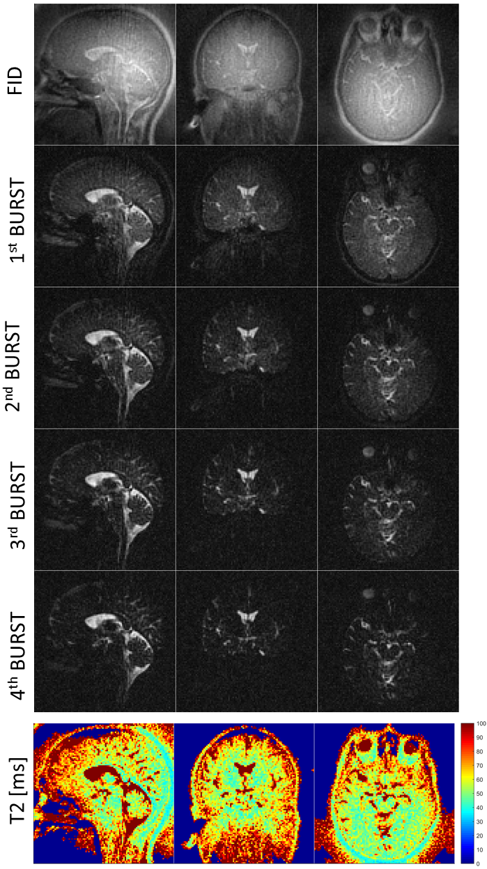
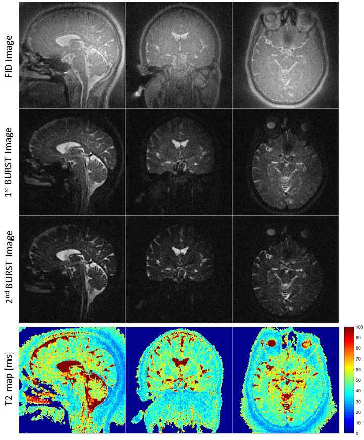
Figures 1 and 2 show exemplary spin-echo ZTE-BURST images for 2mm and 1.5mm isotropic resolution, respectively. The corresponding T2 maps were extracted by fitting the images acquired at the different echo times to an exponential decay. The T2-weighted images exhibit a mixed contrast, because magnetisation is only partially excited into the transverse plane, while a considerable about of magnetisation is constantly flipped to ±Mz, hence imposing some T1 weighting.
The SAR was within limits, but at the higher end of the range. Reducing SAR is possible by increasing the recovery time at the end of each ZTE/BURST segment at the expense of increasing overall acquisition duration. However, this would also increase SNR and reduce T1 weighting.
The spin-echo ZTE-BURST is relatively quiet, but not completely silent, due to the necessity of including crusher gradients. Maximum gradient strength and slewrate were derated by a factor of 1.5 and 5, respectively, hence the resulting sequence is much quieter than traditional spin-echo sequences.
Conclusion
It is possible to acquire good quality T2 weighted images in reasonable scan times using the single spin-echo ZTE-BURST sequence.Acknowledgements
No acknowledgement found.References
[1] Fast imaging in liquids and solids with the back-projection low angle shot (BLAST) technique. Hafner S. Magn Reson Imaging 1994;12:1047-1051.
[2] Ultra-fast imaging using low flip angles and fids. Madio DP, Lowe IJ. Magn Reson Med 1995;34:525–529.
[3] Density of organic matrix of native mineralized bone measured by water- and fat-suppressed proton projection MRI. Wu Y, Ackerman JL, Chesler DA, Graham L, Wang Y, Glimcher MJ. Magn Reson Med. 2003;50:59-68.
[4] Burst imaging. Hennig J, Hodapp M. MAGMA. 1993; 1:39-48.
[5] Silent T2* and T2 Encoding using ZTE Combined with BURST. Schulte RF, Buonincontri G, Costagli M, Menini A, Wiesinger F, Solana AB. Magn Reson Med. In press.
Figures