4572
Rapid High Resolution T1-Weighted Hippocampus Imaging with Yarn-Ball Acquisition1University of Alberta, Edmonton, AB, Canada
Synopsis
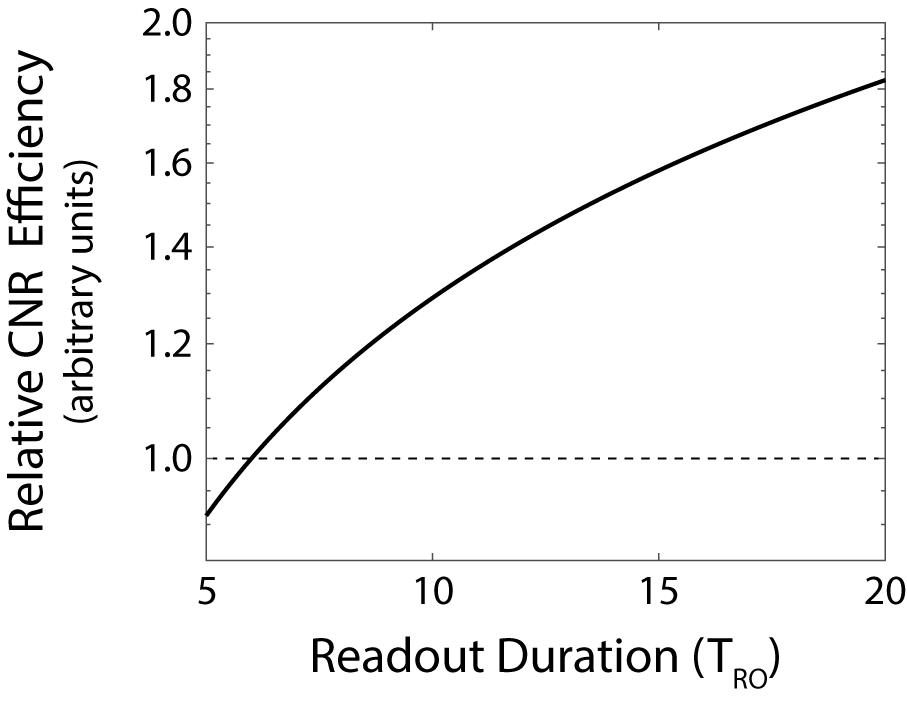
For spoiled steady-state T1-weighted imaging, readout duration (TRO) and repetition time (TR) increase result in greater contrast-to-noise ratio (CNR) efficiency. Novel 3D-twisting Yarnball acquisition realizes this advantage without scan-time penalty (more of k-space sampled with increased TRO), but increased TRO results in greater point-spread-function smearing. Following TRO optimization, Yarnball is used to produce whole-brain 0.36x0.36x1.08 mm3 coronal (defined by 1/(2kmax)) images in 10 minutes (with 2 averages). Compared to 3D-MP-RAGE (same scan time and voxel volume) Yarnball images have greater resolution and grey-white CNR, facilitating sharper depiction of internal hippocampus architecture.
Introduction
High resolution imaging is required to distinguish hippocampal architecture, but this can be time consuming. Here, novel 3D-twisting Yarnball1 is considered as a T1-weighted k-space acquisition alternative to previously considered 3D-MP-RAGE with voxels of 0.38x0.38x0.8 mm3 and 0.6x0.6x0.6 mm3 and long scan times of 37 minutes2 and 33 minutes3 respectively at 3T. Following readout duration (TRO) optimization for contrast-to-noise ratio (CNR) and resolution, 0.36x0.36x1.08 mm3 (defined by 1/(2kmax)) Yarnball was compared to 0.45x0.45x1.35 mm3 MP-RAGE (1/kvolume=0.27 mm3 for both) for patient friendly 10 minute scanning of the whole brain at 3T.Methods
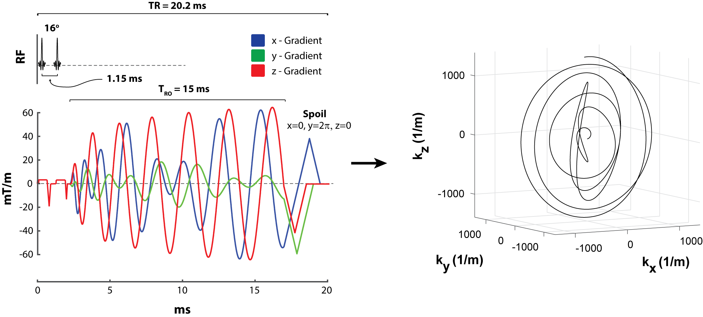
Simple T1-based spoiled steady-state simulation suggests grey-white CNR efficiency increase with longer TRO and TR (Figure 1), but while longer TRO and TR are constrained by scan duration for GRE, that is not the case for Yarnball1 (see Figure 2 for an example gradient waveform and k-space trajectory) which exhibits even greater sampling efficiency with longer readouts (more of k-space sampled per acquisition). For Yarnball, TRO and TR can be arbitrarily increased to maximize CNR efficiency without scan time penalty. However, longer readouts result in greater point-spread-function (PSF) smearing, a consequence of both T2* decay and off-resonance.
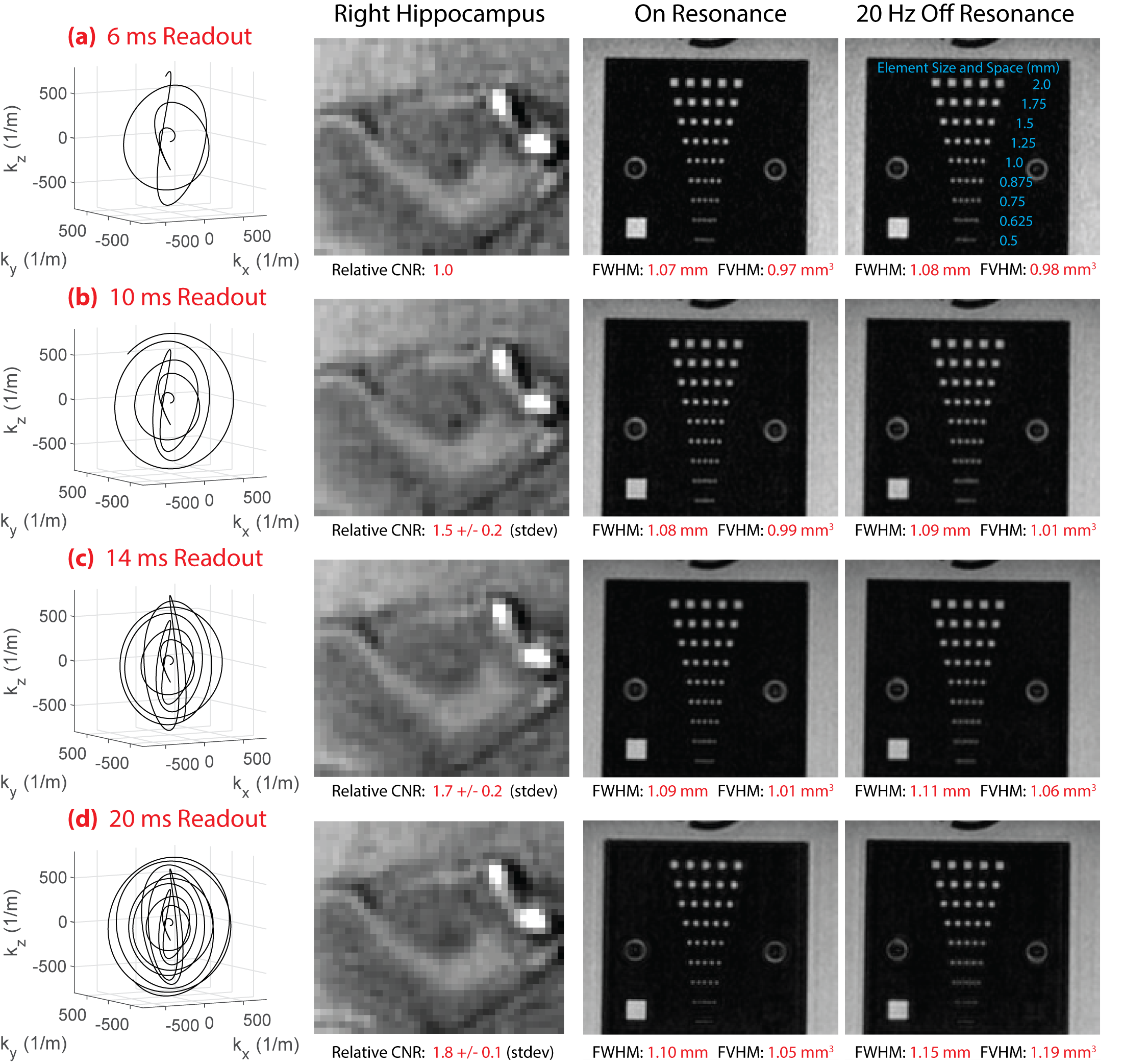
To aid TRO selection for high-resolution hippocampus imaging, TRO = 6, 10, 14, and 20 ms Yarnball waveforms fully sampling the human head were tested with TR = 11, 16, 20, and 25 ms, prescribed flip-angle = 12o, 14o, 16o, and 18o, and number of trajectories = 9537, 6694, 5211, and 4260. In each case voxels were 0.63x0.63x0.95 mm3 coronal (defined by 1/(2kmax)) and scan time was 1:45 minutes. Images were acquired on a Siemens 3T Prisma (64-channel coil) from three healthy volunteers (41, 24, 23 years). All images included 1-1 water excitation and were reconstructed with a β = 2 Kaiser filter. Relative (between scan) CNR was assessed between the inferior longitudinal fasciculus and the collateral sulcus. Images were also acquired from a resolution phantom on-resonance and 20 Hz off-resonance (selected from Bo mapping observation in the hippocampus). Further analysis included calculation of the PSF Full-Width and Full-Volume Half-Max (FWHM/FVHM) with T2*=45 ms both on-resonance and 20 Hz off-resonance.
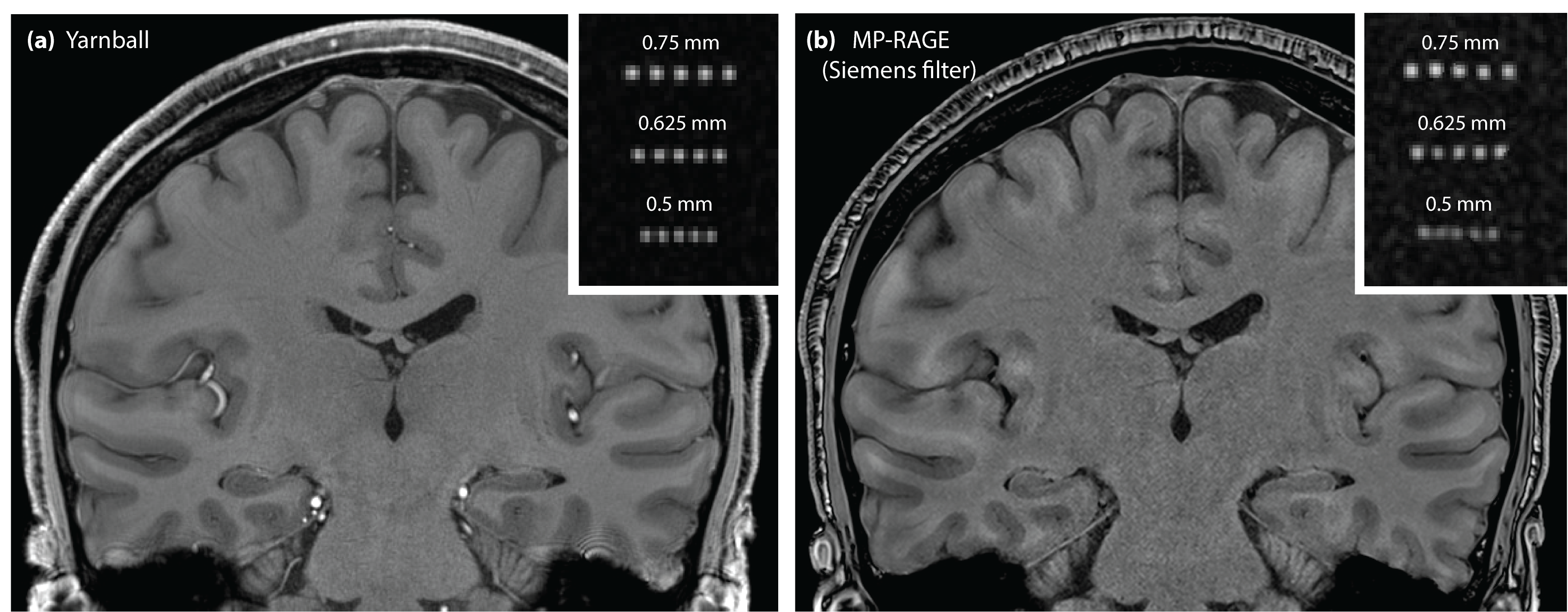
From the experiment above (and explained in Results), TRO=15 ms was selected for testing high-resolution 3D-T1-weighted Yarnball imaging in vivo (Figure 2 pulse sequence). With TR=20 ms, a total of 15801 trajectories provided full head sampling for 0.36x0.36x1.08 mm3 (coronal) voxels in only 5:19 min. Two sequential images were acquired and realigned before averaging to reduce motion-related smearing (total 10:38 min). Note that 1/kvolume=0.27 mm3. Voxel anisotropy was chosen to aid in-plane hippocampus architecture detection. For comparison, 0.45x0.45x1.35=0.27 mm3 3D-MP-RAGE was acquired coronally in 10:51 minutes: FoV=216x183x238 mm3, matrix=406x480x176, BW=200 Hz/px, flip-angle=11o, TI=810 ms, TR=1600 ms, no under-sampling. Sequence parameters were selected from T1-based simulation, and images with and without Siemens (medium, edge-enhancement 3, smoothing 2) filtering compared. High resolution images were acquired from a male volunteer (25 years).
Results
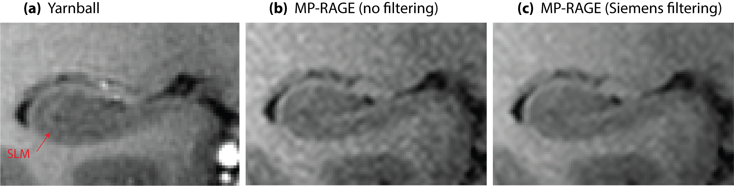
Representative human brain (zoomed-in on hippocampus body) and resolution phantom images are shown in Figure 3 for the four different readout durations. Grey-white CNR increases 80% from TRO=6 ms to 20 ms, similar to simulation (Figure 1). For on-resonance, 0.75 mm resolution element distinction remains similar in the phantom from TRO=6 ms to 20 ms. This is predicted from calculated FWHM and FVHM measures that increase by only 2% and 7%, respectively, over this TRO range. However, resolution element distinction is worsened to 1.0 mm for TRO=20 ms when 20 Hz off-resonance; calculated FWHM and FVHM measures increase by 6% and 22% respectively over TRO=6 ms. Given the detrimental effect of off-resonance, TRO was limited to 15 ms for high-resolution imaging. Yarnball offers both higher CNR and superior resolution element distinction when compared with MP-RAGE (Figure 4). Zoomed-in slices of the right hippocampus more clearly depict the stratum lacunosum-moleculare (SLM) on Yarnball than either unfiltered (very noisy) or Siemens-filtered MP-RAGE images (Figure 5). Note that the SLM is often used to help define hippocampal subfields4.Discussion and Conclusion
Efficient Yarnball acquisition facilitates the use of longer TRO and TR for greater steady-state T1-weighted CNR efficiency without increasing scan duration. However, practical TRO is limited by off-resonance related PSF smearing. Robust shimming over the hippocampus may improve Yarnball utility in this regard. Comparing patient friendly 10 minute scans, Yarnball provides superior images of the hippocampus in terms of both CNR and resolution and enables greater internal hippocampus architecture distinction than MP-RAGE.Acknowledgements
Canadian Institutes of Health ResearchReferences
1. Stobbe RW, Beaulieu C. Rapid 3D spoiled steady-state imaging with yarn-ball acquisition. ISMRM, Abstract 2442. 2015 (Toronto).
2. Van Leemput K, Bakkour A, et al. Model-based segmentation of hippocampal subfields in ultra-high resolution in vivo MRI. Med Image Comput Comput Assist Interv. 2008;11(Pt1):235-243.
3. Kaluga-Yoskovitz J, Bernhardt BC. Multi-contrast submillimetric 3 Tesla hippocampal subfield segmentation protocol and dataset. Sci Data. 2015;2:150059.
4. Steve TA, Yasuda CL et al. Development of a histologically validated segmentation protocol for the hippocampal body. Neuroimage. 2017;157:219-232.
5. Stobbe RW, Beaulieu C. Assessment of averaging spatially correlated noise for 3D radial imaging. IEEE Trans Med Imag. 2011;30(7):1381-1390.
Figures