4570
Predicting pathological subtypes and stages of thymic epithelial tumors using DWI: value of combining ADC and texture parameters1Tangdu Hospital, Department of Radiology,Fourth Military Medical University, Xi’an, China, 2GE Healthcare China, Xi'an, China
Synopsis
To explore the value of combining apparent diffusion coefficients (ADC) and texture parameters from diffusion-weighted imaging (DWI) in predicting the pathological subtypes and stages of thymic epithelial tumors (TETs). In this study, Fifty-seven patients with TETs confirmed by pathological analysis were retrospectively enrolled. The results showed combination of ADC and DWI texture parameters improved the differentiating ability of TET grades, which could potentially be useful in clinical practice regarding the TETs evaluation before treatment.
Introduction
Methods
This retrospective single-center study was approved by the local Ethics Committee, and informed consent was waived. Fifty-seven patients with TETs confirmed by pathological analysis were retrospectively enrolled. ADC values and optimal texture feature parameters were compared for differences among low risk thymoma (LRT), high risk thymoma (HRT) and thymic carcinoma (TC) by one-way ANOVA, and between early and advanced stage of TETs were tested using the independent sample t test. Receiver operating characteristic (ROC) curve analysis was performed to determine the differentiating efficacy.Results
The ADC values in LRT and HRT were significant higher than the value in TC (P = 0.004 and 0.001, respectively), also in early stage were significant higher than ones in advanced stage of TETs (P < 0.001). Among all texture parameters analyzed in order to differentiate LRT from HRT and TC, the V312 achieved higher diagnostic efficacy with an AUC of 0.875, and combination of ADC and V312 achieved the highest diagnostic efficacy with an AUC of 0.933, for differentiating the LRT from HRT and TC. Furthermore, combination of ADC and V1030 achieved a relatively high differentiating ability with an AUC of 0.772, for differentiating early from advanced stages of TETs.Conclusion
Combination of ADC and DWI texture parameters improved the differentiating ability of TET grades, which could potentially be useful in clinical practice regarding the TETs evaluation before treatment.Discussion
As a crucial big data source for the mining of large information, digital medical images are routinely acquired for almost every patient with tumor, and texture analysis is rapidly becoming a noninvasive means of lesion characterization and classification for improved decision support [7] . In this study, the results showed that several DWI texture parameters were significantly different among various subtypes or stages of TETs, which could potentially be useful in clinical practice regarding the TETs evaluation before treatment.Acknowledgements
We would like to thank Dr. Xiao-cheng Wei in GE Healthcare China for providing technical support regarding the application of Analysis-Kit software. This work was supported by the Science and Technology Innovation Development Foundation of Tangdu Hospital (No. 2017LCYJ004).References
1. Engels EA. Epidemiology of thymoma and associated malignancies. Journal of thoracic oncology : official publication of the International Association for the Study of Lung Cancer 2010;5(10 Suppl 4):S260-265.
2. Weis CA, Yao X, Deng Y, et al. The impact of thymoma histotype on prognosis in a worldwide database. Journal of thoracic oncology : official publication of the International Association for the Study of Lung Cancer 2015;10(2):367-372.
3. Ried M, Marx A, Gotz A, Hamer O, Schalke B, Hofmann HS. State of the art: diagnostic tools and innovative therapies for treatment of advanced thymoma and thymic carcinoma. European journal of cardio-thoracic surgery : official journal of the European Association for Cardio-thoracic Surgery 2016;49(6):1545-1552.
4. Priola AM, Priola SM, Giraudo MT, et al. Diffusion-weighted magnetic resonance imaging of thymoma: ability of the Apparent Diffusion Coefficient in predicting the World Health Organization (WHO) classification and the Masaoka-Koga staging system and its prognostic significance on disease-free survival. Eur Radiol 2015;26(7):2126-2138.
5. Asselin MC, O'Connor JP, Boellaard R, Thacker NA, Jackson A. Quantifying heterogeneity in human tumours using MRI and PET. European journal of cancer 2012;48(4):447-455.
6. Choi MH, Lee YJ, Yoon SB, Choi JI, Jung SE, Rha SE. MRI of pancreatic ductal adenocarcinoma: texture analysis of T2-weighted images for predicting long-term outcome. Abdominal radiology 2018.
7.
Hoang UN, Mojdeh Mirmomen S, Meirelles O et
al (2018) Assessment of multiphasic contrast-enhanced MR textures in
differentiating small renal mass subtypes. Abdom Radiol (NY).
10.1007/s00261-018-1625-x
.
Figures
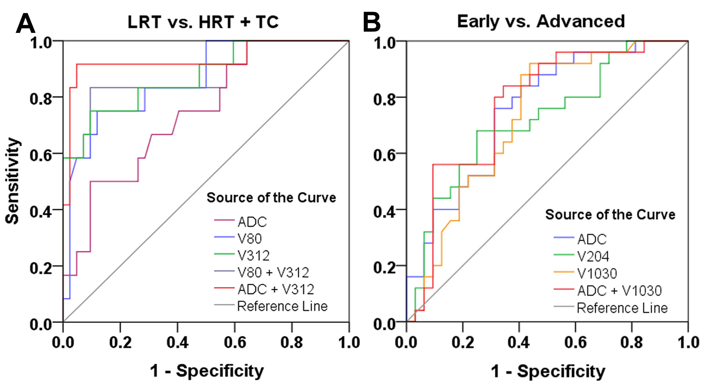
Receiver operating characteristic curves for the differentiating performance of the ADC and DWI texture parameters among the defined groups of thymic epithelial tumors based on the WHO classification and Masaoka stage. (A) LRT vs. HRT and TC with the ADC, V80 and V312 value; (B) Early vs. advanced stage with the ADC, V204 and V1030 value.
Note: LRT = low-risk thymoma; HRT = high-risk thymoma; TC = thymic carcinoma; ADC = apparent diffusion coefficient; V80 = Cluster Shade_angle90_offset1; V312= Cluster Shade_angle45_offset6; V204 = GLCM Entropy_angle0_offset4; V1030 = Maximum 3D Diameter