4565
Accelerating Bi-exponential T1ρ mapping using SCOPE1Paul C. Lauterbur Research Center for Biomedical Imaging, Shenzhen Institutes of Advanced Technology, Chinese Academy of Sciences, Shenzhen, China, 2Department of Imaging and Interventional Radiology, The Chinese University of Hong Kong, Shatin, Hong Kong, China, 3Research center for Medical AI, Shenzhen Institutes of Advanced Technology, Chinese Academy of Sciences, Shenzhen, China
Synopsis
Mono-exponential T1ρ mapping requires 4 or 5 T1ρ-weighted images with different spin lock times (TSLs) to obtain the T1ρ maps, while bi-exponential T1ρ mapping requires a larger number of TSLs, which further prolongs the acquisition time. In this work, we develop a variable acceleration rate undersampling strategy to reduce the total scan time. A signal compensation strategy with low-rank plus sparse model was used to reconstruct the T1ρ-weighted images. We provide the reconstructed images and the estimated T1ρ maps at an acceleration factor up to 6.1 in fast bi-exponential T1ρ mapping.
INTRODUCTION
T1ρ relaxation is normally described by a mono-exponential model1-5. However, compartmentation of tissues may lead to bi-exponential or multi-exponential T1ρ relaxation behavior in certain tissues. Several previous studies have reported that bi-exponential T1ρ relaxation can potentially provide more information than a mono-exponential relaxation6-9. However,bi-exponential T1ρ mapping requires a larger number of spin-lock times (TSLs), which further prolongs the acquisition time. Compressed sensing has shown significant performance in fast quantitative T1ρ mapping10-14. In this work, we extend our previous fast T1ρ mapping method (SCOPE)14 to bi-exponential model, referred to as bio-SCOPE.METHODS
In bi-exponential T1ρ mapping,the T1ρ parameters can be estimated using the bi-exponential model:
$$M=M_0{((1-\alpha)\exp{(-TSL_k/T_{1\rho s})}+\alpha\exp{(-TSL_k/T_{1\rho l})})}_{k=1,2,...,N}\ \ \ \ \ \ \ \ \ \ \ \ \ [1]$$where M is the image intensity obtained at varying TSLs;M0 is the baseline image intensity ;TSLk is the kth spin-lock time;α is the fraction of long relaxation component;T1ρs and T1ρl denote the short and long bi-exponential T1ρ relaxation times; N is the total TSL number.
The reconstruction model can be expressed as follows:
$$min{||L||_*}+\lambda||S||_1 \ \ \ \ s.t.\ \ C(X)=L+S,E(X)=d\ \ \ \ \ \ \ \ [2]$$
where $$$||L||_*$$$ is the nuclear norm of the low-rank matrix L;$$$||S||_1$$$is the $$$\ell_1$$$-norm of the sparse matrix S; X is the image series; λ is a regularization parameter; d is the undersampled k-space data; C(∙) performs pixel-wise signal compensation; E is the encoding operator15,16.Here, the compensation coefficient for signal compensation is calculated by:
$$Coef=1/{((1-\alpha)\exp{(-TSL_k/T_{1\rho s})}+\alpha\exp{(-TSL_k/T_{1\rho l})})}_{k=1,2,...,N}\ \ \ \ \ \ \ \ \ \ \ \ \ [3]$$
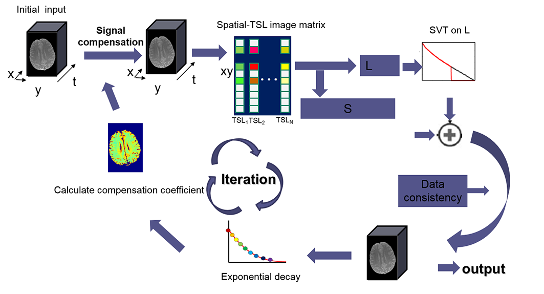
The solving strategy is shown in Figure 1. The image series is first compensated by an initial compensation coefficient calculated from the T1ρ maps estimated from the fully sampled central k-space. Iterative hard thresholding of the singular values of L and a modified soft-thresholding of the entries of S are used to solve the optimization problem in Eq. [2]. T1ρ-weighted images are reconstructed using L+S followed by data consistency. New T1ρ maps are estimated from the reconstructed images using the bi-exponential model described in Eq.[1], and then used to update the compensation coefficient. The reconstruction and signal compensation coefficient updating steps are repeated alternately until convergence.
Evaluation
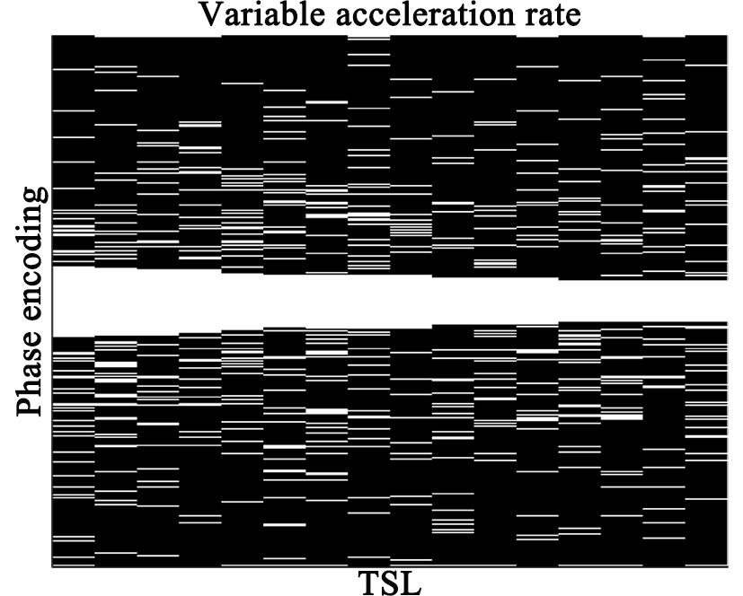
All MR data were acquired on a 3T scanner (Trio, SIEMENS, Germany) using a twelve-channel head coil. Brain T1ρ mapping datasets were acquired from a healthy volunteer (male, age 26, IRB proved, written informed consent obtained) using a spin-lock embedded turbo spin-echo (TSE) sequence. Imaging parameters were: TR/TE=4000ms/9ms, spin-lock frequency 500 Hz, echo train length 16, FOV=230 mm2, matrix size =384 × 384, slice thickness 5 mm, and 16 T1ρ-weighted images were acquired with TSLs =1, 2, 4, 6, 8, 10, 12, 15, 20, 25, 30, 40, 50, 60, 70, and 80 ms. The acquired data was retrospectively undersampled with a variable rate undersampling scheme (shown in Figure 2). T1ρ-weighted images were reconstructed by bio-SCOPE and L+S methods15.
RESULTS and DISCUSSION
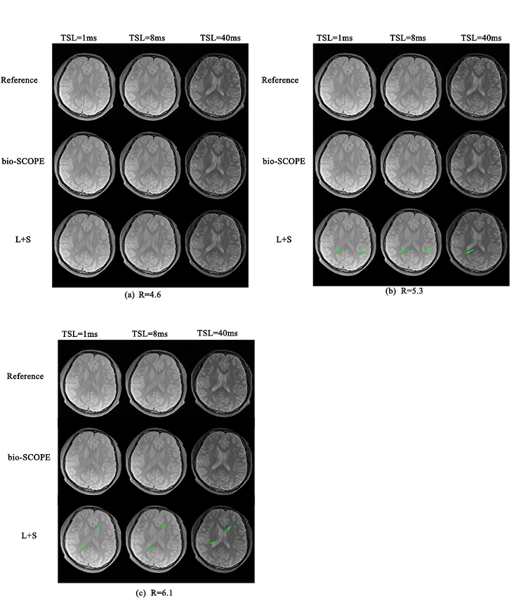
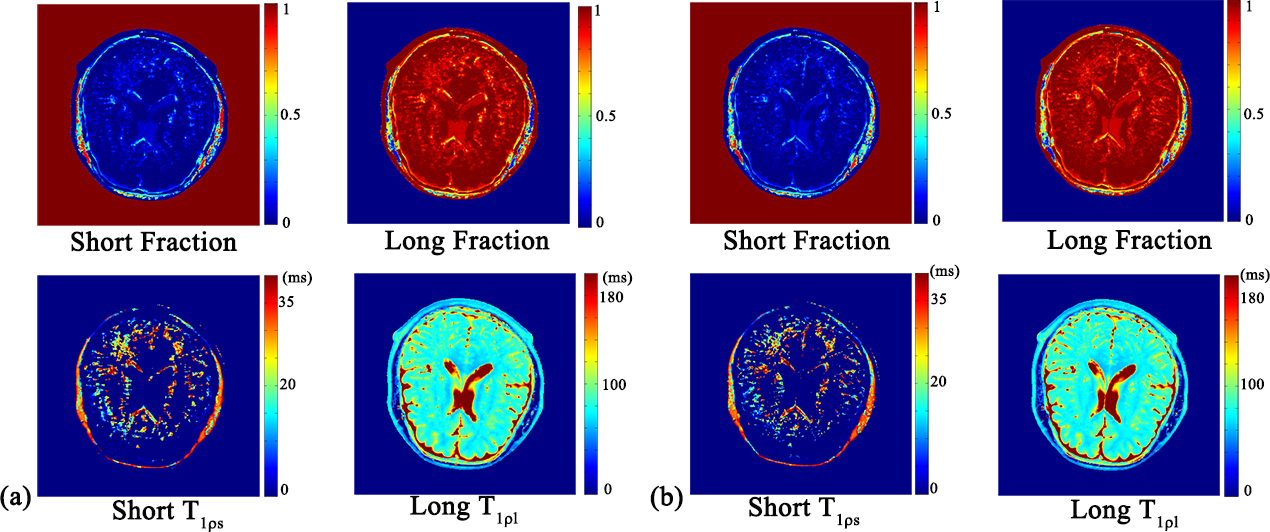
Figure 3 shows the reconstructed T1ρ-weighted images using bio-SCOPE and L+S methods at net acceleration factors (R) of 4.6, 5.3, and 6.1. At R=4.6, all reconstructed images are comparable with the reference, which were reconstructed from the fully sampled k-space data. However, aliasing artifacts (green arrows) are observed on the images reconstructed by the L+S method at higher acceleration factors, i,e, R=5.3 and 6.1. Figure 4 shows the reference T1ρ maps derived from fully sampled k-space data and the T1ρ maps estimated from the reconstructed images using bio-SCOPE at R=4.6. The T1ρ maps derived from bio-SCOPE were comparable to the reference.CONCLUSION
The proposed method, bio-SCOPE can reconstruct the T1ρ-weighted image series from highly undersampled k-sapce data, and thereby significantly reduce the scan time of bi-exponential T1ρ mapping.Acknowledgements
This work is supported in part by the National Natural Science Foundation of China under grant nos. 61771463 and 61471350, National Key R&D Program of China nos. 2017YFC0108802.References
- Allkemper T, Sagmeister F, Cicinnati V, Beckebaum S, Kooijman H, Kanthak C, Stehling C, Heindel W. Evaluation of fibrotic liver disease with whole-liver T1ρ MR imaging: a feasibility study at 1.5 T. Radiology, 2014;271(2):408-415.
- Haris M, Yadav SK, Rizwan A, Singh A, Cai K, Kaura D, Wang E, Davatzikos C, Trojanowski JQ, Melhem ER, Marincola FM, Borthakur A. T1ρ MRI and CSF biomarkers in diagnosis of Alzheimer's disease. Neuroimage Clin., 2015;7:598-604.
- Watts R, Andrews T, Hipko S, Gonyea JV, Filippi CG. In vivo whole-brain T1ρ mapping across adulthood: normative values and age dependence. J. Magn. Reson. Imaging, 2014;40(2):376-382.
- Duvvuri U, Charagundla SR, Kudchodkar SB, Kaufman JH, Kneeland JB, Rizi R, Leigh JS, Reddy R. Human knee: in vivo T1ρ-weighted MR imaging at 1.5 T--preliminary experience. Radiology, 2001;220(3):822-826.
- Regatte RR, Akella SV, Lonner JH, Kneeland JB, Reddy R. T1ρ relaxation mapping in human osteoarthritis (OA) cartilage: comparison of T1ρ with T2. J. Magn. Reson. Imaging, 2006;23(4):547-553.
- Sharafi A, Xia D, Chang G, Regatte RR, Biexponential T1ρ relaxation mapping of human knee cartilage in vivo at 3 T. NMR. Biomed. 2017;30:e3760.
- Yuan J, Zhao F, Chan Q, Wang Y. Observation of bi‐exponential T1ρ relaxation of in‐vivo rat muscles at 3T. Acta. Radiol.,2012;53:675‐681
- Wang N, Xia Y. Dependencies of multi‐component T2 and T1ρ relaxation on the anisotropy of collagen fibrils in bovine nasal cartilage. J. Magn. Reson., 2011;212:124‐132.
- Zibetti M V W, Sharafi A, Otazo R and Regatte R R, Compressed sensing acceleration of biexponential 3D- T1ρ relaxation mapping of knee cartilage. Magn. Reson. Med., 2018; doi: 10.1002/mrm.27416.
- Zhu Y, Zhang Q, Liu Q, Wang YX, Liu X, Zheng H, Liang D, Yuan J. PANDA-T1ρ: Integrating principal component analysis and dictionary learning for fast T1ρ mapping. Magn. Reson. Med., 2015;73(1):263-272.
- Zhou Y, Pandit P, Pedoia V, Rivoire J, Wang Y, Liang D, Li X, Ying L. Accelerating T1ρ cartilage imaging using compressed sensing with iterative locally adapted support detection and JSENSE. Magn. Reson. Med., 2016;75(4):1617-1629.
- Pandit P, Rivoire J, King K and Li X. Accelerated T1ρ acquisition for knee cartilage quantification using compressed sensing and data-driven parallel imaging: A feasibility study. Magn. Reson. Med., 2016;75 1256-6.
- Zibetti M V W, Sharafi A, Otazo R and Regatte R R , Accelerating 3D-T1r mapping of cartilage using compressed sensing with different sparse and low rank models. Magn. Reson. Med., 2018;80(4):1475-1491.
- Zhu
Y, Liu Y, Ying L, Peng X, Wang YJ, Yuan J, Liu X, Liang D. SCOPE: signal compensation
for low-rank plus sparse matrix decomposition for fast parameter mapping. Phys. Med. Biol.,2018;63(18):185009.
- Otazo R, Candes E, Sodickson DK. Low-rank plus sparse matrix decomposition for accelerated dynamic MRI with separation of background and dynamic components. Magn. Reson. Med., 2015;73(3):1125-1136.
- Pruessmann K P, Weiger M, Bornert P and Boesiger P. Advances in sensitivity encoding with arbitrary k-space trajectories. Magn. Reson. Med., 2001; 46:638-51.
Figures