4558
Computational method for T2-weighted images based on polynomial approximation using 3D MR parameter mapping with RF-spoiled gradient echo1Research & Development Group, Hitachi, Ltd., Tokyo, Japan
Synopsis
We propose a computational method for obtaining T2-weighted images from maps of proton density, T1, and T2* acquired by 3D RF-spoiled gradient echo. The proposed method uses a predetermined polynomial that approximates the relationship between the MR parameters and the intensity of T2WI on the basis of datasets of other subjects. Similarities between computed images and actually scanned images were improved compared with a computation method using T2* instead of T2 in the theoretical equation of the spin echo signal.
Introduction
Previously, we developed a method for simultaneously quantifying multiple MR parameters (proton density, B1, T1, and T2*) by fitting a signal equation to RF-spoiled gradient echo images obtained with various scan parameters.1, 2 Quantitative MR parameter mapping is expected to enable fast examinations by calculating multiple types of weighted images3, which are calculated using a theoretical signal equation. However, T2-weighted images (T2WI), which are acquired by spin echo sequences, are difficult to theoretically calculate from the maps of PD, T1, and T2*. In this study, we propose a computational method for obtaining T2WI from the MR parameter maps obtained by using RF-spoiled gradient echo with a predetermined polynomial that approximates the relationship between the MR parameters and the intensity of T2WI.Method
A three-dimensional RF-spoiled gradient echo sequence was performed on two healthy volunteers using a 3T MRI system (Hitachi, Ltd., Japan) and a 32-channel head coil. Seventeen images were obtained with different scan parameters (FA, TR, TE, and phase increment of RF (θ)), as shown in Table 1. The measurement resolution was 0.9×1.4×2.0 mm3, and the total acquisition time was five minutes. Data from the volunteers were obtained according to the standards of the internal review board of the Research & Development Group, Hitachi, Ltd., following receipt of written informed consent.
Maps of proton density, B1, T1, and T2* were obtained from the scanned images using a previously developed method1, 2 that uses the method of least squares to fit a signal equation based on a Bloch simulation. A T2WI was calculated by the following steps, also shown in Figure 1. (a) Coefficients of a 7-order polynomial that calculates intensity from PD, T1, and T2* were determined by using a dataset of parameter maps and a T2WI actually scanned at the same position of a subject by using a fast spin echo (FSE) sequence. The least squares method was used to calculate the coefficient of the polynomial. (b) The T2WI of the other subject was calculated using the predetermined polynomial and parameter maps of the subject.
We compared the proposed method with other calculation method that calculates the theoretical intensity of spin echo by considering T2* as T2 in the following signal equation.
$$I_{T2W}=PD\cdot\{1-\exp(-TR/T1\ )\}\cdot\exp(-TE /T2)$$
Here, TR and TE were the same as in the actually scanned (reference) T2WI (TE = 84 ms, TR = 4.3 s). The similarity between each computational T2WI and reference T2WI by using an FSE sequence were evaluated by analyzing the zero mean normalized cross correlation (ZNCC) of mean intensity in 24 manually settled regions of interest (ROI) and by analyzing the ZNCC of intensity in all voxels of a slice that include principal tissues in the brain (ZNCCROI and ZNCCvoxel, respectively).
Results
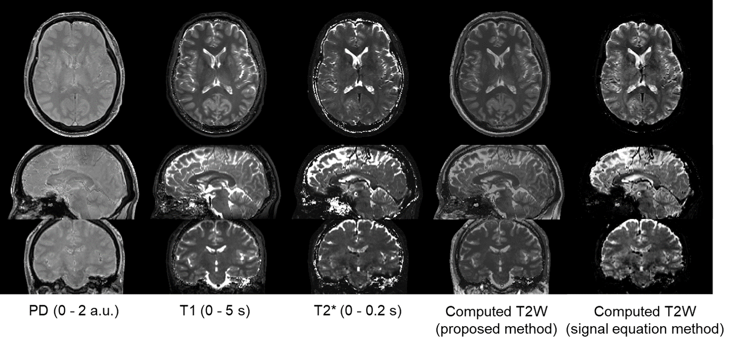
Figure 2 shows the parameter maps and computational T2WI. In comparison with the signal equation method, the visibility of skins and the contrast of brain tissue were improved in the image of the proposed method. Figure 3 shows the reference T2WI with ROIs, the computational T2WI (reconstructed as 5-mm thickness image), and the mean intensity of ROIs. Several ROIs showed low intensity in the signal equation method. Table 2 shows the ZNCCROI and ZNCCvoxel of the proposed method and the signal equation method. Both ZNCCROI and ZNCCvoxel of the proposed method were higher than those of the signal equation method.Discussion
Computational T2WI by using T2* instead of T2 to calculate theoretical intensity were different to the reference image because of the difference between T2 and T2*, especially in skins. The proposed method improved the similarities because the polynomial used in the proposed method could approximate the relationship between PD, T1, and T2* and the intensity of T2WI. Further study is required to make more precise functions for calculating the intensity of T2WI for applications with many subjects including patients with brain diseases.Conclusion
We proposed a computational method for obtaining T2-weighted images from maps of proton density, T1, and T2* acquired by 3D RF-spoiled gradient echo using a predetermined polynomial. Similarities between computed images and actual images were improved compared with a computation method using T2* instead of T2 in the theoretical equation of the spin echo signal. The proposed method would enable fast MR examination by obtaining T2-weighted images in addition to other weighted images at the same time by calculation.Acknowledgements
No acknowledgement found.References
1. Taniguchi Y, Yokosawa S, Bito Y. Simultaneous T1, T2, and B1 Mapping Using Partially RF-Spoiled Gradient Echo. Proc. ISMRM, 2011; 4560.
2. Yokosawa S, Taniguchi Y, Amemiya T, et al. Fast fitting method for simultaneously quantifying multiple MR parameters using local optimization method with predetermined initial values. Proc. ISMRM, 2017; 1441.
3. Warntjes JBM, Leinhard OD, West J, Lundberg P. Rapid magnetic resonance quantification on the brain: optimization for clinical usage. Magn Reson Med, 2008; 60; 320–329.
Figures