4555
EPI based Dual-stage MR Fingerprinting for T1, T2, and T2* mapping1Department of Radiology and Research Institute of Radiological Science, Severance Hospital, Yonsei University College of Medicine, Seoul, Korea, Republic of, 2Center for NanoMedicine, Institute for Basic Science (IBS), Seoul, Korea, Republic of, 3Yonsei-IBS Institute, Yonsei University, Seoul, Korea, Republic of, 4Erwin L. Hahn Institute for Magnetic Resonance Imaging, University of Duisburg-Essen, Essen, Germany
Synopsis
We propose an improved MR fingerprinting which can generate T1, T2, and T2* maps simultaneously. This method is based on single-shot EPI and signal acquisition consists of dual-stage divided by fixed and variable echo time. Dictionary generation and pattern matching were also modified in accordance with acquisition scheme. The feasibility of proposed method was demonstrated by phantom study and the MRF results are well correlated with the conventional T1, T2, and T2* maps. In-vivo brain MRF was also performed with a healthy volunteer.
Introduction
In this paper, we simultaneously acquired T1, T2, and T2* maps using single-shot EPI based dual-stage MR fingerprinting(MRF). Conventional MRF [1] and many followed researches have focused on T1 and T2 mapping. But T2* relaxation time is also valuable parameter to indicate intracranial hemorrhage, calcification, tumor and iron overload. EPI-MRF was proposed previously [2]. However, due to the long readout duration and late echo time, T2 map could not be obtained. Some previous methods proposed for T1, T2, and T2* mapping [3, 4] but they also have limitations. In our study, to achieve both T2 and T2* weighted effects, we divided data acquisition into dual-stage by using fixed and variable echo time and reconstruction algorithm was modified in ac cordance with acquisition scheme. The feasibility of proposed method was demonstrated by phantom experiments. Relaxation time maps from MRF were compared with results from conventional mapping methods. In-vivo brain MRF was also performed with a healthy volunteer.Methods
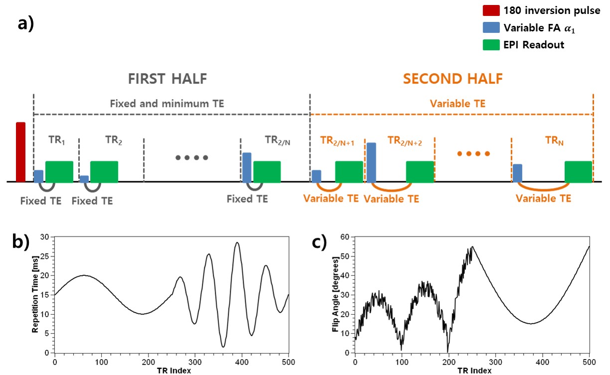
Figure 1 shows a sequence diagram, pseudo random TR and FA patterns. The proposed MRF sequence was divided into two parts. In the first stage which uses fixed echo time, TE and readout duration were minimized to about 1.8/10ms. By reducing those time, we were able to achieve T2 weighted effects in signal evolution, although using the EPI readout. In the second stage, variable echo time was applied according to the TR pattern and signal evolution was considered as having T2* weighted effects. In terms of reconstruction, T1 and T2 maps were matched using the simulated and acquired signal evolution of the first stage. Once the initial T1 and T2 values for each voxel were obtained, total signal evolution was simulated again using obtained T1, T2 and unknown T2* parameter for the second stage matching. In here, T1 and T2 were simulated more precisely than initial values with fine simulation parameter. For the verification of proposed method, phantom experiments are performed. Phantom was made using agarose and contrast agent and had a varied relaxation time. MRF data were acquired at 3T Philips clinical MRI system. The phantom was also scanned with conventional mapping method including IR-SE for T1, multi-echo SE for T2, and multi-echo GRE for T2*. Same MRF protocol and MRI system were used for in-vivo brain experiment with a healthy volunteer.Results
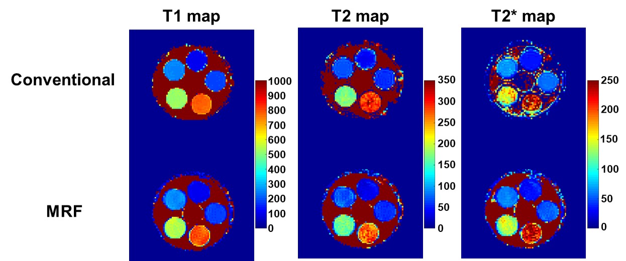
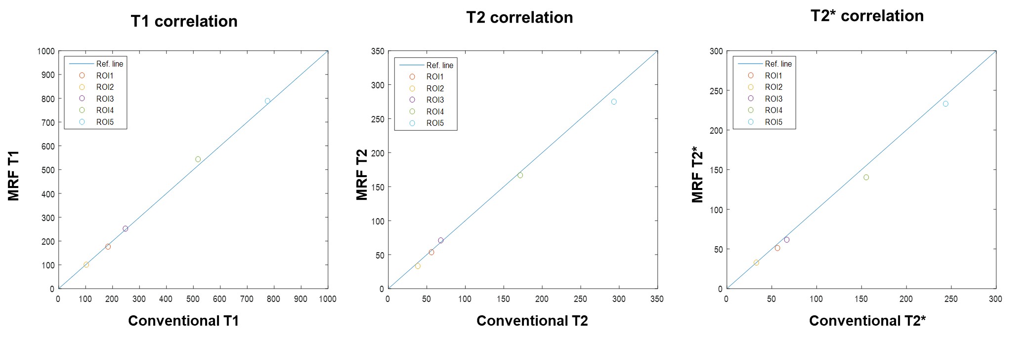
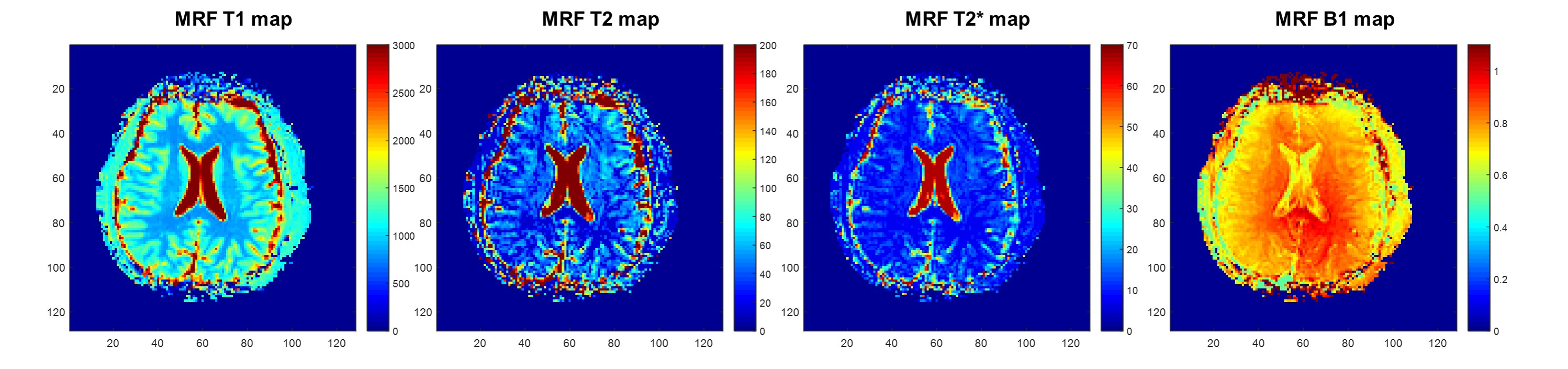
The phantom experiment results of the conventional and MRF are shown in Fig. 2. The mean value correlations of two measurements is plotted in Fig. 3. T2 and T2* under-estimation was shown in which have high T2 and T2* values. Figure 4 shows the results of In-vivo experiments. Using proposed method, T1, T2, T2*, and B1 maps are generated simultaneously. A slight image artifact related to the EPI was seen in the frontal lobe region.Discussion/Conclusion
We demonstrate the ability of the proposed MRF to map the multiple relaxation parameter simultaneously. Results of phantom experiment are well correlated with conventional method's results. We reduce the TE and readout duration of MR sequence, to solve the problem of EPI readout. And we split the acquisition and reconstruction into dual-stage to achieve both of the T2 and T2* weighted effects. Additionally, our method has potential to obtain the fine resolution map from the pre-defined sparse dictionary by applying iterative reconstruction approach. Further research will improve the limitation including under-estimation of T2 and T2*, and EPI artifact.Acknowledgements
This work was supported by the National Research Foundation of Korea (RF) grant funded by the Korea government (MISP) (No. 2016R1C1B1013837)References
[1] Ma, Dan, et al. "Magnetic resonance fingerprinting." Nature 495.7440 (2013): 187.
[2] Rieger, Benedikt, et al. "Magnetic resonance fingerprinting using echo‐planar imaging: Joint quantification of T1 and relaxation times." Magnetic resonance in medicine 78.5 (2017): 1724-1733.
[3] Wang, C., et al. "Magnetic resonance fingerprinting with quadratic rf phase for simultaneous measurement of δf, T1, T2, and T2*." Proceedings of the Intl. Soc. Mag. Reson. Med (ISMRM) (2017).
[4] Wyatt, Cory R., et al. "Multi‐parametric T2* magnetic resonance fingerprinting using variable echo times." NMR in Biomedicine 31.9 (2018): e3951.
Figures