4550
High resolution 3D magnetic resonance fingerprinting with hybrid radial cartesian-EPI acquisition1Yonsei University, Seoul, Korea, Republic of
Synopsis
A high resolution (0.5x0.5x1mm3) 3D MRF method was proposed using a hybrid radial cartesian-EPI acquisition with both segmented & interleaved EPI strategy. For the reconstruction, k-space SVD compression and CG-SENSE were applied. An in vivo brain results were presented.
Introduction
Magnetic resonance fingerprinting introduced a novel fast PD, T1 and T2 mapping strategy [1]. Thereafter, various MRF researches were proposed based on MRF’s nature characteristic, high degree of freedom in MR acquisition [2,3]. Several 3D MRF techniques were also developed with an isotropic resolution or larger than isotropic resolution [4-6]. In this study, a high resolution (0.5x0.5x1mm3) 3D MRF method was proposed using a hybrid radial cartesian-EPI acquisition.Method
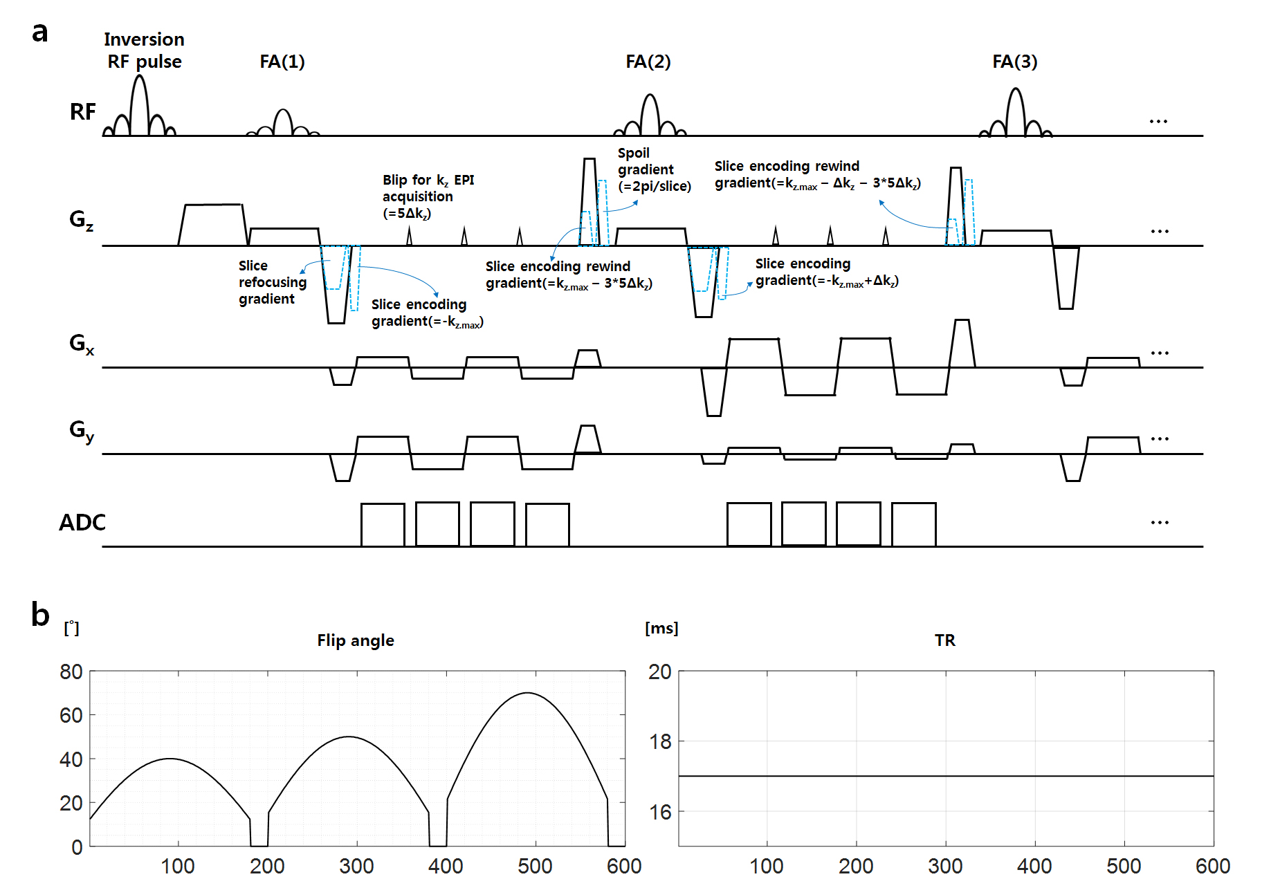
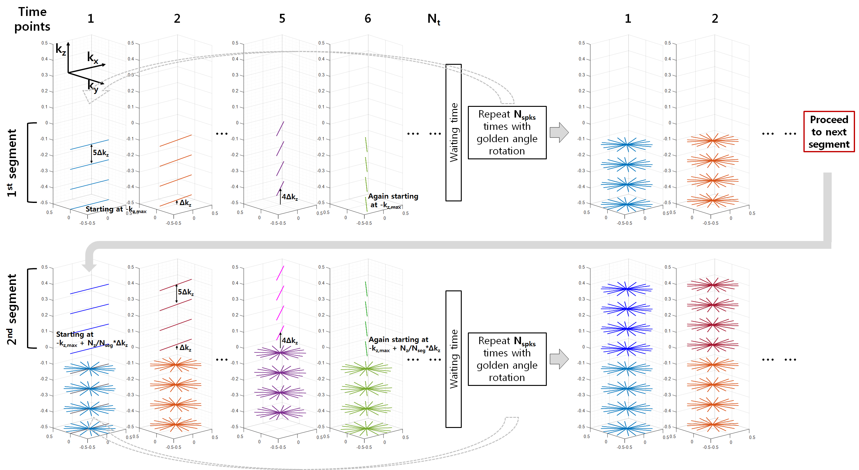
Figure 1 shows the propose pulse sequence diagram and the flip angle, TR patterns. A hybrid radial cartesian-EPI trajectory [7] was applied in 3D MRF acquisition to minimize the scan time while obtaining under-isotropic resolution (0.5x0.5x1mm3). Furthermore, to mitigate many artifacts caused by the EPI, both segmented & interleaved EPI acquisition though the kz-axis were used. Total 120 slices were acquired and 120 kz lines (=Nz) were segmented to 6 segments (=Nseg). Therefore, each segment has 20 kz lines and then interleaved EPI acquisition was applied with acceleration factor 5 (kz EPI blip has 5Δkz momentum shown in Fig 1a). Therefore, only 4 kz lines were acquired during one TR as shown in Fig 1a. For the next TR, the slice encoding gradient was increased as 1 Δkz momentum, and then same segmented & interleaved EPI acquisition was applied while radial spoke rotation with golden angle along the kxy direction [8]. The momentum of the slice encoding gradients were increased until 5 consequtive TRs (Δkz*(acceleration factor-1)=4Δkz). After then, the partition number was started from the bottom of the kz segment. This manner was applied until 600 TRs (=Nt), then waiting time (5 sec) was applied. Then repeated 8 times (=Nspks) to fill the kxy plane. A graphical example with reduced Nz=40 and Nseg=2 were presented in Figure 2.
Brain data of healthy volunteer was acquired using 20ch head coil, 3T Siemens Prisma scanner with IRB approval. Scan parameters : resolution : 0.5x0.5x1mm3, FOV : 192x192x120 mm3, Nspks=8 (number of radial spokes, inplane acceleration factor was 72 with respect to Nyquist limit), Nz=120 (number of slices), Nseg=6, REPI=5 (EPI reduction factor), Nt=600 (MRF time points). Total scan time=12min 10sec. Dictionary was generated with T1=[50:10:2000, 2050:50:3500] ms and T2=[20:1:250,260:10:350] ms using Bloch simulation. The dictionary was compressed using SVD [9].
For the reconstruction, the acquired data S was compressed using SVD to $$$\widetilde{S}$$$ in k-space domain [9]. After then, the following problem was solved using CG-SENSE [10].
$$\widetilde{x}(n)=\arg \min_{\widetilde{x}(n)}\sum_c\parallel G\cdot F \cdot E_c \cdot \widetilde{x}(n) - \widetilde{S}(n) \parallel_2^2 , n=1, 2, ... ,N_{sv}$$
G is the gridding matrix, F is the Fourier matrix and Ec is the coil sensitivity maps. Ec was calculated from 1st singular k-space data using ESPIRiT [11]. Nsv is the compressed singular numbers in SVD. L2 regularization was also applied with λ=1.5. CG-SENSE and ESPIRiT was performed using BART tool box [12]. After the reconstruction, dictionary matching was performed.
Results
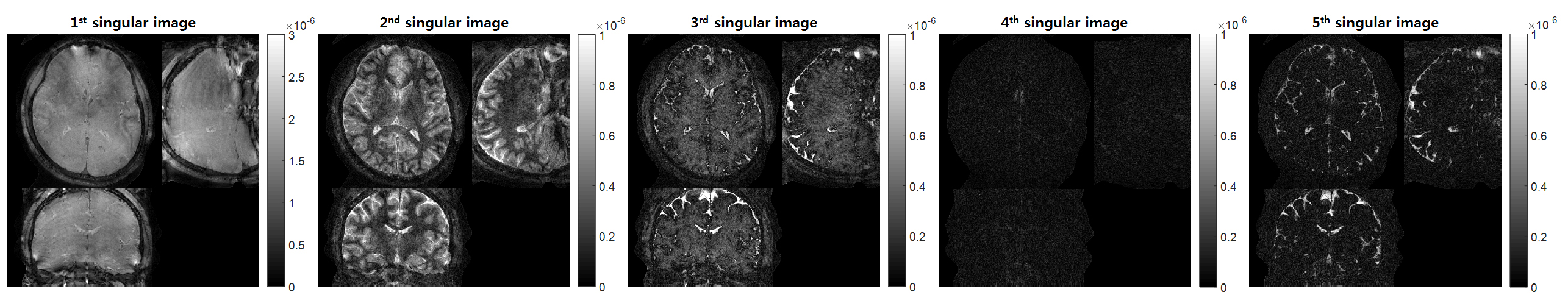
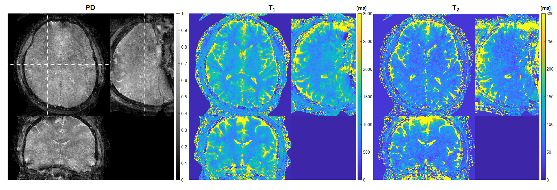
Figure 3 shows the 5 singular images after the k-space SVD compression and CG-SENSE reconstruction process. The 600 extremely highly under sampled 3D MRF data were compressed to 5 singular images with few under-sampling artifacts. Figure 4 presents the PD, T1 and T2 map results of the proposed method. Brain structures were clearly shown in T1 maps. Also the artifacts due to the EPI acquisition, such as aliasing and distortion, were hard to find in the sagittal and coronal view of the three maps, because both segmented & interleaved EPI acquisition was applied (minimize the number of acquired kz line during TR). However, some estimation errors of T2 values caused by B1+ inhomogeneity was shown in T2 maps.Discussion
The B1+ field inhomogeneity affects estimation accuracy in MRF [13,14]. Therefore, acquiring additional B1+ map and including B1 effect in dictionary might be needed. Bloch-Siegert with spiral acquisition, used in previous study [5], is an effective way to acquire 3D B1+ map within short scan time.
Waiting time used in this study was set to be 5sec. This waiting time is quite long compared to other 3D MRF studies [5,6]. Therefore, if we reduce the waiting time to 2sec, the total scan time was also reduced to 9min 46sec from 12min 10sec.
Changing the number of segments and reduction factor of EPI will affect the quality of quantitative maps and scan time. Thus, further optimization and analysis would be required and its our future work.
Conclusion
We have developed a method to achieve high resolution (0.5x0.5x1mm3) PD, T1, T2 map using the hybrid radial cartesian-EPI 3D MRF. This technique would be useful not only in the brain but also in the knee cartilage.Acknowledgements
This research was supported by Basic Science Research Program through the National Research Foundation of Korea(NRF) funded by the Ministry of Science, ICT and future Planning (NRF-2016R1A2B3016273)References
[1] Ma D, Gulani V, Seiberlich N, Liu K, Sunshine JL, Duerk JL, Griswold MA. Magnetic resonance fingerprinting. Nature 2013;495(7440):187-192.
[2] Jiang Y, Ma D, Seiberlich N, Gulani V, Griswold MA. MR fingerprinting using fast imaging with steady state precession (FISP) with spiral readout. Magn Reson Med. 2014
[3] Jiang Y, Ma D, Jerecic R, Duerk J, Seiberlich N, Gulani V, Griswold MA. MR fingerprinting using the quick echo splitting NMR imaging technique. Magn Reson Med. 2016
[4] Ma D, Pierre EY, Jiang Y, Schluchter MD, Setsompop K, Gulani V, Griswold MA. Music-based magnetic resonance fingerprinting to improve patient comfort during MRI examinations. Magn Reson Med. 2016;75(6):2303-2314.
[5] Ma D, Jiang Y, Chen Y, McGivney D, Mehta B, Gulani V, Griswold MA. Fast 3D magnetic resonance fingerprinting for a whole-brain coverage. Magn Reson Med. 2018 Apr;79(4):2190-2197.
[6] Liao C, Bilgic B, Manhard MK, Zhao B, Cao X, Zhong J, Wald LL, Setsompop K. 3D MR fingerprinting with accelerated stack-of-spirals and hybrid sliding-window and GRAPPA reconstruction. Neuroimage. 2017 Nov 15;162:13-22.
[7] Chiew M, Graedel NN, McNab JA, Smith SM, Miller KL.Accelerating Functional MRI Using Fixed-Rank Approximations and Radial-Cartesian Sampling. Magn Reson Med. 2016 Dec;76(6):1825-1836.
[8] Winkelmann S, Schaeffter T, Koehler T, Eggers H, Doessel O. An optimal radial profile order based on the Golden Ratio for time-resolved MRI. Medical Imaging, IEEE Transactions on 2007;26(1):68-76.
[9] McGivney DF, Pierre E, Ma D, Jiang Y, Saybasili H, Gulani V, Griswold MA. SVD compression for magnetic resonance fingerprinting in the time domain. IEEE Trans Med Imaging 2014;33(12):2311-2322.
[10] Pruessmann KP, Weiger M, Bornert P, Boesiger P. Advances in sensitivity encoding with arbitrary k‐space trajectories. Magn Reson Med 2001;46:638–651.
[11] Uecker M, Lai P, Murphy MJ, Virtue P, Elad M, Pauly JM, Vasanawala SS, Lustig M. ESPIRiT--an eigenvalue approach to autocalibrating parallel MRI: where SENSE meets GRAPPA. Magn Reson Med 2014;71(3):990-1001.
[12] The Berkeley Advanced Reconstruction Toolbox (BART) toolbox (https://mrirecon.github.io/bart/) 2015.
[13] Ma D, Coppo S, Chen Y, McGivney DF, Jiang Y, Pahwa S, Gulani V, Griswold MA. Slice profile and B1 corrections in 2D magnetic resonance fingerprinting. Magn Reson Med 2017;78(5):1781-1789.
[14] Hong T, Han D, Kim MO, Kim DH. RF slice profile effects in magnetic resonance fingerprinting. Magnetic resonance imaging 2017;41:73-79.
Figures