4541
Accelerated R1 or R2 Mapping with Geometric Relationship Constrained Reconstruction Method1Biomedical Engineering, Yale University, New Haven, CT, United States, 2Diagnostic Radiology, Yale University, New Haven, CT, United States, 3Radiology and Biomedical Imaging, Yale University, New Haven, CT, United States, 4Department of Radiology and Biomedical Imaging, Yale University, New Haven, CT, United States, 5Department of Neurosurgery, Yale University, New Haven, CT, United States
Synopsis
In this work we present a constrained reconstruction method that can produce either an R2- or R1- weighted image series, in tandem with the parameter map, from undersampled data. The method has been demonstrated in vivo for radial TSE, and radial TSE augmented with nonlinear encoding (O-space), and inversion recovery (IR) datasets. The algorithm iteratively calculates the entire series of T2 or T1 weighted images while enforcing the exponential decay posed as a geometric relationship between the images. Experimental brain images generated with these maps are in excellent agreement with the fully sampled images and show less undersampling artifact than images reconstructed from individual undersampled datasets.
Introduction
Since parameter mapping is a powerful tool for tissue characterization, various fast imaging techniques and algorithms have been developed to accelerate parameter mapping acquisitions(1,2). Radial acquisitions are popular since repeated sampling of the center of k-space provides contrast data with each line acquired(3). Radial TSE accelerates R2 mapping by acquiring multiple T2 weighted lines in each TR. Without special reconstruction techniques, the mixing of contrast information results in highly blurred and contrast averaged images. Accelerated R1 mapping techniques suffer similar problems. In this work we introduce a reconstruction algorithm that reduces blur and undersampling artifacts in T2 and T1 weighted images while simultaneously generating R2 or R1 maps.Theory
The algorithm incorporates the entire data equally in the reconstruction by minimizing the following objective function: $$$∑_{i=1}^{C}‖S_i-E_i x_i ‖^2 +λ∑_{i=1}^C‖x_{(i-1)}-αx_i-β‖$$$, where $$$β$$$ is zero for T2 weighted data processing. The data at the ith echo time, $$$S_i$$$, is acquired with the encoding function $$$E_i (x,t)=C_q (x)e^{(j Φ(x,t) )}$$$, where $$$Φ(x,t)=k^T (t)ψ(x)$$$. In the contrast weighted image series, $$$x_i$$$ is the ith image out of $$$C$$$ total images. The variables $$$α$$$ and $$$β$$$ have the dimensions of a single T2w or T1w image. In the constraint term $$$α(x)=e^{(Δt/(T2(x)))}$$$ or $$$α(x)=e^{(Δt/(T1(x)))}$$$ as appropriate such that the exponential T2 or T1 decay relationship between echoes is enforced. For initialization, $$$m_i$$$ is reconstructed using the undersampled data corresponding to each contrast, and $$$α$$$ and $$$β$$$ are derived from these by conventional means. From there, $$$x_i$$$ is iteratively updated to minimize the objective function using fixed $$$α$$$ and $$$β$$$. Once this converges, $$$α$$$ and $$$β$$$ are updated according to closed form solutions that minimize the objective function.Methods
In vivo imaging experiments were performed on a 3T MRI scanner (MAGNETOM Trio Tim, Siemens Healthcare, Erlangen, Germany) with a 4 channel for radial experiments and 8 channel RF head coil for O-space and Cartesian TSE experiments. Radial 250mm2 data with TR=4s and BW=1500Hz/pix using either a TSE acquisition with ETL=4 and echo spacing 25ms or an IR sequence with inversion time 600ms was acquired. Additionally, we acquired TR=2000s ETL=8 and BW=470Hz/pix Cartesian TSE and O-space data with Z2 strength= 41.6mT/m2. The Human Investigation Committee granted Institutional Review Board approval to image healthy human volunteers. After obtaining informed consent the brains of two volunteers were imaged. All calculations were performed in MATLAB (MathWorks Inc, Natick, Massachusetts, USA). Reconstructions were performed via a CG algorithm with 10 iterations using GPU processing.
Discussion
By defining a single minimization which incorporates all the data and all the contrasts, this approach simultaneously reduces artifacts due to competing contrasts and those due to k-space undersampling. Importantly, posing the regularization in image space and as a geometric relationship, unlike previous approaches, provides a closed form solution for the parameter map and a truly quadratic problem well suited to CG reconstruction. Unlike previous reconstruction methods for O-space TSE, this method is applicable to a wide range of acquisition methods and contrasts, does not require any parameters to be tuned, and does not require a special excitation order. Compared to simple regridding of the undersampled data, this iterative algorithm does have higher computational load and reconstruction time, though it results in much sharper images.Results
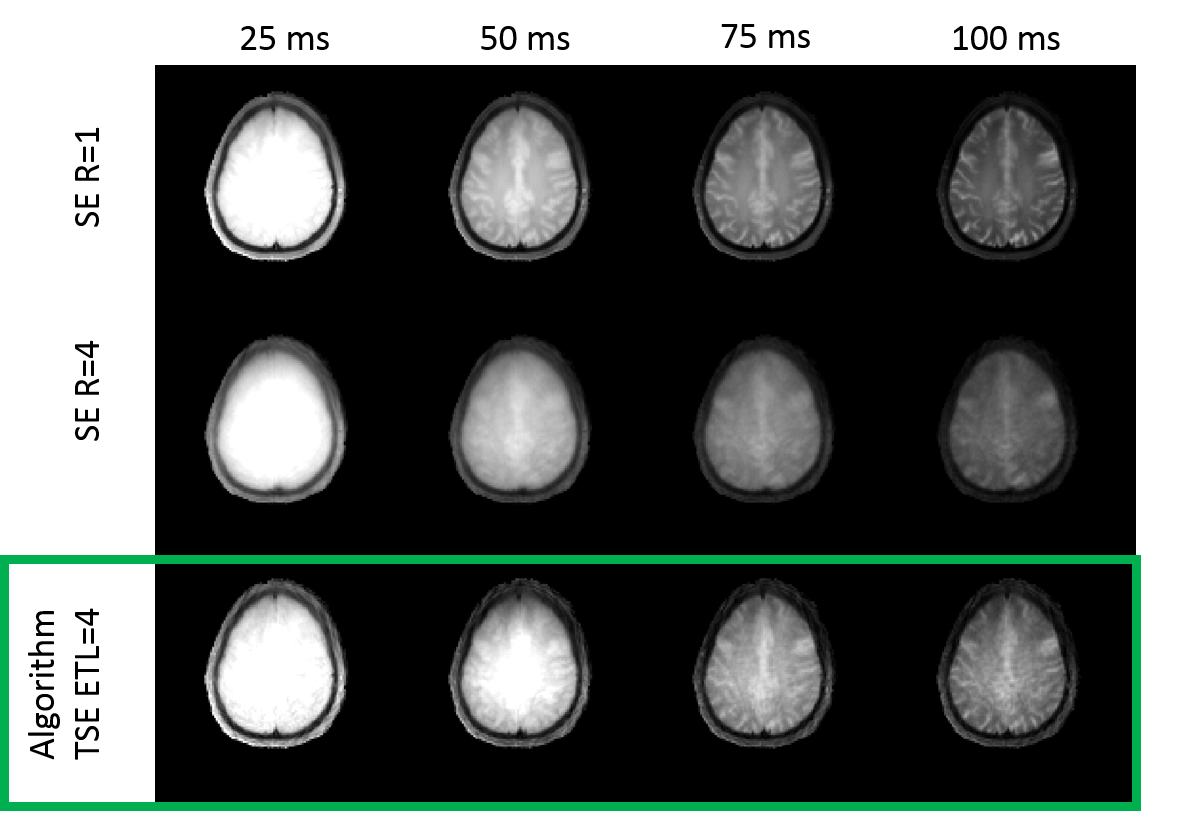
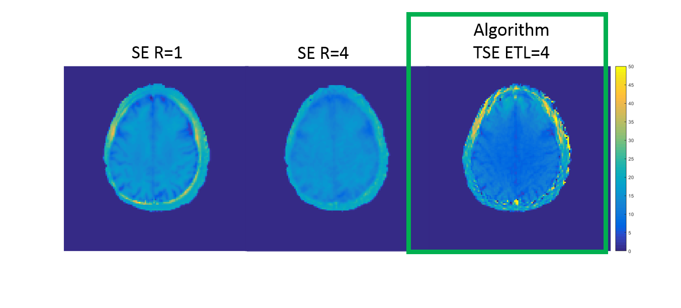
The first row of Figure 1 shows fully sampled T2w radial images taken at different echo times. The 2nd and 3rd rows of Figure 1 use data from a TSE acquisition with ETL 4, so only ¼ of the spokes are available at each echo time. Individual reconstruction of each undersampled dataset (2nd row) shows considerable blur compared to the holistic reconstruction algorithm (3rd row) despite using the same data. Figure 2 shows that the R2 map generated from the algorithm matches the reference R2 map generated conventionally from fully sampled radial data. It also shows reduced blur when compared to a map generated from the undersampled radial data.
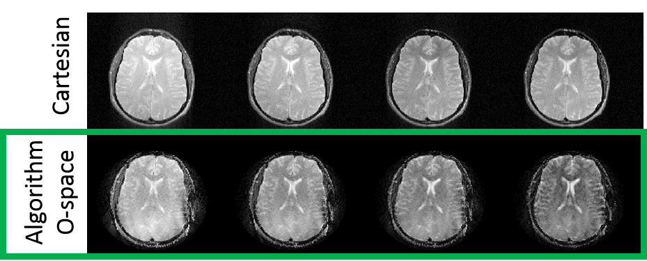
Figure 3 shows that the algorithm can also be applied to nonlinear gradient imaging techniques such as O-space to generate T2w images from a single dataset. The acquisition to generate the O-space images is 8 fold faster than the acquisition for the datasets to generate the Cartesian images.
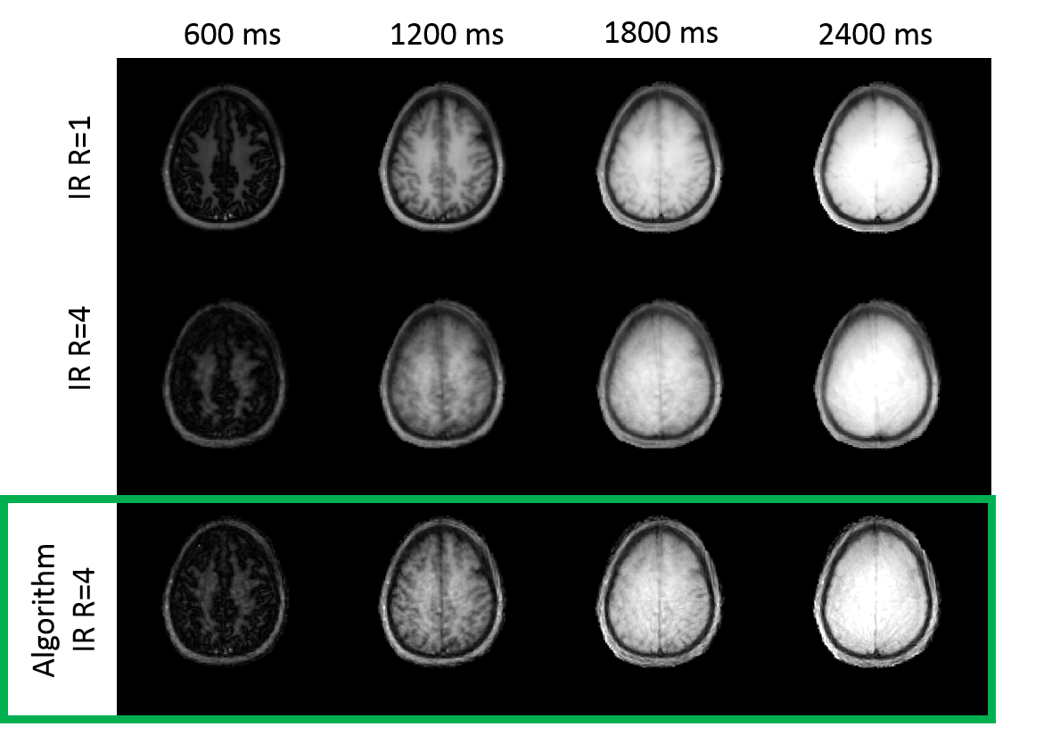
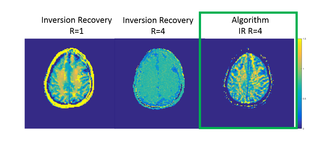
Figure 4 shows the algorithm applied to fit inversion recovery contrast. Similar to Figure 1, the columns show images with different inversion times, and the rows correspond to full sampling, undersampling in k-space to maintain uniform contrast, and application of the proposed algorithm. Fig. 5 shows the R1 maps generated conventionally compared to the R1 map generated with the algorithm.
Conclusion
Using a reconstruction method that imposes a geometric relationship between contrast images, we simultaneously reduce effects from undersampling and competing contrast for accelerated R1 or R2 mapping.Acknowledgements
No acknowledgement found.References
I. TJ, Martin U, Weitian C, Peng L, T. AM, S. VS, Michael L. T2 shuffling: Sharp, multicontrast, volumetric fast spin-echo imaging. Magnetic Resonance in Medicine 2017;77(1):180-195.
2. J. ST, Martin U, Susann B, Jens F. Model-based nonlinear inverse reconstruction for T2 mapping using highly undersampled spin-echo MRI. Journal of Magnetic Resonance Imaging 2011;34(2):420-428.
3. Song HK, Dougherty L. k-Space weighted image contrast (KWIC) for contrast manipulation in projection reconstruction MRI. Magnetic Resonance in Medicine 2000;44(6):825-832.
Figures