4511
Routine B0 eddy current measurements with TOPPE for more robust spiral imaging1University of Michigan, Ann Arbor, MI, United States, 2Memorial Sloan Kettering Cancer Center, New York, NY, United States
Synopsis
Spiral imaging is an SNR- and time-efficient alternative to conventional cartesian MRI, but is relatively sensitive to gradient system imperfections. Unfortunately, measuring the k-space trajectory and B0 eddy currents for a particular spiral readout is cumbersome and not routinely performed. We propose to leverage the TOPPE development environment for rapid pulse sequence prototyping to easily measure both k-space trajectory and B0 eddy currents using “pencil-beam” acquisitions. To demonstrate this setup, we obtained k-space and B0 measurements of a pair of spiral-in and spiral-out readouts. We show that compensating for B0 eddy currents can improve image quality, and that TOPPE provides a convenient platform for these types of measurements.
INTRODUCTION
Spiral imaging is an SNR- and time-efficient alternative to conventional cartesian MRI, but is relatively sensitive to object- and hardware-dependent non-idealities such as off-resonance and eddy currents. One such factor that is often overlooked is B0 eddy currents, i.e., spatially invariant phase offsets that vary temporally during data readout. To first order, B0 eddy currents produce a linear phase modulation across k-space, resulting in a spatial shift in the image domain that is often tolerated [Jim Pipe, personal communication]. However, when combining spiral-out and spiral-in images such spatial shifts must be corrected. Furthermore, any unknown higher-order B0 offsets can produce image blurring or other artifacts. B0 eddy currents for a particular spiral can be measured using the “pencil-beam” method [1], however this is generally not done since it typically requires time-consuming pulse programming. Another alternative is specialized field mapping hardware [2], but that is not yet widely available. Here we propose to leverage the TOPPE development environment for rapid pulse sequence prototyping [3,4] to measure both k-space trajectory and B0 eddy currents for use in a dual-echo state state (DESS), multi-shot stack-of-spirals 3D acquisition; such an acquisition is useful for, e.g., T2 mapping [5] and myelin water fraction (MWF) imaging [6]. We show that compensating for B0 eddy currents can improve image quality, and that TOPPE provides a convenient platform for these types of measurements.METHODS
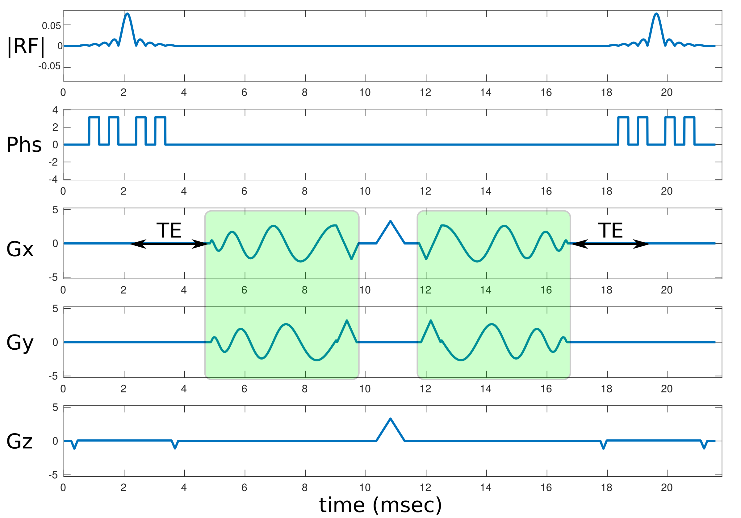
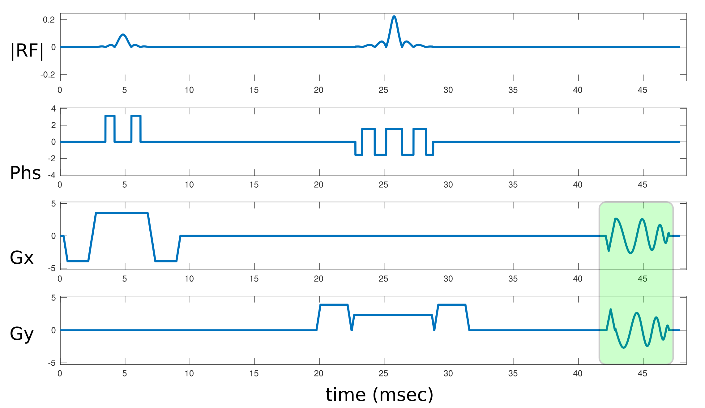
Imaging: All sequences were implemented in TOPPE and executed on a GE Discovery MR750 3T scanner equipped with a 32-channel receive array. Figure 1 shows the stack-of-spirals DESS sequence, where spiral-out and spiral-in readouts are needed to ensure equal echo time (TE) for the first and second echo, respectively. The readout “modules” (shaded in green) were inserted into the pencil-beam sequence shown in Fig. 2; in this way the exact same spiral waveform was used in both the DESS and pencil-beam sequences. We excited a 1 mm wide square beam in a uniform Agar ball (FBIRN) phantom using slice-selective excitation and refocusing (180o) radiofrequency (RF) pulses, and measured the phase of the FID signal during playback of the spiral waveform. Pencil-beams were excited at the corners of a 4 cm wide square. For each pencil-beam FID, we subtracted the phase from a separate reference acquisition without any gradients (to correct for off-resonance and other sources of phase). Combining the resulting FID signals produced measured k-space trajectory (kx and ky) and B0 offsets for each of 36 spiral leafs. We smoothed the observed k-space and B0 waveforms using a spline fit (in Matlab). We also imaged a resolution phantom with a spoiled gradient-echo (SPGR) sequence, using the same spiral-out and spiral-in readouts as in the DESS sequence.
Image reconstruction: Prior to reconstruction we demodulated the acquired (raw) data with the (smoothed) observed B0 waveforms. We then reconstructed images using nuFFT, based on the measured k-space trajectory.
RESULTS
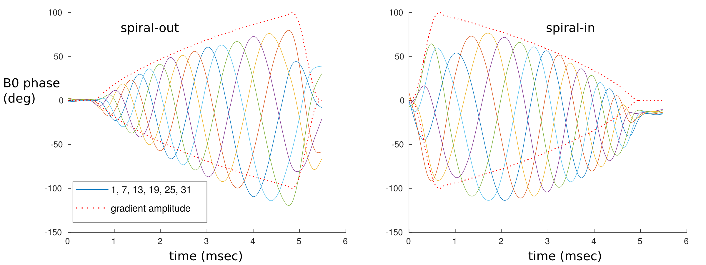
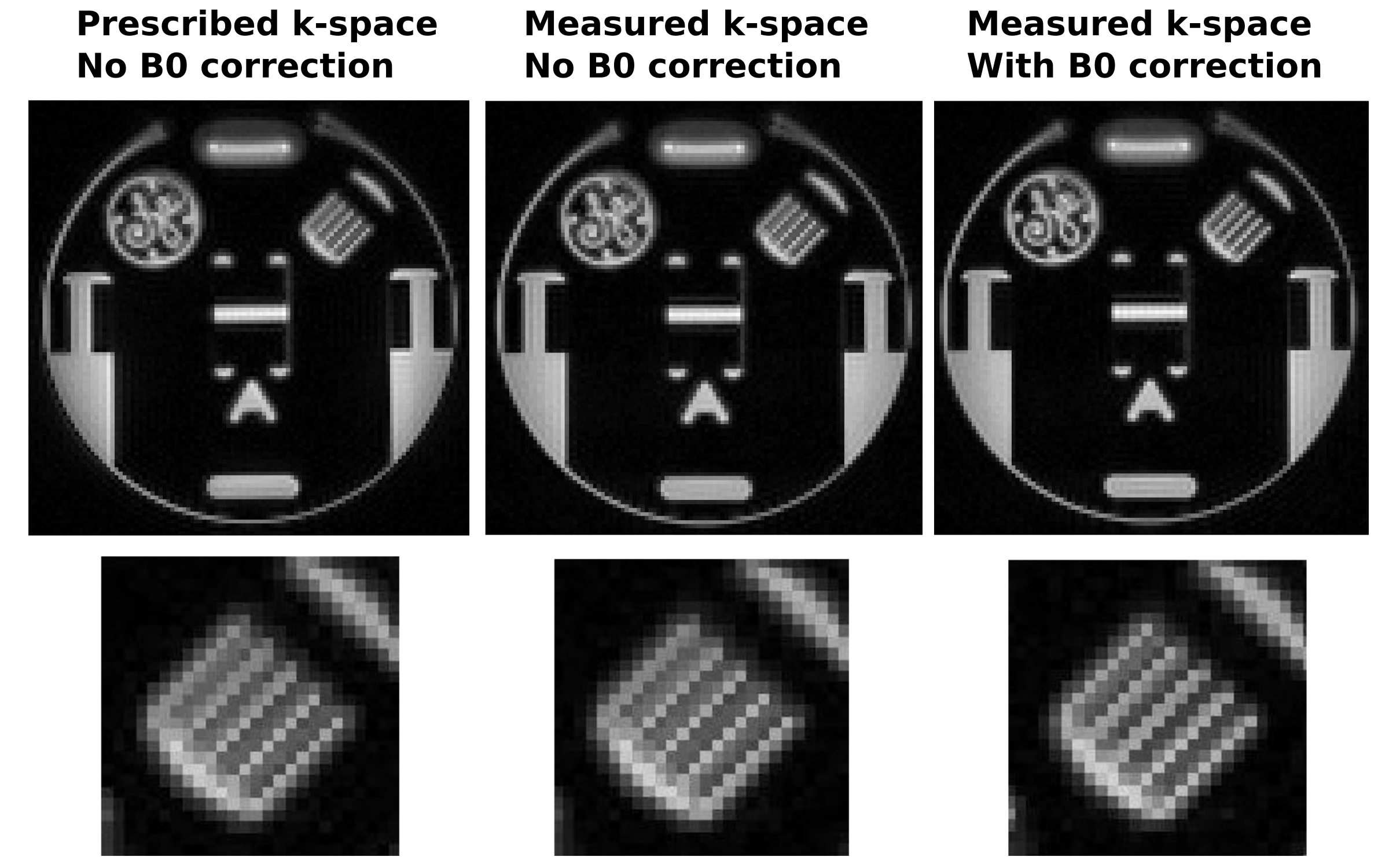
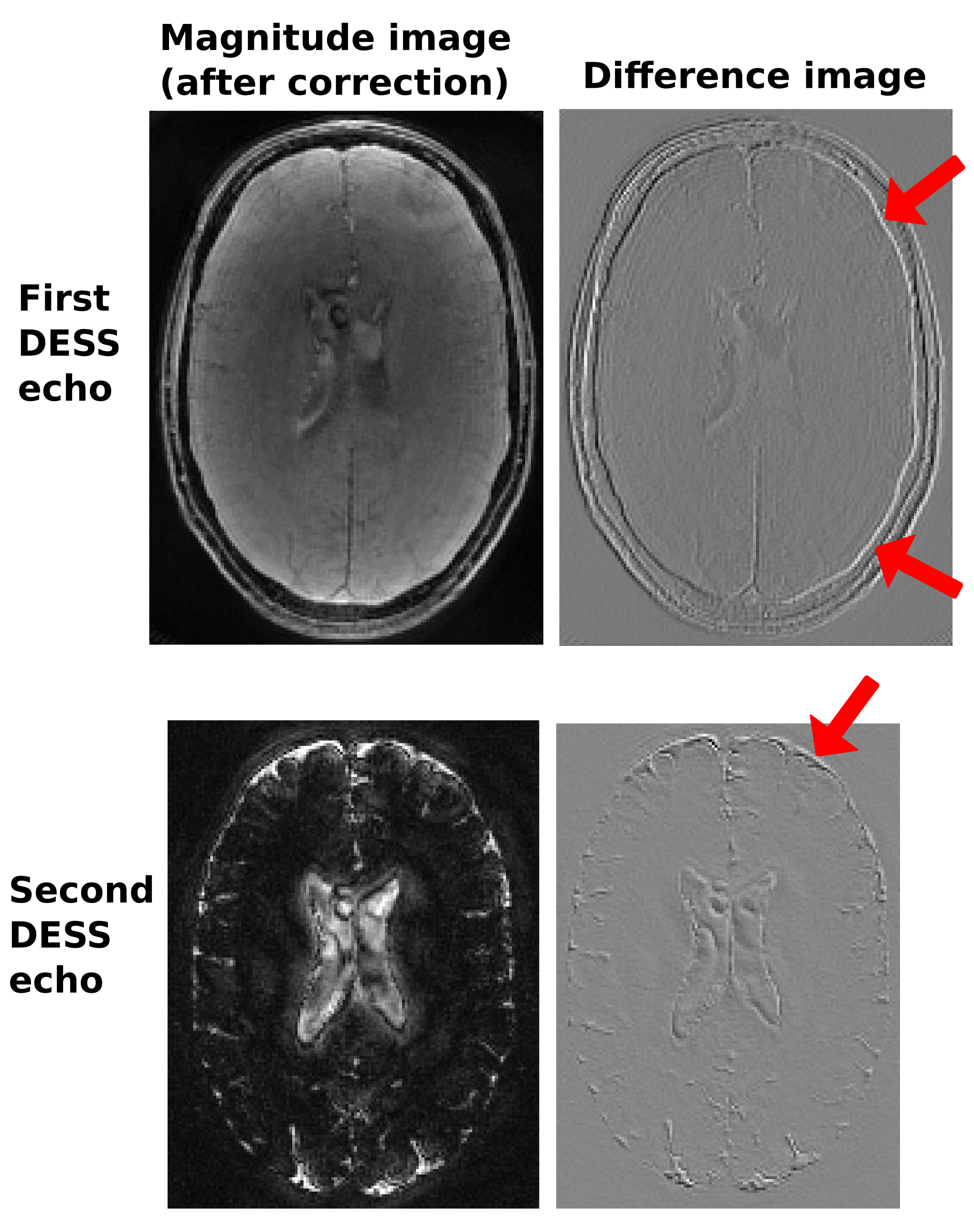
Figure 3 shows the measured B0 waveforms for a subset of the 36 leafs. Although the B0 phase offset tends to “follow” the spiral turns (giving rise to approximately linear phase in k-space and hence a spatial shift in image space), significant deviations from this pattern are also observed. Figure 4 shows spiral-out phantom imaging results, before and after the proposed B0 correction. Although significant off-resonance artifact can be seen, particularly near the top of the phantom, we observe a subtle improvement in the “comb” pattern after B0 correction. Finally, Fig. 5 shows representative DESS imaging results in the brain of a healthy volunteer. B0 correction produces a small shift in both the first (spiral-out) and second (spiral-in) DESS echo, but in different directions.CONCLUSION
TOPPE provides a convenient framework for gradient trajectory and B0 eddy current measurements that should permit routine use of such measurements in MR research labs. B0 eddy current correction can improve spiral image quality, and is particularly important when combining spiral-in and spiral-out images. In future work we will incorporate off-resonance correction for further improvements in image quality.Acknowledgements
This work was supported by NIH grant R01EB023618References
- Duyn JH, Yang Y, Frank JA, van der Veen JW. Simple correction method for k‐space trajectory deviations in MRI. J Mag Res 1998;132:150–153.
- Dietrich et al. A field camera for MR sequence monitoring and system analysis . Magn Reson Med 2016; 75(4); 1831-1840.
- Nielsen JF, Noll DC. TOPPE: A framework for rapid prototyping of MR pulse sequences. Magn Reson Med 2018; 79(6):3128-3134.
- https://toppemri.github.io/
- Chaudhari et al. Five-minute knee MRI for simultaneous morphometry and T2 relaxometry of cartilage and meniscus and for semiquantitative radiological assessment using double-echo in steady-state at 3T. J Magn Reson Imaging, 47, 5:1328-1341.
- Nataraj et al. Fast, Precise Myelin Water Quantification using DESS MRI and Kernel Learning. arXiv:1809.08908 [physics.med-ph]
Figures