4506
Evaluation of the Uniform Combined Reconstruction (UNICORN) Algorithm for Improving 7T Knee MRI Uniformity1Siemens Healthineers, Rochester, MN, United States, 2Siemens Healthineers, Austin, TX, United States, 3Siemens Healthineers, Portland, OR, United States, 4Department of Radiology, Mayo Clinic, Rochester, MN, United States, 5Siemens Healthineers, Erlangen, Germany
Synopsis
MR image intensity non-uniformity is often observed at 7T. A novel algorithm termed ‘Uniform Combined Reconstruction’ (UNICORN) was developed recently to correct for intensity non-uniformity in MR images without the use of a calibration/reference scan. In this work, 3 fellowship trained musculoskeletal radiologists with cumulative experience of 42 years evaluated the efficacy of UNICORN in 33 7T musculoskeletal MRI volumes. The uniformity, contrast, signal-to-noise-ratio and overall image quality were evaluated. Without the use of a reference scan, UNICORN was rated to provide better image uniformity, contrast and overall image quality than the N4 bias-field correction algorithm at 7T.
Introduction
MR image intensity non-uniformity is often observed at 7T (1–3). Reference scans from body-coil used for uniformity correction at lower field-strengths are typically not available at 7T. Recently, a novel algorithm termed ‘Uniform Combined Reconstruction’ (UNICORN) was developed to correct for intensity non-uniformity in MR images without the use of a calibration/reference scan (4). Preliminary results showed promise for improvement in image uniformity at 7T (4). In this work, 3 fellowship trained musculoskeletal radiologists evaluated the uniformity, contrast, signal-to-noise-ratio and overall image quality for images with no post-processing, images processed with N4 bias-field correction algorithm (5), and the UNICORN algorithm.Methods
20 subjects’ knees were imaged at 7T on a MAGNETOM Terra (Siemens Healthineers, Erlangen, Germany) with a single-channel transmit, 28-channel phased-array receive knee coil (QED, Quality Electrodynamics, Mayfield Village, OH, USA) under the guidelines of an Institutional Review Board.
A 2D turbo-spin-echo (TSE) sequence without fat-suppression was used to acquire knee images. The imaging parameters for the non-fat-saturated TSE sequence included: TR=4000-5500ms, TE=20-45ms, receive-BW=350-1042Hz/pixel, slice-thickness=1.5-2mm, slice-gap=0.5-1mm, echo-train length=6-8 echoes, field-of-view=150-160mm, in-plane acquisition voxel size=0.35×0.35mm2-0.45×0.45mm2, in-plane reconstructed voxel size=0.2×0.2mm2-0.25×0.25 mm2 and refocusing flip-angle=135°-180°.
A total of 33 non-fat-suppressed image series were acquired from 20 subjects. The 33 series were processed using both the N4 and UNICORN algorithms. UNICORN improves image uniformity by enhancing spatial frequency in the coil-combined images and decoupling the non-uniformity induced by the local receive-coils (assuming the coil-sensitivities to be of low spatial frequency).
3 fellowship trained musculoskeletal radiologists with cumulative experience of 42 years evaluated the uniformity, contrast, signal-to-noise-ratio (SNR) and overall image quality of the images.
Intraclass correlation-coefficient (ICC) was used for measuring the inter-rater reliability. ICC and 95% confidence-intervals (CI) were calculated using the R statistical package employing a two-way mixed-effects model based on a mean-rating (k=3) for absolute-agreement. Wilcoxon signed-rank test with continuity correction was used for analyzing the overall image quality scores.
Results
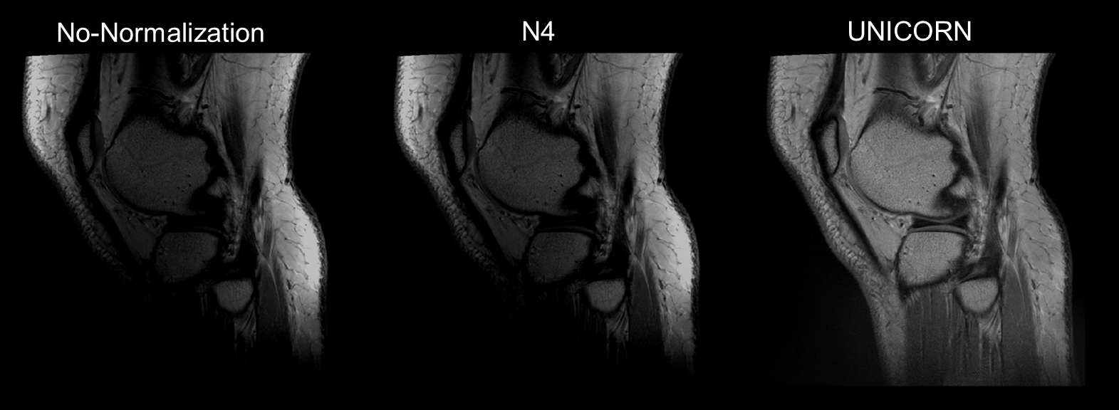
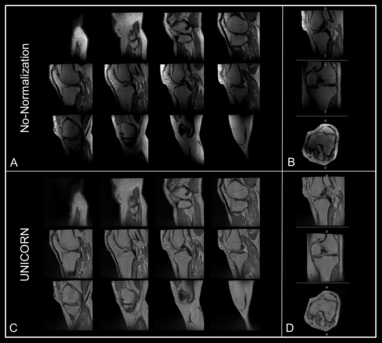
Figure 1 demonstrates the improvement in image uniformity achieved by UNICORN as compared to images with no-normalization and N4 normalization on a representative sagittal non-fat-saturated 7T knee MRI. Figure 2 shows the consistent through-slice profile and uniformity across a 3D imaged volume achieved by the UNICORN algorithm.
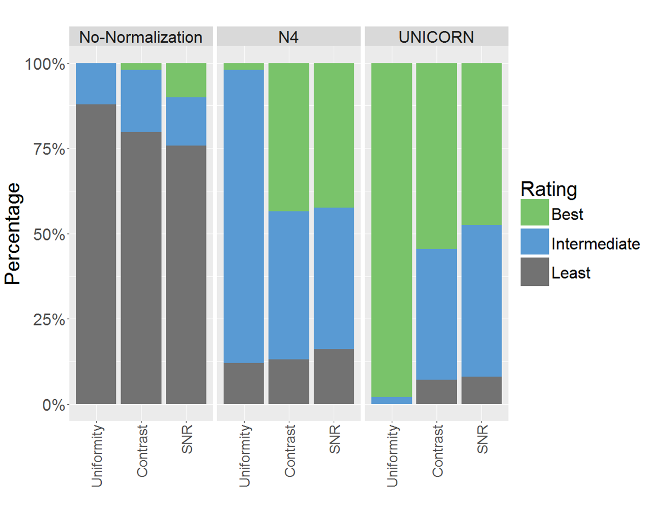
Figure 3 shows stacked bar plots illustrating the percentage of best, intermediate and least preferred ratings received by the images without normalization and with N4 and UNICORN normalization for evaluation image metrics of uniformity, contrast, and SNR. UNICORN was rated best among the three methods evaluated for uniformity in 97.9% of the cases with excellent inter-rater agreement (ICC of 0.98, CI 0.97-0.99). Images with no, N4 and UNICORN normalization were rated best for contrast in 2%, 43.4% and 54.6% of the cases respectively with good inter-rater agreement (ICC of 0.80, CI 0.72-0.86). In other words, UNICORN was rated better than N4 in 11.2% cases with good inter-rater agreement. SNR ratings had moderate inter-rater agreement with an ICC of 0.67 (CI 0.54-0.77). Images with no, N4 and UNICORN normalization were rated best for SNR in 10.1%, 42.4% and 47.5% cases respectively. In other words, SNR ratings for N4 and UNICORN were equivalent.
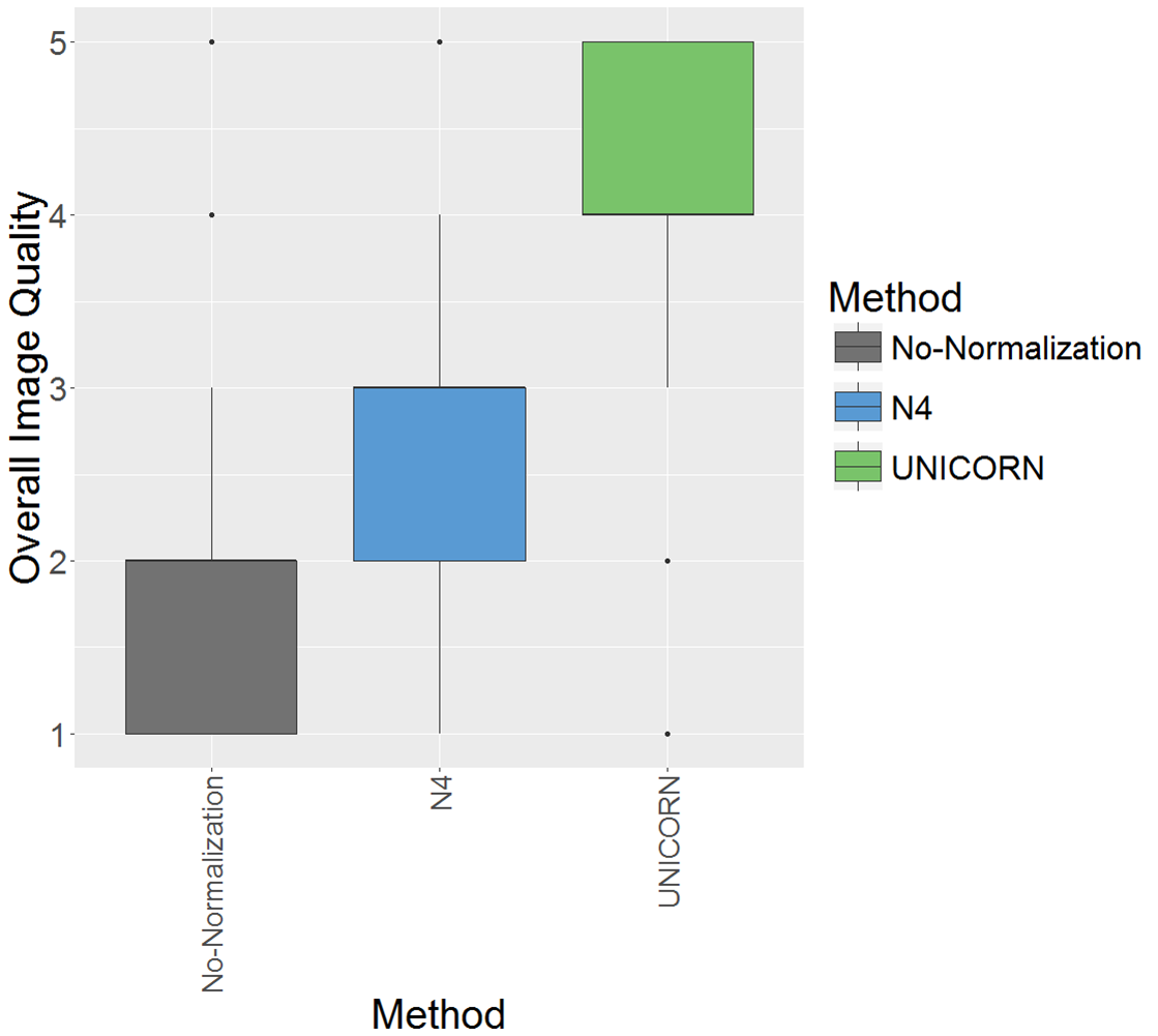
Figure 4 shows the box-plot of scores for overall image quality for images with no, N4 and UNICORN normalization. The overall image quality scores for UNICORN were significantly higher than N4 (p < 6×10-13), with good to excellent inter-rater agreement (ICC 0.90, CI 0.86-0.93).
Discussion and Conclusion
Efficacy of the UNICORN and N4 algorithms in improving 7T musculoskeletal MRI uniformity was evaluated. Furthermore, the effect of both algorithms on overall image quality, inter-tissue contrast and SNR was compared.
The UNICORN algorithm is able to simultaneously reduce hyper-intensity adjacent to the receive-coils and increase intensity near the center of the field-of-view and the anterior-inferior regions of the knee without the use of reference scan. Image contrast, another critical factor for image quality, was evaluated and it was found that an increase in image uniformity following application of the UNICORN algorithm did not adversely affect tissue contrast.
UNICORN uses the reciprocal of cumulative coil-sensitivity maps to improve the image uniformity. This also enhances areas of low SNR, such as background regions, which might result in noise enhancement in those areas. However, the overall image quality is significantly higher with UNICORN compared to N4, suggesting that background enhancement with UNICORN is a minor factor and does not affect the overall image quality.
Without the use of a reference scan, UNICORN provides better image uniformity, contrast and overall image quality at 7T compared to the N4 bias-field correction algorithm.
Acknowledgements
No acknowledgement found.References
1. Uwano I, Kudo K, Yamashita F, Goodwin J, Higuchi S, Ito K, et al. Intensity inhomogeneity correction for magnetic resonance imaging of human brain at 7T. Med Phys [Internet]. 2014 Jan 14;41(2):022302. Available from: http://doi.wiley.com/10.1118/1.4860954
2. Ganzetti M, Wenderoth N, Mantini D. Quantitative Evaluation of Intensity Inhomogeneity Correction Methods for Structural MR Brain Images. Neuroinformatics. 2016 Jan 26 [cited 2018 Mar 8];14(1):5–21. Available from: http://link.springer.com/10.1007/s12021-015-9277-2
3. Uğurbil K. Imaging at ultrahigh magnetic fields: History, challenges, and solutions. Neuroimage. 2017 Jul 8; Available from: http://www.ncbi.nlm.nih.gov/pubmed/28698108
4. Chebrolu VV, Kollasch P, Deshpande V, Grinstead J, Benner T, Heidemann R, Spence D, Felmlee J, Frick M, Amrami KA. Uniform Combined Reconstruction (UNICORN) of Multi-channel Surface-coil Data at 7T without use of a Reference Scan. Proceedings Joint Annual Meeting ISMRM-ESMRMB. Paris, France; 2018. P. 3524.
5. Tustison NJ, Cook PA, Gee JC. N4ITK: Improved N3 Bias Correction. IEEE Trans Med Imaging. 2010;29(6):1310–20.
Figures