4500
Removing bias and increasing dynamic range in DREAM flip angle mapping at 7T1Clinical Sciences Lund, Medical Radiation Physics, Lund University, Lund, Sweden, 2National 7T Facility, Lund University, Lund, Sweden, 3Philips Danmark A/S, Philips Healthcare, Copenhagen, Denmark
Synopsis
DREAM is an ultra-fast multi-slice B1+-mapping technique based on the single-shot STEAM sequence. To study noise and bias related to slice-profiles, DREAM B1+-maps at 3.75mm resolution were acquired at 7T in phantoms and in human brain with nominal flip angles (FA) between 20° and 90° of the two STEAM preparation pulses. B1+ was decreasing at actual FAs above 50°; noise became apparent at actual FAs below 20° reducing dynamic range. By varying the preparation FA, this reliable range (20°<FA<50°) is shifted over a B1+ range from 20% to 250%. The FA map is constructed from overlapping B1+ maps after thresholding.
Introduction
At high and ultra-high B0, reproducible and bias-free measurement of the flip angle (FA) is crucial for T1 determination by the variable flip angle approach [1]. The DREAM method [2] calculates the actual FA from a single-shot STEAM image [3] normalized by a gradient echo image created by the same RF pulse train. Thus, multi-slice FA mapping with whole brain coverage at 7T can be performed in less than half a minute [4]. However, the DREAM approach limits the range of flip angles to 0° to 90°, with errors increasing towards the limits as one or both signal approach zero. More importantly, non-linear slice profile effects may introduce bias in 2D multi-slice B1+ mapping [5].
In this 7T study, we i) experimentally assessed the influence of the slice profile in DREAM, in order to ii) set up a scan protocol that minimizes slice-profile bias, and iii) increases the dynamic range of B1 mapping while limiting SNR losses. The presented strategy is readily applicable with a minimum of post-processing.
Methods
Measurements were performed on an actively shielded 7T MR system (Achieva, Philips Healthcare, Best, NL) using a dual transmit head coil with 32 receive elements (Nova Medical, Wilmington MA). Healthy adult volunteers gave informed written consent.
i) DREAM B1 mapping was performed at 3.75 mm isotropic resolution on interleaved transversal slices (240x180 mm FoV). To minimize sequence timing related bias, [4] the number of slices in vivo was increased to 80 (300 mm FoV) to achieve 10 ms shot-duration to avoid cross-talk. The preparation interval TS=2.39 ms is defined by two slice selective pulses of “STEAM angle” α. The gradient-echo FIDs and the stimulated echos (STE) were acquired at the shortest TEs yielding water and fat signals in-phase (0.99 ms and 1.4 ms, respectively). Thus, the T2- and T2*-losses of the STE (3.79ms) and FID (0.99ms) are kept small. The flip angle angle of the centric read-out (TR=2.4ms) was 12° to keep the point-spread-function of centric-ordered images smaller than voxel size (2.7 mm FWHM in WM of T1 = 1200 ms), [3] while maximizing SNR.
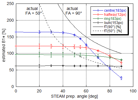
ii) Slice-profile bias was assessed on an 18cm Gadolinium-doted phantom (T1 = 400ms) by increasing α from 20° to 90°. Estimated flip angles were evaluated at 4 different ROIs (Figure 1).
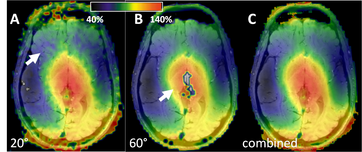
iii) In vivo, α was increased from 20° to 60° in steps of 5° (Figure 2) and used correct
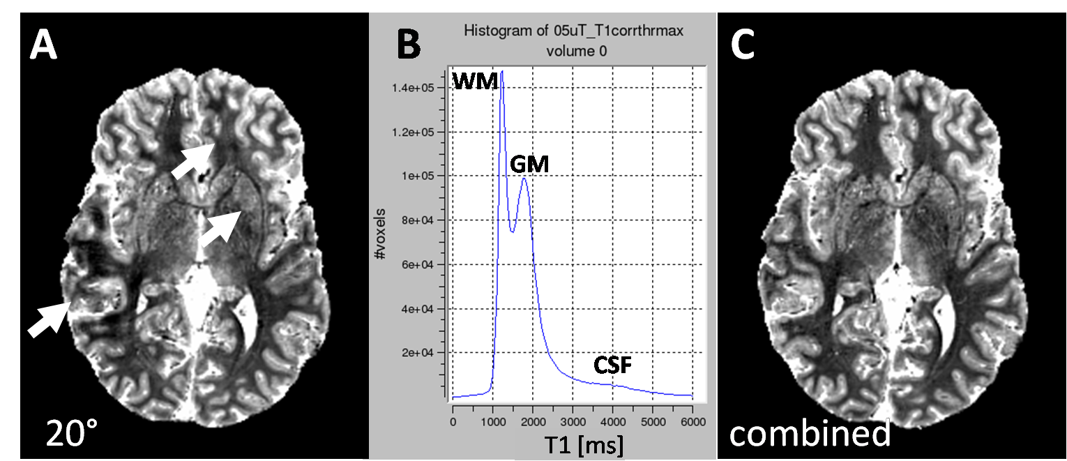
T1-maps from a non-selective dual flip angle experiment [6] (TR=18ms, TE=n∙1.97ms (n=1,…8), FA=4°,
18°) (Figure 3). Each B1+ map was ceilinged at 50° actual
flip angle to exclude bias. Before combining subsets, each B1+
map was thresholded to exclude large errors, creating a 5% overlap with
the ceilinged B1+ map of the next higher FA.
Results and Discussion
Above an actual FA of 50° of the preparation pulses, DREAM showed decreasing B1+ estimates (Figure 1). This bias is caused by non-linearities in the preparation pulses, creating broader excitation and flip-back profiles, but narrower saturation profiles above the small flip angle regime [7]. The actual STE and FID signals thus deviate from the theoretical sin2α and cos2α dependence [2]. The typical range of B1+ at 7T (50%-150%) will thus limit α to below 33° when using only one DREAM map, which yields low SNR in regions of low B1+ (Figure 2). This, in turn, introduces artifacts in the B1+-corrected T1 maps (Figure 3A) and limits spatial resolution. The SNR at low B1+ is governed by the cotangent of the actual FA [2] and thus suffering from high noise levels. This can be overcome by obtaining several DREAM B1+ maps at increasing α. These are then ceilinged at 50° actual FA to avoid bias and a composite B1+ map is combined from overlapping regions, typically between 30° and 50°. Thus, the dynamic range of DREAM enhanced while curtailing bias and avoing low SNR, which is important to cover the B1+ inhomogeneities in brain imaging at 7T or to map coil sensitivities for parallel transmit applications. For human brain at 7T, a DREAM series of 30°, 45° and 60° can be performed in about one minute.Conclusion
DREAM B1+ mapping exhibits slice-profile-related bias at actual FA>50°. By combining thresholded maps from STEAM preparation FAs between 20° and 90°, a bias free B1+ range between 20% and 250% can be obtained.Acknowledgements
Vincent O. Boer and Marjolein Piek, Danish Research Center for Magnetic Resonance (DRCMR), Copenhagen, for inspiring discussions on B1-mapping and dynamic range.References
[1] Weiskopf N, Lutti A, Helms G, Novak M, Ashburner J, Hutton C. Unified segmentation based correction of R1 brain maps for RF transmit field inhomogeneities (UNICORT) NeuroImage 2011 54(3):2116-2124.
[2] Nehrke K, Börnert P. DREAM – A novel approach for robust, ultrafast, multislice B1 mapping. Magn Reson Med. 2012 68:1517-1526.
[3] Nolte UG, Finsterbusch J, Frahm J. Rapid isotropic diffusion mapping without susceptibility artifacts: Whole brain studies using diffusion-weighted single-shot STEAM MR imaging. Magn Reson Med. 2000 44:731-736.
[4] Nehrke K, Versluis MJ, Webb A, Börnert P. Volumetric B1+ mapping of the brain at 7T using DREAM. Magn Reson Med. 2014 71:246-256.
[5] Helms G, Finsterbusch J, Weiskopf N, Dechent P. Rapid radio frequency field mapping in vivo using single-shot STEAM MRI. Magn Reson Med. 2008; 60(3):739-743.
[6] Helms G, Dathe H, Dechent P. Quantitative FLASH MRI at 3T using a rational approximation of the Ernst equation. Magn Reson Med 2008; 59:667-672.
[7] Helms G. Radiofrequenz-Pulse in der lokalisierten STEAM MR Spektroskopie. PhD thesis, Göttingen 1994.
Figures