4495
MR-assisted PET motion correction improves tumor-to-background and contrast-to-noise ratios in a phantom study with ground truth reference1Washington University in St. Louis, Saint Louis, MO, United States
Synopsis
Respiratory motion leads to signal blurring and reduced tumor-to-background (TBR) and contrast to noise (CNR) ratios. As a result, it can severely affect the detectability of lesions in PET imaging.1,2 Simultaneous PET/MR imaging uniquely allows for MR assisted motion correction in PET imaging.3 In this study, we have demonstrated that the MR assisted PET motion correction significantly improves both tumor-to-background and contrast-to-noise ratios, leading to better lesion detection.
Introduction
Respiratory motion leads to signal blurring and reduced tumor-to-background (TBR) and contrast to noise (CNR) ratios. As a result, it can severely affect the detectability of lesions in PET imaging.1,2 Simultaneous PET/MR imaging uniquely allows for MR assisted motion correction (MoCo) in PET imaging.3 In this study, we evaluated the impact of motion correction on TBR and CNR using a deformable motion phantom. Static and motion scans are performed in an interleaved way. The static scan is used as a ground truth reference to evaluate the performance of MR assisted PET MoCo.Methods
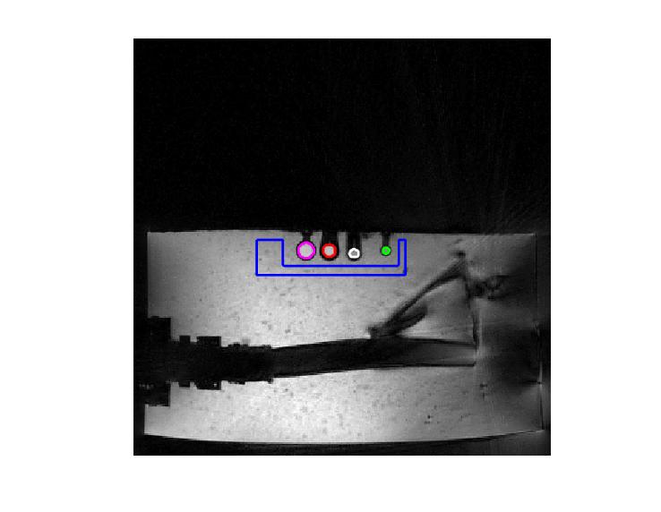
A recently published self-navigated free breathing MR motion correction method (CAPTURE: Consistently acquired projections for tuned and robust estimation), is utilized to derive deformable motion.4 The acquisition parameters were as follows: TE/TR=1.99ms/4.12ms, FOV= 240mm × 240mm, voxel size = 0.75 × 0.75 × 1.7mm3, acquisition time per measurement = 170 sec. A custom deformable motion phantom was made to generate respiratory-like motion by pumping air into a bellows immersed in gel using a physiological pump. Four spheres filled with [18F]2-fluoro-2-deoxyglucose (FDG) were placed inside the gel to mimic lesions. The diameters of the four spheres were 9 mm, 7.5 mm, 5.75 mm and 4,75mm, and their respective motion ranges were 10.8mm, 11.1mm, 11.9mm and 12.8mm. The motion phantom was filled with gel mixed with 11C tracers. The initial radioactivity in both the spheres and the gel was roughly 0.11mCi for 18F and 0.14mCi for 11C, resulting in an initial TBR of ~0.8. Since [11C] has a shorter half-life (20 min) compared to [18F] (109.7 min). TBR increases as time elapsed. This design allowed us to evaluate PET MoCo using a variety of TBRs in a single scan. 15 pairs of static (Motion-free) and motion states were induced by turning off and on physiological pump interleavely. PET listmode data were continuously acquired throughout the entire 15 pairs of static and motion states. Meanwhile, the MR CAPTURE scans were acquired simultaneously with PET. All images were acquired on a Siemens Biograph mMR system. Motion was detected by the MR CAPTURE sequence. Two types of PET listmode rebinning were used to reconstruct MoCo and motion compromised PET images. In the MoCo PET reconstruction, the MR-derived motion was used to re-bin the simultaneously acquired PET listmode data into 5 motion phases (MoCo rebining). In the motion compromised images, consecutively acquired listmode data were grouped into 5 bins (temporal rebinning) without motion correction. Moreover, the static PET listmode data were separated into 5 bins and reconstructed. The static reconstruction was considered as the ground truth for evaluating the performance of motion correction. All reconstructed PET images were fused with the MR images and MR images were used to define several ROIs on the spheres and background as shown in Figure 1. These ROIs were then applied to PET images. TBR was defined as TBR = SUVmedian(lesion ROI) / SUVmean(background). CNR was defined as CNR = (Smedian(lesion ROI)- Smean(background)) / σ(background). S is the signal intensity and σ is the standard deviation of the background noise. TBR and CNR were computed from all static, motion corrected, and motion compromised PET images in all four spheres.Results
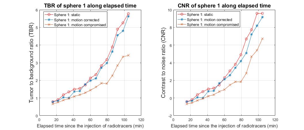
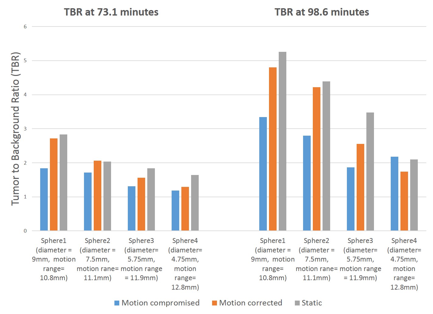
Figure 2 shows that MR assisted PET MoCo significantly and almost completely recovers reduced TBR and CNR in sphere 1. The performance of PET MoCo depends on both baseline TBR and lesion size (Figure 3).Discussion and Conclusion
Our MR-based motion correction method significantly improves TBR and CNR, leading to better lesion visibility and detectability. The detectability of lesions is dependent on the lesion size, motion range and background activity. Higher noise, larger respiratory motion and smaller TBR make it challenging to detect smaller lesions on PET images. Our MR-assisted PET motion correction makes it possible to detect lesions with significantly higher visibility and accuracy.Acknowledgements
No acknowledgement foundReferences
1. Nehmeh, S. A., et al. "Effect of respiratory gating on reducing lung motion artifacts in PET imaging of lung cancer." Medical physics 29.3 (2002): 366-371.
2. Bai, Wenjia, and Michael Brady. "Motion correction and attenuation correction for respiratory gated PET images." IEEE transactions on medical imaging 30.2 (2011): 351-365.
3. Würslin, Christian, et al. "Respiratory motion correction in oncologic PET using T1-weighted MR imaging on a simultaneous whole-body PET/MR system." J Nucl Med 54.3 (2013): 464-71.
4. Eldeniz, Cihat, et al. "CAPTURE: Consistently Acquired Projections for Tuned and Robust Estimation: A Self-Navigated Respiratory Motion Correction Approach." Investigative radiology 53.5 (2018): 293-305.
Figures