4493
Respiratory Motion Corrected GROG based L+S Reconstruction for Free Breathing Golden-Angle Radial MRI1Electrical Engineering, Comsats University Islamabad, Islamabad, Pakistan
Synopsis
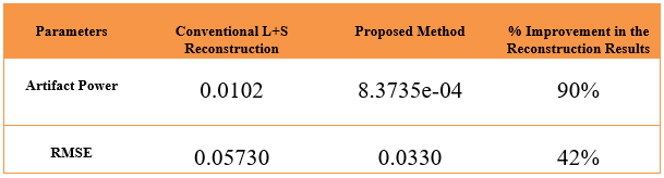
Respiratory motion during MRI scan causes inconsistencies in the acquired k-space data providing strong blurring artifacts in the reconstructed images. In this work, a new method ( respiratory motion corrected GROG followed by L+S reconstruction for free breathing Golden-Angle Radial DCE-MRI) is presented.The proposed method is tested on 3-T free-breathing Golden angle radial DCE liver MRI data. The proposed method is compared with the conventional L+S reconstruction model. The proposed method provides 90% improvement in Artefact Power and 42% in RMSE as compared to conventional L+S reconstruction at acceleration factor 8.
Introduction:
In free breathing dynamic MRI,k-space data is acquired in different respiratory positions, which results in strong blurring artefacts in the reconstructed images 1.Simplest approach to avoid respiratory motion is: (i) breath-hold data acquisition (ii)use of navigator signals that increases the patient discomfort and prolongs scan time 2,3. Low-Rank plus Sparse (L+S) reconstruction with effective separation of the background and dynamic components proposed by Otazo et.al. to reconstruct the unalised dynamic MR images 4.The L+S technique combines the idea of parallel MRI(pMRI),Compressed sensing(CS) and Principal Component Analysis(PCA) to reconstruct under-sampled MR images. L+S reconstruction is mathematically formulated as 4:
$$ min L,S 1/2 ||G.E (L+S) -y||2 + λL||L||* +λs||S||1 $$
where y is the under-sampled k-space data acquired from the MRI scanner, T is the temporal Total-Variation operator, E is the multi coil Encoding operator, λL and λs are the regularization parameters.G represents the gridding operation which converts non-Cartesian data onto Cartesian grid. Conventional L+S uses Fessler gridding to convert the radial data to a Cartesian grid 5.
In the proposed method, Golden-angle radial liver perfusion data is first sorted into time frames. Motion signal is extracted by using self-navigator properties of this Golden angle radial data 6. This data is gridded onto a Cartesian grid using SC GROG gridding 7,8.The resulting gridded data contains incoherent artifacts that are removed using L+S reconstruction.
Methods:
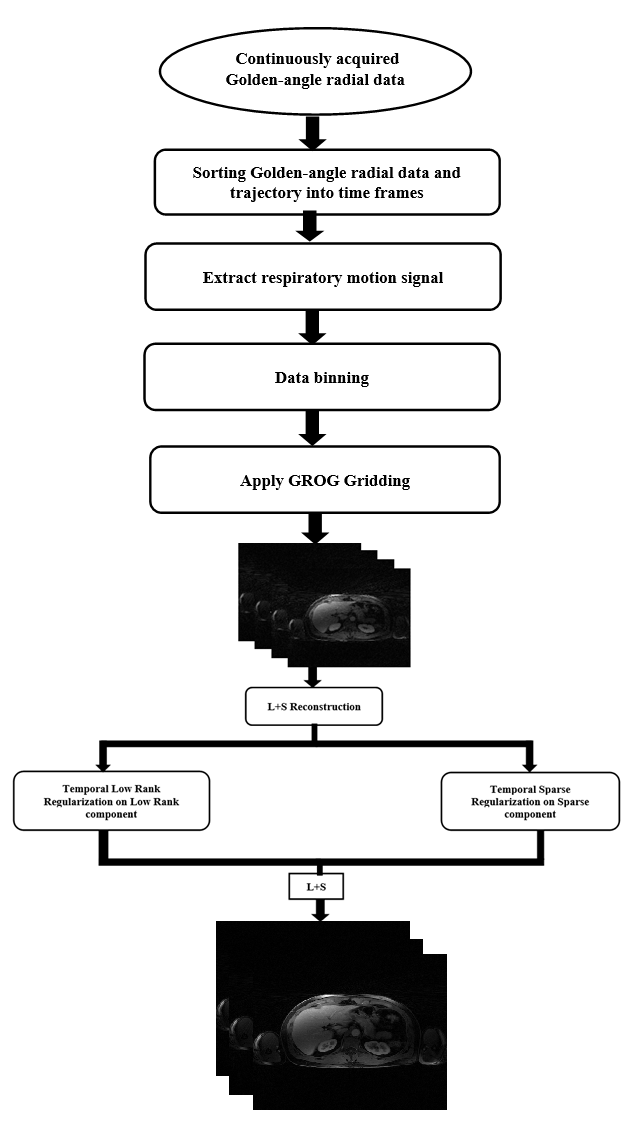
This work proposes a respiratory motion corrected GROG based L+S reconstruction with effective separation of the background and dynamic components for free breathing Golden-Angle Radial DCE-MRI data. In the proposed method, initially a motion signal is directly extracted from the center of the Golden angle radial k-space data 6. Golden-angle radial data is first sorted into time frames and binned into multiple motion states. The self-calibrated GROG (SC-GROG) gridding is applied to map the motion compensated Golden angle radial data on a Cartesian grid 9. Finally, L+S reconstruction is applied on the motion corrected GROG gridded data to get the final solution image.Figure 1 shows a flow chart of the proposed method.Results and Discussions:
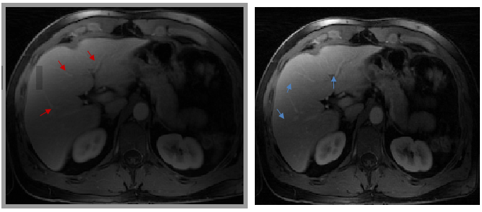
The proposed method is tested on free-breathing Golden angle radial DCE data acquired with 3-T Siemens scanner at New York University 2.The data acquisition parameters are: 512 readouts, 1144 radial spokes and 12 receiver coils. In order to generate a dynamic series, 96 adjacent spokes are grouped into one time point, these frames are further divided into 4 respiratory states. This data is gridded onto a Cartesian grid by using SC-GROG, corresponding Cartesian data size is : .The Nyquist sampling rate for this case is 512×π/2 =800,corresponding simulated Acceleration factor (AF) is 8.3 2. Figure 2 shows the reconstruction results. Left, middle and right hepatic veins are not clearly visible in the conventional L+S reconstruction (Figure 2a), while they can be clearly seen in the reconstruction results of the proposed method(Figure 2b).
AP and RMSE values of the reconstructed images for both the standard L+S and proposed methods are given in Table 1. The results show that the proposed method by incorporating motion correction frame work in L+S reconstruction outperforms conventional L+S method. For example there is 90% improvement in AP and 42% improvement in RMSE in the reconstruction results of the proposed method for Golden angle radial data at AF=8.
Conclusion:
In this work a respiratory motion corrected GROG based L+S reconstruction model with effective separation of Land S components for free breathing Golden-Angle Radial DCE-MRI data is proposed. The results show good quality reconstructed images with motion correction as compared to the conventional L+S approach.Acknowledgements
No acknowledgement found.References
1. Usman M, Atkinson D, Odille F, et al. Motion corrected compressed sensing for free-breathing dynamic cardiac MRI. Magn Reson Med. 2013;70(2):504-516.
2. Feng L, Grimm R, Block KT, et al. Golden-angle radial sparse parallel MRI: Combination of compressed sensing, parallel imaging, and golden-angle radial sampling for fast and flexible dynamic volumetric MRI. Magn Reson Med. 2014;72(3):707-717. doi:10.1002/mrm.24980.
3. Feng L, Axel L, Chandarana H, Block KT, Sodickson DK, Otazo R. XD-GRASP: Golden-angle radial MRI with reconstruction of extra motion-state dimensions using compressed sensing. Magn Reson Med. 2016;75(2):775-788. doi:10.1002/mrm.25665.
4. Otazo R, Candès E, Sodickson DK. Low-rank plus sparse matrix decomposition for accelerated dynamic MRI with separation of background and dynamic components. Magn Reson Med. 2015;73(3):1125-1136.
5. Fessler JA. On NUFFT-based gridding for non-Cartesian MRI. J Magn Reson. 2007;188(2):191-195.
6. Feng L, Huang C, Shanbhogue K, Sodickson DK, Chandarana H, Otazo R. RACER-GRASP: Respiratory-weighted, aortic contrast enhancement-guided and coil-unstreaking golden-angle radial sparse MRI. Magn Reson Med. 2018;80(1):77-89.
7. Aslam I, Najeeb F, Omer H. Accelerating MRI Using GROG Gridding Followed by ESPIRiT for Non-Cartesian Trajectories. Appl Magn Reson. September 2017:1-18.
8. Seiberlich N, Breuer FA, Blaimer M, Barkauskas K, Jakob PM, Griswold MA. Non-Cartesian data reconstruction using GRAPPA operator gridding (GROG). Magn Reson Med. 2007;58(6):1257-1265.
9. Wright KL, Hamilton JI, Griswold MA, Gulani V, Seiberlich N. Non-Cartesian parallel imaging reconstruction. J Magn Reson Imaging. 2014;40(5):1022-1040.
Figures