4492
Iterative Static Motion Compensated(IS-MoCo) Reconstruction: application to high resolution lung imaging1Radiology, University of California San Francisco, San Francisco, CA, United States, 2Bioengineering, University of California San Francisco, San Francisco, CA, United States, 3Department of Medical Physics, University of Wisconsin, Madison, Madison, WI, United States, 4Department of Electrical Engineering and Computer Sciences, UC Berkeley, Berkeley, CA, United States
Synopsis
High resolution 3D MRI thoracic and abdominal MRI is always challenging, due to long acquisition time and susceptibility to subject motion. We proposed a novel reconstruction method, named Iterative Static Motion Compensated(IS-MoCo) reconstruction, to compensate motion affects during the reconstruction instead of gating. The proposed method is applied to high resolution free breathing lung imaging, outperforms widely used motion correction strategies with higher SNR and less residual motion artifacts.
Introduction
Long scan time and body motion, especially respiratory motion make 3D high spatial resolution thoracic and abdominal MRI very challenging1. To avoid the respiratory motion effect, external respiratory monitor or navigator sequence are used to trigger the MRI acquisition at the same motion state. However, these gating methods would significantly reduce the scan efficiency and prolong the scan time2.To reduce the motion effect without sacrificing the acquisition efficiency, lots of work from both acquisition and reconstruction aspects have been done. One solution2,3 is to give acquired data different weightings based on the respiratory signal to compromise between motion artifacts and undersampling artifacts, called soft-gated based method. The other option is to group data with similar motion state and reconstruct different motion states images by using the mutual information among different states, such as XD-GRASP4. However, both methods did not fully utilize the motion information within the data. To improve that, motion compensated(MoCo) reconstruction could be used. The compensation could combine motion resolved images to one high SNR image5–7.In this work, we proposed a new reconstruction method to iteratively reconstruct a single motion state high resolution motion free image incorporating motion compensation, named Iterative Static Motion Compensated(IS-MoCo) reconstruction, and apply to high resolution lung imaging study. We evaluated our method via both volunteer and patient studies, and compared the reconstruction results with other state-of-art reconstruction methods.Methods
Acquisition
3D UTE sequence with free-breathing, golden angle ordering, and variable density readout acquisition was used for all the scans. The data were acquired with FOV varying from 28cm to 32cm, image matrix size 256x256x256, flip angle=4°, TE=80μs, sampling bandwidth=125kHz.
Coarse reconstruction and Motion estimation
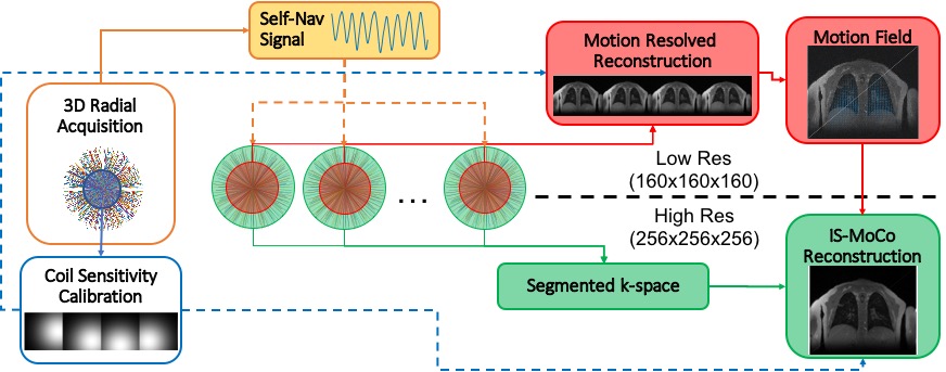
The proposed method consists of two main parts, one is motion resolved reconstruction and non-rigid motion estimation, the other is motion compensated reconstruction. The workflow is summed in Figure 1. Firstly, k0 data is used to track respiratory motion and sort the acquired data to different motion states, sensitivity maps are estimated by using the center k-space(blue circle). Then, relatively low resolution, ~2mm isotropic, motion resolved images are reconstructed via XD-GRASP4 method with part of the binned data(red circle). After that, motion fields are calculated by registering all the images to the same motion state. The derived motion fields finally are incorporated in the IS-MoCo reconstruction with all motion states data(green circle).
IS-MOCO reconstruction
Multi-channel sensitivity maps $$$S_i$$$, motion states sorted multi-channel data $$$d_{ik}$$$, and derived motion fields $$$M_k$$$ are all fed into the proposed reconstruction model.
$$ \underset{X}{\operatorname{argmin}} \sum_{i,k}^{N,m}||W(GS_iM_k-d_{i,k}||_2^2+\lambda_sTGV_s(X) $$
In the data consistency term(left), is sampling density compensation weights, is the gridding operator $$$G$$$, $$$F$$$ is FFT operator. $$$X$$$ is the final reconstructed image. A sparse penalty term(right) is added in the model, spatial total generalized variation(TGV)8. The problem is solved by using alternating direction method of multipliers(ADMM) framework. Then the problem is splitted into two sub-problems. As high resolution non-cartesian reconstruction with is very memory and computation intense, subproblems are solved by using first-order primal-dual algorithm.
Result
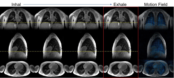
Figure 2 shows low resolution(~2.25mm isotropic) motion resolved images, the exhale state image is selected as the reference motion state, other motion states images are registered to the reference image via hierarchical demons registration9.
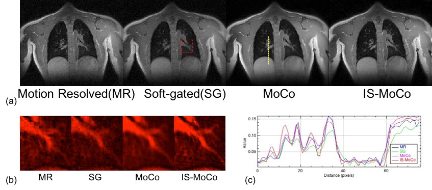
The proposed method is evaluated by comparing to other widely used motion correction reconstruction strategies, motion resolved reconstruction, soft-gated method and standard MoCo based method. Motion resolved(MR) reconstruction method is described in XD-GRASP. Soft-gated method sets the exhale state as reference, weighs data based on the respiratory signal. For standard MoCo based method, motion resolved images are reconstructed, then combined to single state image via registration. Figure 3 shows comparison on a volunteer study, the same coronal slice image of all the reconstruction methods are shown in (a), a small area(red dash rectangle) are zoomed in (b), and the slice profiles(yellow dash line) are plotted in (c). Compared to MR, IS-MoCo has higher SNR as all the data are used in IS-MoCo reconstruction, and some small airway could be told from the noise. Compared to SG, IS-MoCo has higher SNR and less motion corruption. Compared to standard MoCo reconstruction, from (b) and (c), the edge of structure, especially some small airways and vessels are more sharper.
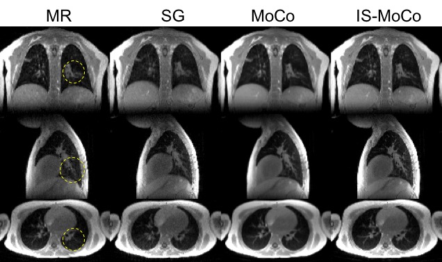
We also evaluate IS-MoCo on a pediatric patient, three orthogonal plane images comparison is shown in Figure 4. Images with IS-MoCo reconstruction has the high SNR, makes small lungs structure distinguishable(yellow dash circles).
Conclusion
In this work, we proposed a novel IS-MoCo reconstruction, and we applied this method to free breathing lung imaging. The proposed method has outperformed three widely used motion correction strategies on both volunteer and patient studies.The method could be extended to other applications in the future.Acknowledgements
The work is supported by NIH grant R01 HL136965.References
1. Feng L, Benkert T, Block KT, Sodickson DK, Otazo R, Chandarana H. Compressed Sensing for Body MRI. J Magn Reson Imaging. 2017;45:966–987. doi:10.1002/jmri.25547.
2. Cheng JY, Zhang T, Ruangwattanapaisarn N, et al. Free-breathing pediatric MRI with nonrigid motion correction and acceleration. J Magn Reson Imaging. 2015;42(2):407-420. doi:10.1002/jmri.24785.
3. Johnson KM, Block WF, Reeder SB, Samsonov A. Improved Least Squares MR Image Reconstruction Using Estimates of k- Space Data Consistency. 2012;1608:1600-1608. doi:10.1002/mrm.23144.
4. Feng L, Axel L, Chandarana H, Block KT, Sodickson DK, Otazo R. XD-GRASP: Golden-angle radial MRI with reconstruction of extra motion-state dimensions using compressed sensing. Magn Reson Med. 2015;00(October 2014):n/a-n/a. doi:10.1002/mrm.25665.
5. Pang J, Bhat H, Sharif B, et al. Whole-Heart Coronary MRA with 100 % Respiratory Gating Efficiency : Self-Navigated Three-Dimensional Retrospective Image-Based Motion Correction ( TRIM ). 2014;74:67-74. doi:10.1002/mrm.24628.
6. Manber R, Thielemans K, Hutton B, et al. MR Image-based PET Respiratory Motion Correction in PET/MR. J Nucl Med. 2015;56(supplement_3):98-. doi:10.2967/jnumed.114.151779.
7. Rank CM, Heußer T, Buzan MTA, et al. 4D Respiratory Motion-Compensated Image Reconstruction of Free-Breathing Radial MR Data With Very High Undersampling. 2017;1183(February 2016):1170-1183. doi:10.1002/mrm.26206.
8. Knoll F, Bredies K, Pock T, Stollberger R. Second Order Total Generalized Variation ( TGV ) for MRI. 2011;491:480-491. doi:10.1002/mrm.22595.
9. Thirion, J. Image matching as a diffusion process : an analogy with Maxwell ’ s demons. Med Image Anal. 2004;2(3):243-260.
Figures