4489
A segmented ultra-short echo (UTE) sequence equipped with robustness to respiratory motion1Mallinckrodt Institute of Radiology, Washington University in St. Louis, St. Louis, MO, United States, 2Siemens Healthineers, St. Louis, MO, United States
Synopsis
Synopsis
Introduction
Solid structures like tumors, bones and implants have traditionally been imaged with computed tomography (CT). However, CT uses ionizing radiation, and also requires breath-holds which can be difficult for some subjects. Recently, ultra-short echo (UTE) magnetic resonance imaging (MRI) has become popular in the way of replacing CT1–4. However, as with other free-breathing MR acquisitions, UTE scans must be coupled with motion correction. Several groups proposed various solutions such as using the k-space center as a navigator5 or using image-based navigation6,7. In this UTE study, we adopted Consistently Acquired Projections for Tuned and Robust Estimation (CAPTURE), the projection-based self-navigation that was recently published and shown to be quite robust8. The results show the promise of the method.Methods
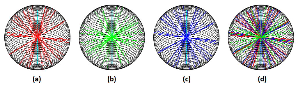
The proposed sequence is a modification the acquisition scheme proposed in2. Figure 1 illustrates the proposed method. Rather than traversing the whole sphere from the north pole towards the south pole in a continuous fashion, the proposed scheme traverses the whole sphere much more sparsely, but repeatedly, yielding numerous small segments. This allows for binning the data in many different ways, but still having a good coverage for each bin, be it for respiratory, cardiac or some other purpose. But more importantly, each segment begins by acquiring a straight line from the north pole towards the south pole and this is the only direction that is consistently being acquired across all segments. The consistency of this navigator spoke substantially cleans the spectrum of the respiratory curve because the gradient delays and eddy currents for all navigators will be similar. A detection scheme similar to proposed in8 was used for detecting the respiratory curves.
Acquisition parameters: TE1=0.07 ms, TE2=2.46 ms, TE3=3.69 ms and TE4=4.92 ms with TR = 6.94 ms. Only the second echo was used for detecting motion and the resulting curve was used for binning the data for the first echo. The two other echoes were not used for this particular study. FlipAngle1 = 3 deg and FlipAngle2 = 15 deg, which ran for 5 minutes each.
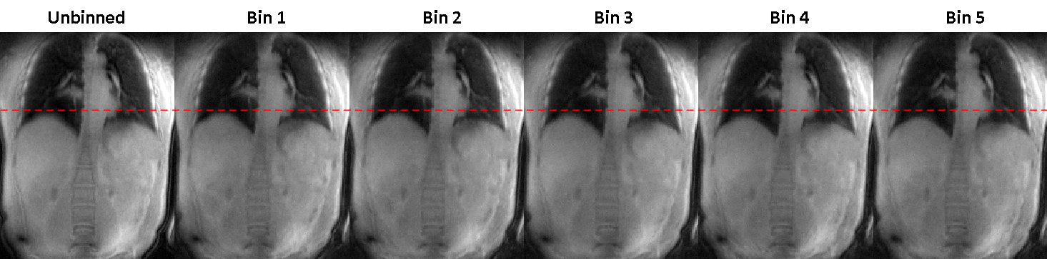
42.9K spokes were binned into 5 respiratory phases. The navigator spokes were also included into the reconstruction, so this self-navigation sequence is 100% efficient.
4 healthy informed subjects were recruited with an approved IRB.
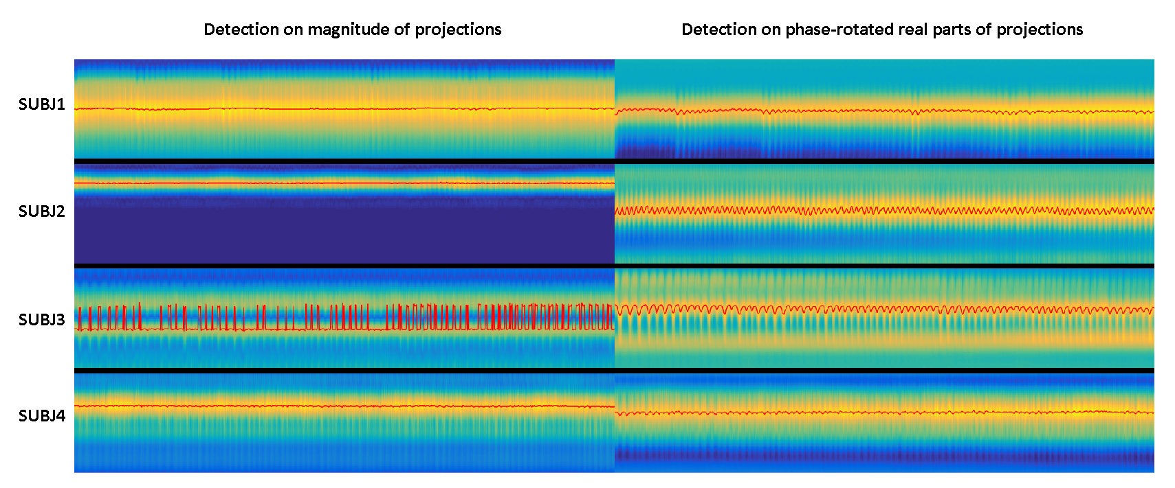
The spectrum of the navigator was compared with that of the point navigator. The phase-tuning-based detection was compared with magnitude-based detection.
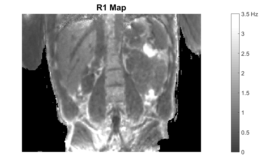
An R1 map was also calculated as a nice application for this variable-flip-angle set.
Results
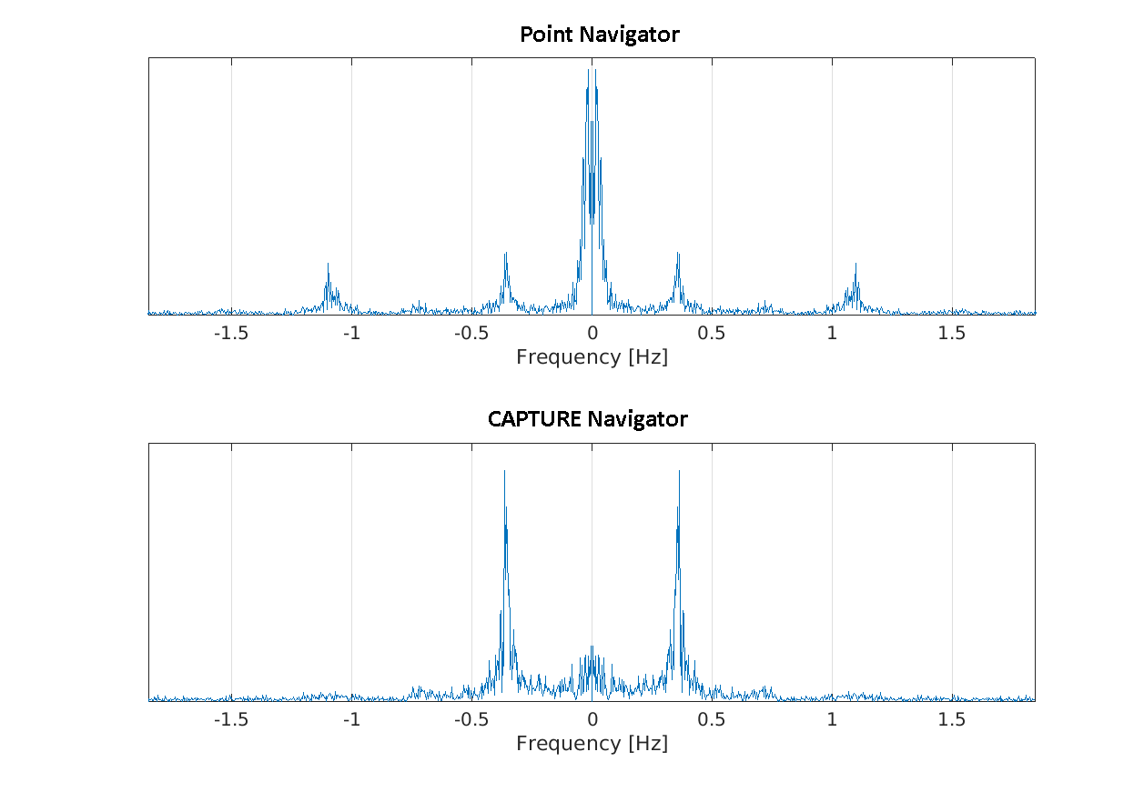
Figure 2 shows the cleanliness of the CAPTURE spectrum when compared with the point navigator.
Figure 3 shows that phase-tuning results in robust detection.
Figure 4 shows the success of binning.
Figure 5 shows the anatomical structures very well, proving the good quality of the reconstruction.
Discussion
Consistency of the navigator is the key for avoiding the severe contamination seen in the point navigator spectrum.
The images produced here can be used for tumor detection and radiation therapy planning. Bone structures that move with respiration [i.e. ribs] can also be imaged with this sequence.
Acknowledgements
No acknowledgement found.References
1. Robson MD, Gatehouse PD, Bydder M, et al. Magnetic resonance: an introduction to ultrashort TE (UTE) imaging. J. Comput. Assist. Tomogr. 2003;27(6):825–846.
2. Nielles‐Vallespin S, Weber M-A, Bock M, et al. 3D radial projection technique with ultrashort echo times for sodium MRI: Clinical applications in human brain and skeletal muscle. Magn. Reson. Med. 2007;57(1):74–81.
3. Takizawa M, Hanada H, Oka K, et al. A robust ultrashort TE (UTE) imaging method with corrected k-space trajectory by using parametric multiple function model of gradient waveform. IEEE Trans. Med. Imaging. 2013;32(2):306–316.
4. Johnson KM, Fain SB, Schiebler ML, et al. Optimized 3D Ultrashort Echo Time Pulmonary MRI. Magn. Reson. Med. 2013;70(5):1241–1250.
5. Higano NS, Hahn AD, Tkach JA, et al. Retrospective respiratory self-gating and removal of bulk motion in pulmonary UTE MRI of neonates and adults. Magn. Reson. Med. 2017;77(3):1284–1295.
6. Jiang W, Ong F, Johnson KM, et al. Motion robust high resolution 3D free-breathing pulmonary MRI using dynamic 3D image self-navigator. Magn. Reson. Med. 2018;79(6):2954–2967.
7. Tibiletti M, Paul J, Bianchi A, et al. Multistage three-dimensional UTE lung imaging by image-based self-gating. Magn. Reson. Med. 2016;75(3):1324–1332.
8. Eldeniz C, Fraum T, Salter A, et al. CAPTURE: Consistently Acquired Projections for Tuned and Robust EstimationA Self-Navigated Respiratory Motion Correction Approach. Invest. Radiol. 2018;53(5):293–305.
Figures