4487
Motion Correction Resolved for MRI via Multi-Tasking: A Simultaneous Reconstruction and Registration Approach1Department of Applied Mathematics and Theoretical Physics, University of Cambridge, Cambridge, United Kingdom, 2Department of Pure Mathematics and Mathematical Statistics, University of Cambridge, Cambridge, United Kingdom, 3Wolfson Brain Imaging Centre, Department of Clinical Neurosciences, University of Cambridge, Cambridge, United Kingdom, 4Cambridge University Hospitals, Department of Radiology, University of Cambridge, Cambridge, United Kingdom, 5INSA Rouen, Laboratoire de Mathématiques, Normandie Université, Saint-Étienne-du-Rouvray, France
Synopsis
The prolonged time required to form an MR image continues to impose different challenges at both theoretical and clinical levels. With this motivation in mind, this work addresses a central topic in MRI, which is how to correct the motion problem, through a new multitask optimisation framework. The significance is that by tackling the reconstruction and registration tasks $$$-$$$ simultaneously and jointly $$$-$$$ one can exploit their strong correlation reducing error propagations and resulting in a significant motion correction. The clinical potentials of our approach are reflected in having higher image quality with fewer artefacts whilst keeping fine details. We evaluate our approach through a set of quantitative and qualitative experimental results.
INTRODUCTION
The prolonged time required to form an MR image continues to impose different challenges at both theoretical and clinical levels. In particular, this is negatively reflected during the acquisition as involuntary physiological motion is introduced. This motion is manifested as undesirable artefacts including geometric distortions and blurring, which causes a significant degradation of the image quality and affects the clinical relevance for diagnosis$$$^{1,2,3,4}$$$. With this motivation in mind, this work addresses a central topic in MRI, which is how to correct the motion problem, through a new multitask optimisation framework. The significance is that by tackling the reconstruction and registration tasks $$$-$$$ simultaneously and jointly $$$-$$$ one can exploit their strong correlation reducing error propagations and resulting in a significant motion correction. The clinical potentials of our approach are reflected in having higher image quality with fewer artefacts whilst keeping fine details. We evaluate our approach through a set of quantitative and qualitative experimental results.THEORY AND METHODS
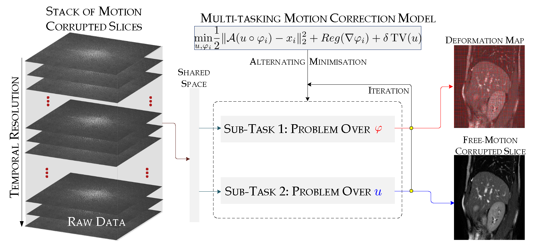
In a MRI setting, a target image $$$u\in \mathbb{R}^{N}$$$ representing a part of the patient body is acquired in spatial-frequency space. The measured samples can be represented in a matrix form as $$$x=\mathcal{A}u+\varepsilon$$$ where $$$x \in\mathbb{C}^{M} (M\ll N)$$$ refers to the $$$\mathbf{k},t-$$$space measurements, $$$\mathcal{A}:\mathbb{R}^N \rightarrow \mathbb{C}^M$$$ is the Fourier operator (neglecting the phase), and $$$\varepsilon$$$ models some noise. For a multiple receiver coil, $$$\mathcal{A}$$$ encodes coils sensitivities and the Fourier transform.In this work, we seek to extract simultaneously from a set of multiple MR acquisitions $$$x_i$$$, corrupted by motion, a mean static and clean reconstructed image $$$u$$$ as well as the deformation maps $$$\varphi_i$$$ aligning each image of the set to the mean image. Combining these two tasks in a unified variational framework, our optimisation problem is the following:
$$ \begin{align}\nonumber \min_{u, \varphi_i} \bigg\{ & \frac{1}{T}\sum_{i=1}^{T} \Big(\underbrace{ \beta ( \| \nabla \varphi_i \|^2 - \alpha)^2 \cdot H_{\epsilon}(\| \nabla \varphi_i \|^2 - \alpha) + \varPsi( \operatorname{det} \nabla \varphi_i)}_{\text{$=Reg(\nabla \varphi_i)$, nonlinear-elasticity-based regularisation}} \label{P} \\ \nonumber & + \underbrace{\frac{1}{2} \|\mathcal{A}(u\circ \varphi_i) - x_i\|_2^2 \Big)}_{\text{fidelity term intertwining registration and reconstruction tasks}} \\ &+ \delta \underbrace{\operatorname{TV}(u)}_{\text{edge preserving regularisation}} \bigg\},\\\nonumber \text{with } \psi : \mathbb{R} \rightarrow \mathbb{R},\,& s\mapsto -\frac{\mu}{2}s^2 + \mu(s-1)^2+\frac{\mu(\lambda + \mu)}{2(\lambda+2\mu)},\\\nonumber H_\epsilon &\text{ is the regularized Heaviside function,}\\\nonumber \mu=800,\text{ and } \lambda=10& \text{ are the Lamé coefficients.}\end{align} $$
This minimisation problem for motion correction is composed of three terms: (i) a nonlinear-elasticity-based regulariser that describes the nature of the deformations $$$-$$$ we model the organs as homogeneous, isotropic, and hyperelastic materials (more precisely, as Saint Venant-Kirchhoff materials) as shown in $$$^{5,6}$$$; (ii) a discrepancy term that enforces the deformed mean to match the acquisitions; (iii) a total variation (TV) type regulariser for edge preservation of the reconstructed image. We obtain an approximate solution by an alternating optimisation scheme. Our approach is summarised in Figure 1.
Results
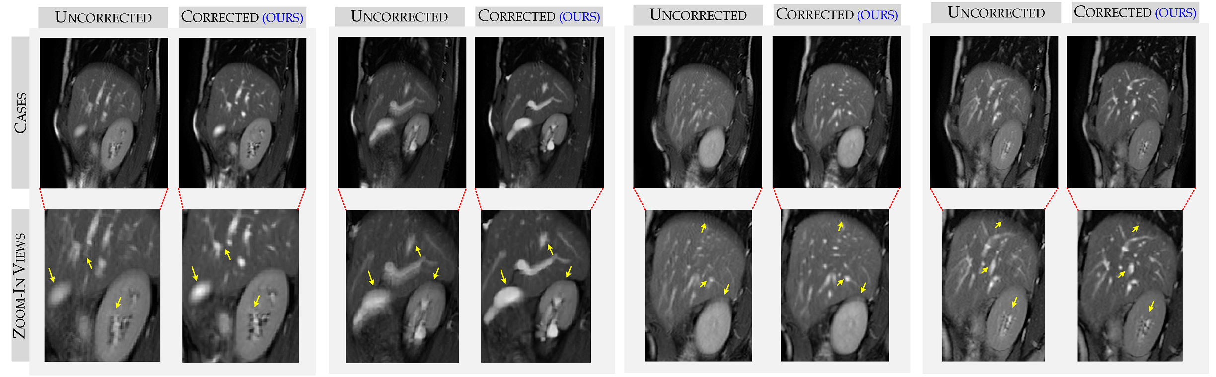
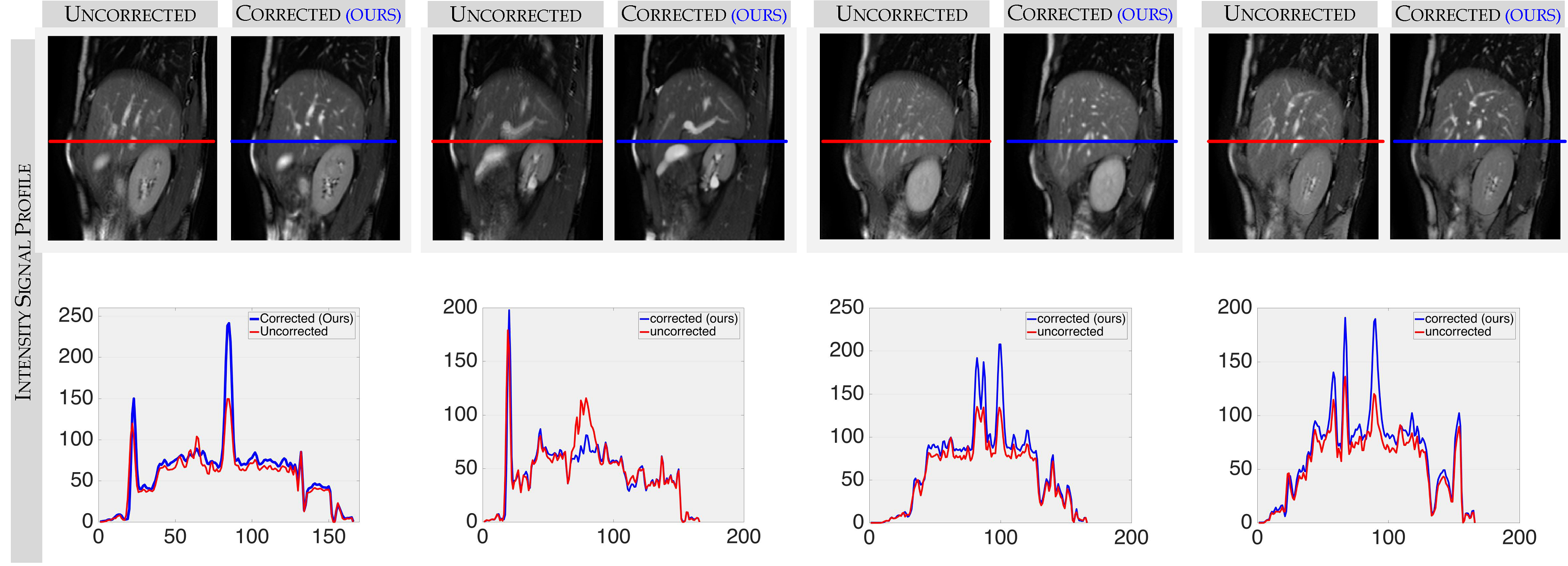
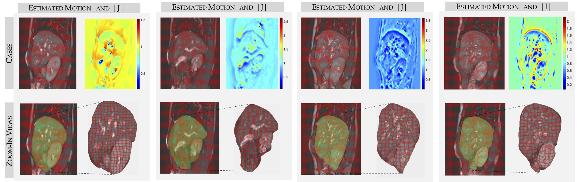
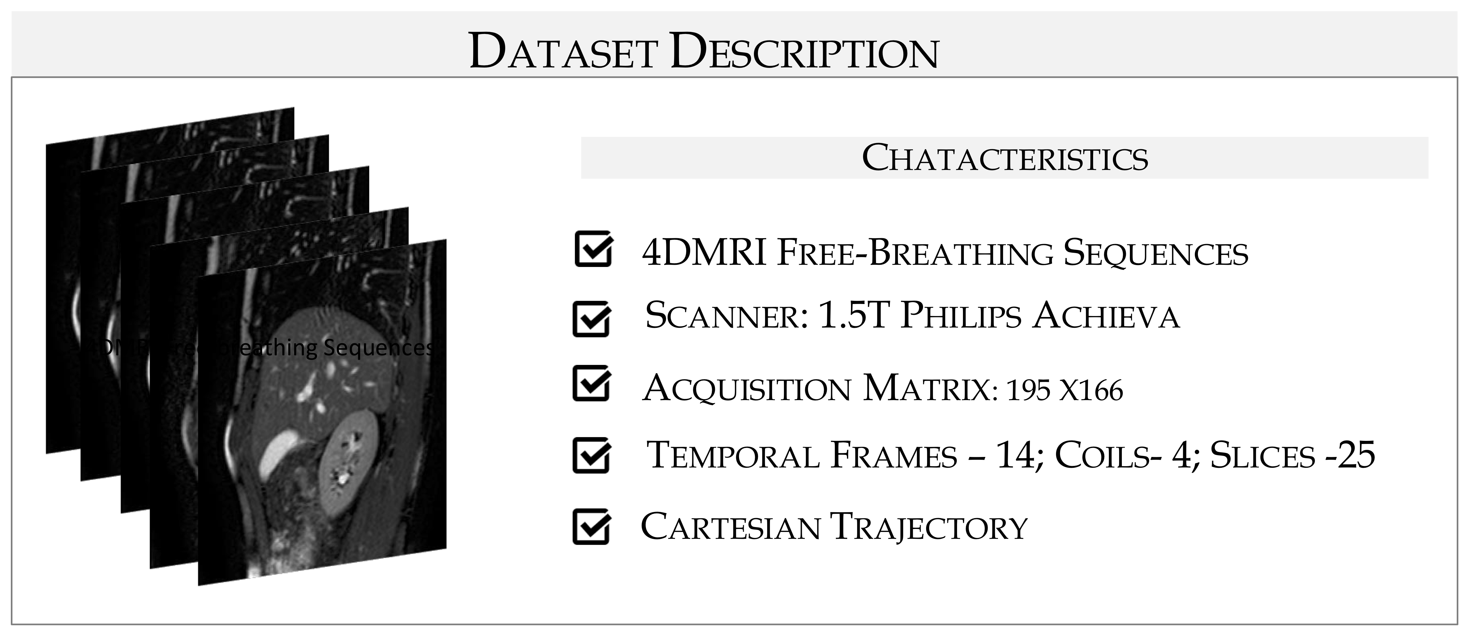
$$${\bf{Data \text{ } description.}}$$$ We evaluate our framework on 4DMRI sequences$$$^7$$$ acquired during free-breathing of the right liver lobe. A detailed description is given in Figure 5. $$${\bf{Results.}}$$$ We test our method for the full temporal resolution of the dataset and show our results for some sample frames. Figure 2 displays a comparison between uncorrected and motion-corrected mean of the samples. In a closer look at the zoom-in views, we observe that our approach allows for better reconstructions in terms of contrast, shape and fine details preservation whilst reducing blur artefacts (see yellow arrows). These results are further supported by the signal intensity profiles in Figure 3. In particular, we see that the amplitude values of the fluctuations in the uncorrected samples are smaller which can be translated into more blurry artefacts. For a more detailed analysis, we display in Figure 4, the deformation grids overlaying the sample frames along with the Jacobian determinant of the deformations to show critical details of the estimated motion. In a closer inspection, it is to be noticed that our framework achieves a plausible computation of the breathing dynamics of the liver lobe (positivity of the determinant everywhere).Conclusion
Our multi-task approach exploits redundancy in the temporal resolution to correct for motion artefacts due to breathing. Our framework provides better quality reconstructions, showing promising potential to improve diagnostic in clinical practice.Acknowledgements
The authors acknowledge the financial support of the Cancer Research UK, Cambridge Cancer Centre, the Cantab Capital Institute for the Mathematics of Information and the Centre of Mathematical Imaging in Healthcare.References
[1] Pipe JG. Motion correction with PROPELLER MRI: application to head motion and free‐breathing cardiac imaging. Magnetic Resonance in Medicine 1999; 42(5), 963-969.
[2] Zaitsev M, Maclaren J and Herbst M. Motion artifacts in MRI: a complex problem with many partial solutions. Journal of Magnetic Resonance Imaging 2015; 887-901.
[3] Aviles-Rivero AI, Williams G, Graves M and Schonlieb CB. CS+M: A Simultaneous Reconstructionand Motion Estimation Approach for Improving Undersampled MRI Reconstruction. In Proceedings of the 26th Annual Meeting ISMRM 2018.
[4]Aviles-Rivero AI, Williams G, Graves M and Schonlieb CB. Compressed Sensing Plus Motion (CS+M): A New Perspective for Improving Undersampled MR Image Reconstruction. arxiv:1810.10828, 2018
[5] Derfoul R, Le Guyader C, A relaxed problem of registration based on the Saint Venant-Kirchhoff material stored energy for the mapping of mouse brain gene expression data to a neuroanatomical mouse atlas, SIAM Journal on Imaging Sciences 2014; 7, 2175-2195.
[6] Ozeré S, Gout C, Le Guyader C, Joint segmentation/registration model by shape alignment via weighted total variation minimisation and nonlinear elasticity, SIAM Journal on Imaging Sciences 2015; 8, 1981-2020.
[7] von Siebenthal M, Skékely G, Gamper U, Boesiger P, Lomax A, Cattin Ph. 4D MR imaging of respiratory organ motion and its variability. Physics in Medicine and Biology 2007; 52(6), 1547-1564. (\url{http://www.vision.ee.ethz.ch/~organmot/chapter_download.shtml})
Figures