4479
Free breathing & Ungated Multi-Slice cardiac cine MRI using spiral-SToRM1Electrical Engineering, University of Iowa, Iowa city, IA, United States, 2Biomedical Engineering, University of Iowa, Iowa city, IA, United States, 3Radiology, University of Iowa Hospitals and Clinics, Iowa city, IA, United States, 4Healthcare, GE, Munich, Germany
Synopsis
The advantages of cardiac cine MRI are often limited by its long acquisition and breath-held requirement. To overcome these limitations, we have introduced a navigator based spiral SToRM to acquire free breathing and ungated cardiac cine MRI in a short acquisition time. Our algorithm is fully automated and does not depend on explicit binning. It gives improved image quality compared to the existing self-gated methods. Post-reconstructions, the time series can be processed to extract cardiac cycles at different respiratory phases, facilitating the estimation of anatomical and functional evaluation of the heart.
Background
Many subject groups (e.g pediatric & CoPD patients) are often excluded from current breath-held cardiac cine MRI protocols due to their inability in holding breath. Recently, several free breathing acquisition schemes that explicitly bin the data to a few cardiac and respiratory phases, followed by compressed sensing recovery have been introduced to overcome this difficulty [1-3]. We have recently introduced an implicit binning strategy termed as smoothness regularization on manifold (SToRM) approach [4], which enables the recovery of ungated cardiac cine imaging data in the free breathing mode, where we relied on radial single slice acquisition schemes.Methods
The main focus of this study is to demonstrate the ability of SToRM to enable multi-slice free breathing and ungated cine data using a navigated spiral. SToRM formulates the recovery of the image time series as the optimization algorithm:
$$\mathbf X^{*} =\arg \min_{\mathbf X} \|\mathcal A(\mathbf X)-\mathbf B\|^{2}_{F} + \lambda~ \mathrm{{\rm trace}}(\mathbf X {\mathbf L} \mathbf X^{H})$$
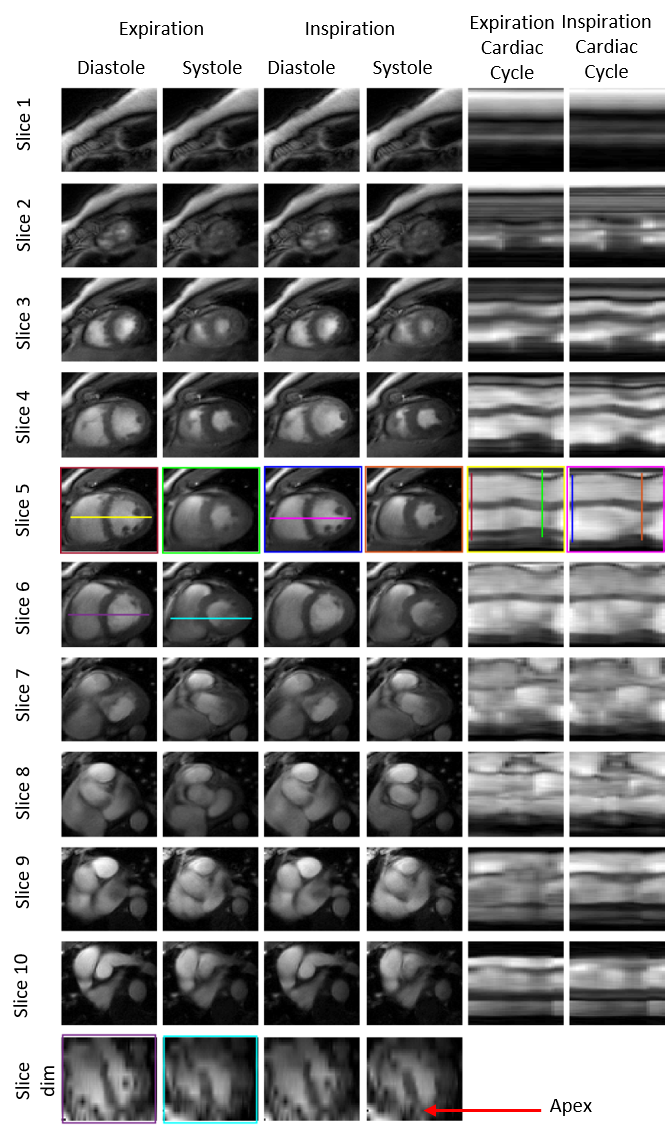
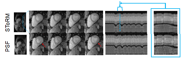
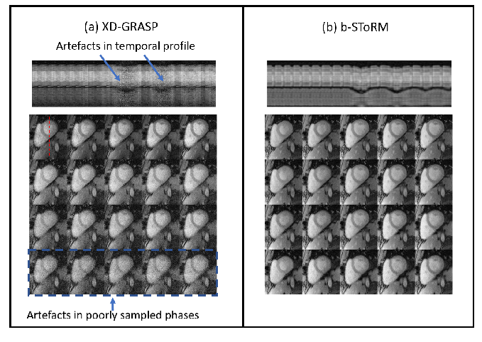
where L is the manifold Laplacian matrix that is estimated from the k-space navigators using an iterative kernel low-rank method. Multislice spiral data was collected on a 3T GE scanner using a golden-angle rotated gradient echo spiral pulse sequence with a navigator after every six spirals. Sequence parameters include: TR 8ms, slice thickness 10mm, resolution 1.25mm2, number of slices 10 covering the whole heart, starting from apex to base of the heart, total acquisition time 4 mins. The image time-series from each slice is reconstructed using SToRM as shown in Fig. 1. Following reconstruction, matching cardiac cycles corresponding to expiration and inspiration are identified automatically by an image-based comparison, and shown in Fig. 2; this facilitates the estimation of cardiac function parameters from the data. We also compare the algorithm against existing low-rank (PSF) and XD-GRASP using single slice radial GRE sequences on a 1.5T Siemens Aera scanner, as shown in Fig. 3 and 4.
Results
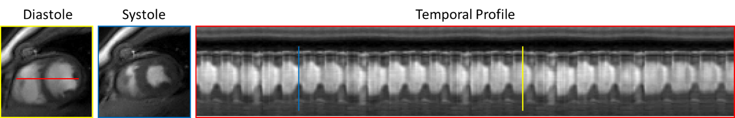
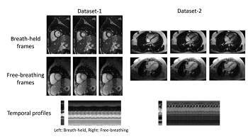
Fig. 1 shows two frames of a slice and its time profile. Note that we recover the entire time series similar to real-time acquisitions. The images from two cardiac cycles per slice are shown in Fig. 2. Each row shows the diastole and systole frames at the end of expiration and inspiration states of a slice and the last two columns show their respective cardiac cycles. The stacked image time series can also be reformatted to obtain a four chamber view of the heart from the apex to base, as shown in the bottom row. The comparison with PSF in Fig. 3 shows the improved image quality offered by SToRM. Similarly, the comparisons against XD-GRASP in Fig. 4 shows the ability of SToRM to yield good reconstructions in subjects with large respiratory motion. The comparisons in Fig. 5 show that SToRM can yield similar image quality as breath-held acquisitionsDiscussion
In this work, we have acquired and reconstructed multislice spiral cine MRI using navigator-based SToRM algorithm. The data from four minute multislice acquisition can provide three-dimensional coverage of the myocardium. Post-recovery, the data can be reformatted to evaluate the cardiac functional parameters such as stroke volume and ejection fraction, in different respiratory phases. The comparison of SToRM against state of the art methods demonstrate the benefits. Note that the approach does not require explicit binning, and hence it may be used in subjects with limited respiratory breath-holding capability (e.g COPD subjects) as well as patients with arrythmias.Conclusion
Navigator based spiral SToRM offers a reliable option to acquire free breathing and ungated data from the entire heart in a short acquisition time. The proposed algorithm is fully automated and does not require any explicit binning. It also offers improved image quality compared to state of the art self-gated methods. Post-reconstructions, the time series can be processed to extract cardiac cycles at different respiratory phases, facilitating the estimation of anatomical and functional evaluation of the heart.Acknowledgements
No acknowledgement found.References
1. Zhou R, Yang Y, Matthew R, Salerno M. “Cardiac and Respiratory Self-Gated Motion-Corrected Free-Breathing Spiral Cine Imaging, Proceedings of the 26th ISMRM 2018
2. Otazo, Ricardo, Emmanuel Candès, and Daniel K. Sodickson. "Low‐rank plus sparse matrix decomposition for accelerated dynamic MRI with separation of background and dynamic components." Magnetic Resonance in Medicine 73.3 (2015): 1125-1136.
3. Feng, Li, Leon Axel, Hersh Chandarana, Kai Tobias Block, Daniel K. Sodickson, and Ricardo Otazo. "XD‐GRASP: golden‐angle radial MRI with reconstruction of extra motion‐state dimensions using compressed sensing." Magnetic resonance in medicine 75, no. 2 (2016): 775-788.
4. Poddar, Sunrita, and Mathews Jacob. "Dynamic MRI using smoothness regularization on manifolds (SToRM)." IEEE transactions on medical imaging 35.4 (2016): 1106-1115.
5. Liang, Zhi-Pei. "Spatiotemporal imagingwith partially separable functions." In Biomedical Imaging: From Nano to Macro, 2007. ISBI 2007. 4th IEEE International Symposium on, pp. 988-991. IEEE, 2007.
Figures