4478
Self-navigation Liver Respiratory Motion Correction Based on Deep Learning1Department of Biomedical Engineering, School of Medicine, Tsinghua University, Beijing, China, 2School of Biomedical Engineering and Imaging Sciences, King's College London, London, United Kingdom
Synopsis
Correction of respiratory motion with 100% acquisition efficiency is of great significance for clinical abdominal imaging. In this study, we propose a novel self-navigation liver respiratory motion correction method for 3D radial sampling. This new approach is based on the fact that radial acquisition enables oversampled k-space center to extract motion-state signal and neural network can be used for data dimensionality reduction. Both regular and irregular hepatic breathing experiments were conducted and the proposed method has shown similar reconstruction image quality with bellow.
Introduction
In liver MR imaging, the limitation of the encoding speed will lead to artifacts and image blurring since k-space lines may be acquired in different respiratory motion states for free-breathing acquisition1. There are some traditional methods to reduce motion corruption such as breath-holding2, navigator echoes3 and auxiliary hardware gating. Nevertheless, these approaches will either restrict total scan time or cause inefficient acquisition. Therefore, self-navigation respiratory motion correction method is of great need, and motion binning may also provide extra motion information1. Since neural network uses nonlinear data modeling to discover latent and complex relationships superior than conventional machine learning methods, this study used convolutional neural network (CNN) to acquire motion information directly from k-space center.Methods
Data Acquisition: A total of 24 datasets were acquired from 12 free-breathing volunteers on a 3T scanner (Philips, Achieva) with 16-channel SENSE Torso coil, using a 3D Golden-Angle Radial Stack-of-Stars SPGR sequence4, with TR=4.4 ms, TE=1.93 ms, flip angle=15°, FOV=320×320×120 mm3 with 1.5×1.5 mm2 in-plane resolution and 5 mm slice thickness. During the acquisition, a bellow was used to record respiratory motion. Among 24 datasets, 20 datasets were acquired with required regularly breathing during the scan, while the another 4 datasets were imaged with instructed irregularly breathing including short respiration cessation and deeply breathing.
Date Preparation: The k-space center data is a 3D matrix with each dimension representing spoke, slice and coil. For the purpose of data augmentation, one k-space center was divided into 128 datasets by cutting the spoke dimension into 8 parts and each coil component (16-channel coil) was regarded as separate individuals. At the same time, logs recorded by bellow were divided into the same size corresponding to the input. Before sent into the CNN, the input was also pre-processed by normalization and filtered by hamming window.
Neural Network: The input of the neural network was a 2D matrix with each dimension meaning spoke and slice. The network architecture was composed of three convolutional layers with 3×3 kernel. The output was the desired motion curve and the label was the bellow logging. This PyTorch model was accelerated by NVIDIA Titan Xp GPU with MSE as the cost function, Epoch=20, Batch size=4, lr=0.001.
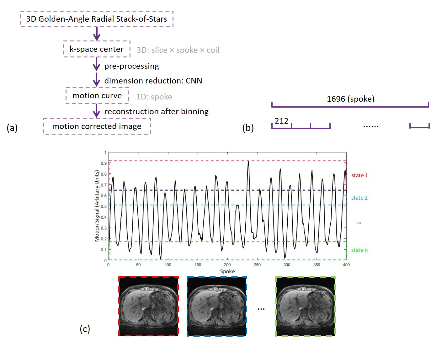
Data Analysis: Motion curves: Desired integrated motion curves which were cut into 8 parts along spoke dimension for data augmentation are jointed together according to the original order. Since PCA1 is a conventional and relatively accurate method for dimensionality reduction, the motion curve derived from deep learning was compared with PCA. Correlation Coefficients (CCs) were calculated between the motion curve derived from deep learning and bellow as well as the motion curve derived from PCA and bellow. Imaging: According to the moving distance indicated by motion signal, radial k-space spokes were sorted into a specific number of respiratory states with each state having the same number of spokes. Binned spokes were reconstructed with iGRASP5 (Fig. 1).
Results
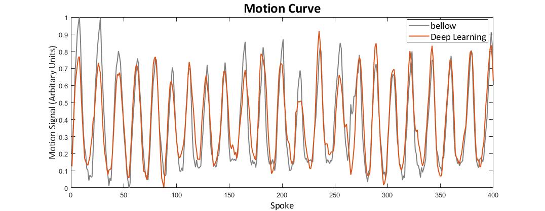
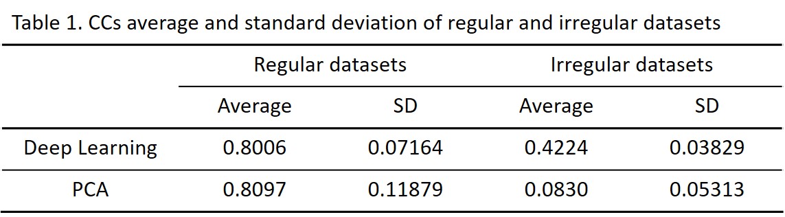
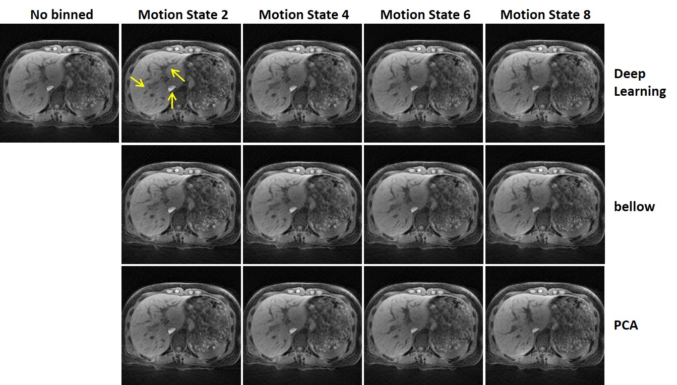
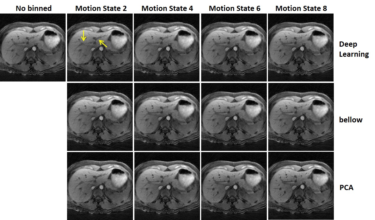
Fig. 2 demonstrates the motion curve derived from deep learning, which is very similar with bellow curve. For 20 regular breathing datasets, both deep learning method and PCA have similar average CCs with bellow (Table 1). But for 4 irregular breathing datasets, the CCs of deep learning results are higher than PCA. For all the datasets, the standard deviation of deep learning is lower than PCA, representing better robustness of deep learning method. As for the reconstructio, improved image quality can be achieved after motion state binning using the motion curve obtained by CNN than no-binned data, especially at details such as blood vessels (Fig. 3, 4). Besides, the quality of reconstructed images using the bellow was similar with that using PCA for regular and irregular breathing datasets.
Discussion and Conclusions
This result demonstrates the feasibility of the proposed CNN method in obtaining respiration motion curve and correct motion artifacts in liver imaging using radial sampling. The motion curve produced by the proposed deep learning method is not inferior to the bellow and conventional PCA for both regular breathing and irregular breathing data. Since CCs of PCA with bellow are much lower than that of deep learning for irregular datasets, it may be possible that deep learning can extract complicated and non-linear relationships from aperiodic irregular data than linear PCA. For future experiments, navigator echoes should be the golden standard for motion curve to achieve better training results because it directly reflects the liver motion other than the chest wall motion measured by bellow.
In conclusion, this proposed deep learning technique, with 100% acquisition efficiency to retrospectively acquire self-navigation motion curve without any auxiliary hardware equipment, is comparable with bellow and may have tremendous potential in outperforming conventional motion correction methods especially for irregular breathing data.
Acknowledgements
No acknowledgement found.References
[1] Feng L, Axel L, Chandarana H, et al. XD-GRASP: Golden-angle radial MRI with reconstruction of extra motion-state dimensions using compressed sensing[J]. Magnetic resonance in medicine, 2016, 75(2): 775-788.
[2] McVeigh E R, Atalar E. Cardiac tagging with breath‐hold cine MRI[J]. Magnetic Resonance in Medicine, 1992, 28(2): 318-327.
[3] Ehman R L, Felmlee J P. Adaptive technique for high-definition MR imaging of moving structures[J]. Radiology, 1989, 173(1): 255-263.
[4] Block K T, Chandarana H, Milla S, et al. Towards routine clinical use of radial stack-of-stars 3D gradient-echo sequences for reducing motion sensitivity[J]. Journal of the Korean Society of Magnetic Resonance in Medicine, 2014, 18(2): 87-106.
[5] Feng L, Grimm R, Kai T B, et al. Golden-Angle Radial Sparse Parallel MRI: Combination of Compressed Sensing, Parallel Imaging, and Golden-Angle Radial Sampling for Fast and Flexible Dynamic Volumetric MRI[J]. Magnetic Resonance in Medicine, 2015, 72(3):707-717.
Figures