4458
Three-dimensional, fast parameter mapping at 7.0T with SSFP MR Fingerprinting: comparison of radial and spiral projections k-space trajectories1Laboratory of Medical Physics and Magnetic Resonance - IRCCS Stella Maris Foundation and IMAGO7 Foundation, Pisa, Italy, 22Department of Computer Science, Technische Universitat Munchen, Germany;, Munich, Germany, 3University of Pisa, Pisa, Italy, 4GE Healthcare, Munich, Germany
Synopsis
When using ultra-high field MRI scanners (UHF, B0>= 7T), quantitative imaging is challenging due to B0 and B1+ non-uniformities. Magnetic resonance fingerprinting (MRF) represents a great opportunity for quantitative imaging at UHF as it can estimate these effects at the same time of the parameters of interest. Here, we compare two novel 3D SSFP MRF approaches, one based on a three-dimensional spiral projection acquisition and one using a radial acquisition in vivo at 7.0T. We estimate M0, T1, T2 and B1+ simultaneously at high resolution (1mm isotropic) within 6.5 minutes acquisition time.
Purpose:
Recent technological advances have improved on the effectiveness of ultra-high field MRI scanners (UHF, B0>=7T). Although new contrasts have been explored at high field strengths, common approaches are inherently non-quantitative, mostly due to the difficulties in dealing with B0 and B1+ non-uniformities, as well as a higher specific absorption rate (SAR). Magnetic resonance fingerprinting (MRF) represents a great opportunity for quantitative imaging at UHF as it does not require fields to be homogeneous. By including nuisance parameters into the signal encoding model it is possible to estimate these at the same time with the parameters of interest.
Here, we compare two novel 3D SSFP MRF approaches based on a three-dimensional radial and spiral projection k-space acquisitions, estimating M0, T1, T2 and B1+ simultaneously, demonstrating it in vivo at 7.0T.
Methods:
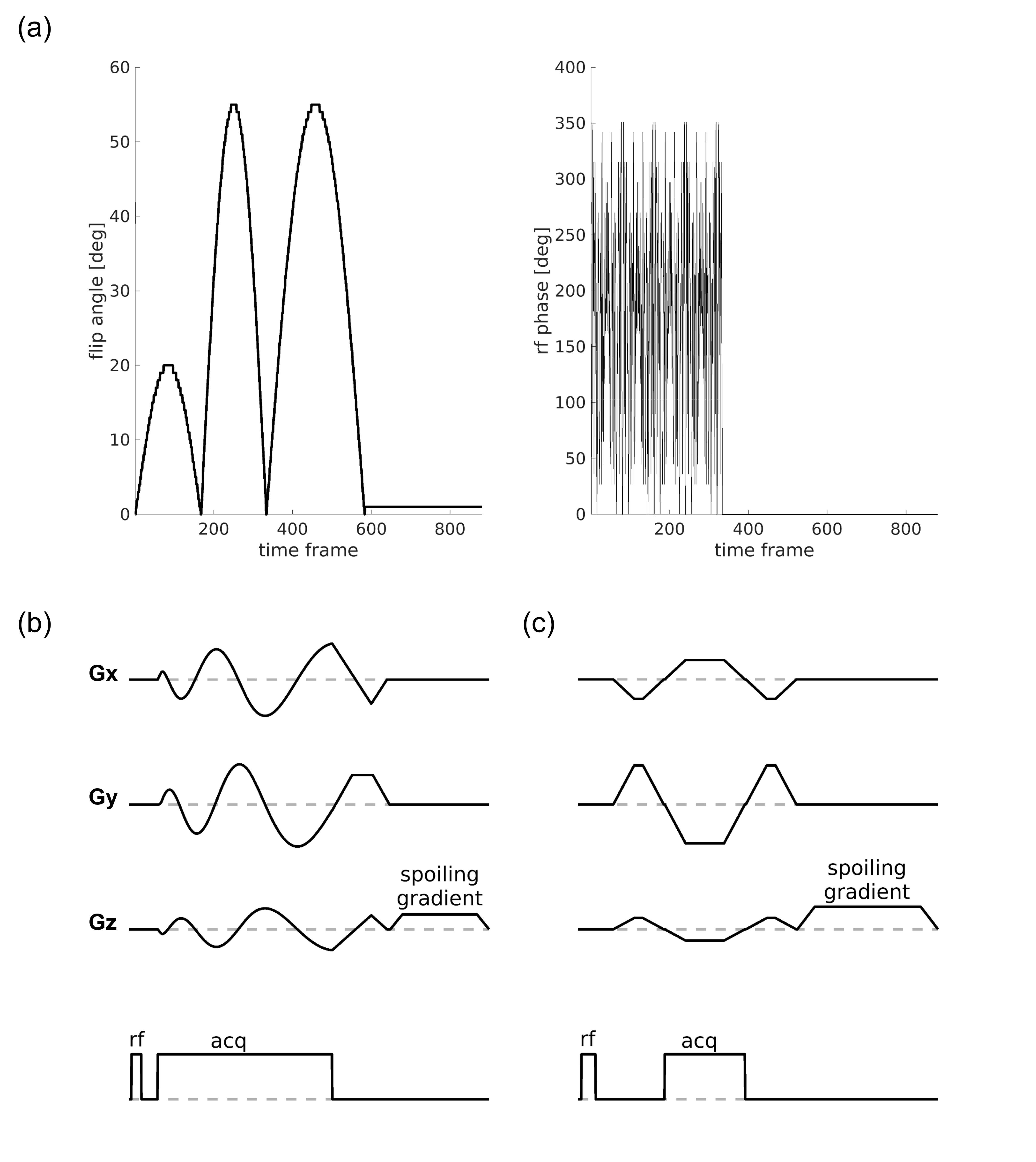
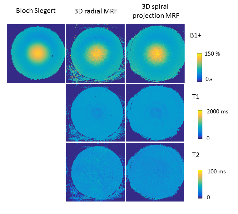
Acquisition parameters: We obtained images of a uniform phantom (T1 600ms, T2 45ms) and a healthy volunteer using a Discovery MR950 7T MRI system (MR950, GE Healthcare, Milwaukee, WI, USA) equipped with a 2-channel transmit / 32-channel receive head coil (Nova Medical, Wilmington, MA, USA). The acquisition sequence was based on SSFP MRF 1 (FOV 19.2x19.2x19.2cm3, 192x192x192 matrix), using hard pulses of 500 us length for excitation. We used a fixed TR of 8ms, acquiring 56 repetitions of 880 frames preceded by a 10ms-long hyperbolic secant adiabatic inversion pulse, using the flip angle and phase lists in Figure 1a. To increase the capability of the sequence to discriminate T2 and B1+ effects, we introduced radiofrequency spoiling in the first part of the acquisition following Cloos et al2. For comparison of the B1+ values, in the phantom we acquired a standard 2D Bloch-Siegert B1 map3 (TR=100ms, TE=13ms, Flip Angle=30°, 4ms Fermi pulse, 2kHz off-resonance).
3D spiral projection trajectory: Three dimensional spiral projections, as recently described by Castets et al4 and by Cao et al5 were obtained rotating a variable density spiral with 704 readout points at 250kHz sampling rate, 55 interleaves in-plane every 880 frames, and rotating the plane by the golden angle for each frame, achieving a total of 49280 interleaves. Each trajectory achieved zero-moment nulling in x, y and z after which a spoiler in z was added achieving 4π dephasing across a 1-mm voxel (see figure 1b).
3D Radial trajectory: For comparison with 3D spiral projections, we designed a matching 3D radial trajectory consisting of 49280 spokes of 358 readout points at 250kHz sampling rate, fully-sampling the 3D kspace for achieving 1mm isotropic resolution. Each spoke was acquired symmetrically with respect to k-space center and achieved zero-moment nulling in x, y and z, after which a spoiler in z was added achieving 4π dephasing across a 1-mm voxel (see figure 1c). To increase spatial and temporal incoherence, we randomly permutated the spoke indexes using a uniform probability distribution.
For both k-space trajectories tested, prior to reconstruction and matching, k-space data were combined using SVD compression and adaptive coil combination was performed in the image domain.
Results
All acquisitions tested at 7.0T achieved a specific absorption rate (SAR) lower than 2 W/Kg, showing that these are safe at ultra-high field. Acquisition time for both MRF acquisitions was 6.5 minutes.
Figure 2 shows the comparison of the MRF B1+ map from our MRF acquisitions with a standard 2D Bloch-Siegert map acquired in 7 minutes, showing good agreement.
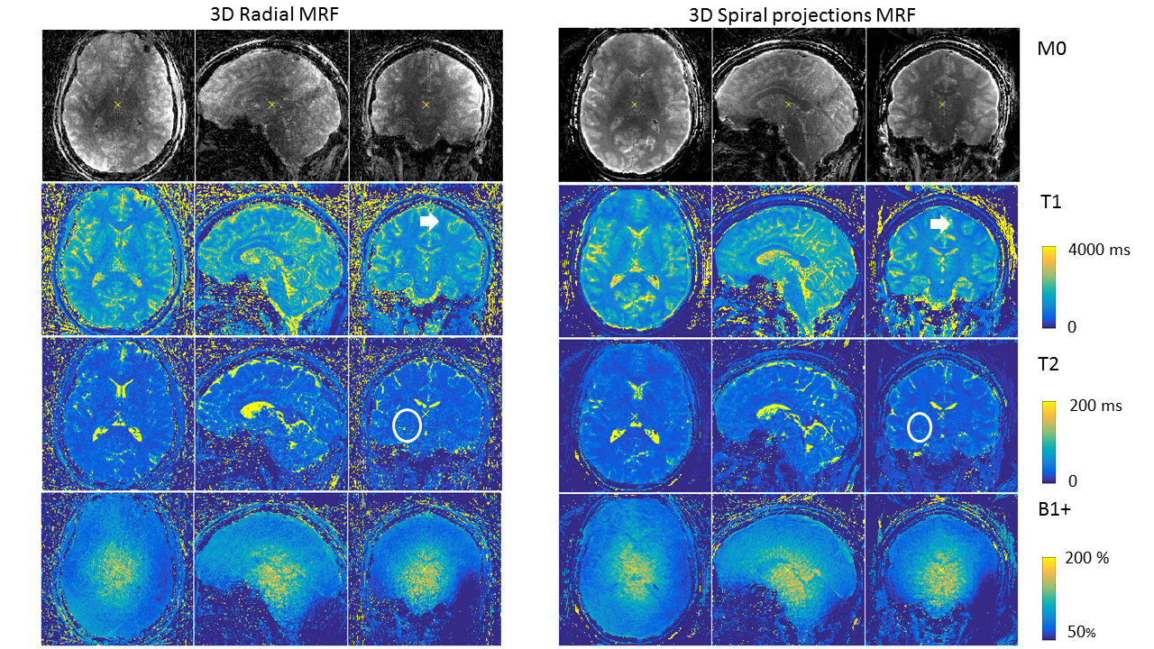
Figure 3 shows representative parametric maps in one subject. It can be noted that T1 and T2 maps are free from B1+ artifacts. Three-dimensional spiral projections obtained clearer images, due to the more efficient sampling of the edges of k-space, when compared to 3D radial trajectories. However, despite the relatively short spiral readouts, the radial images achieved higher robustness to blurring artifacts and a higher anatomical accuracy.
Conclusion
We could obtain an estimate of T1, T2, M0 and B1+ with 1mm isotropic resolution in 6.5 minutes at 7.0T. Further optimised acquisitions can achieve higher spatial resolution and include other biophysical parameters of interest. Spiral projection imaging obtained less undersampling artifacts when compared to 3D radial acquisitions of k-space, while 3D radials achieved a higher anatomical detail.Acknowledgements
No acknowledgement found.References
[1] Jiang Y, Ma D, Seiberlich N, Gulani V, Griswold MA. MR fingerprinting using fast imaging with steady state precession (FISP) with spiral readout; Magn Reson Med. 2015 Dec;74(6):1621-31. doi: 10.1002/mrm.25559. Epub 2014 Dec 9.
[2] Martijn A. Cloos, Florian Knoll, Tiejun Zhao, Kai T. Block, Mary Bruno, Graham C. Wiggins, and Daniel K. Sodickson. Multiparametric imaging with heterogeneous radiofrequency fields. Nat Commun. 2016; 7: 12445.
[3] Sacolick LI, Wiesinger F, Hancu I, Vogel MW. B1 mapping by Bloch-Siegert shift. Magn Reson Med. 2010 May;63(5):1315-22. doi: 10.1002/mrm.22357.
[4] Charles R. Castets William Lefrançois Didier Wecker Emeline J. Ribot Aurélien J. Trotier Eric Thiaudière Jean‐Michel Franconi Sylvain Miraux. Fast 3D ultrashort echo‐time spiral projection imaging using golden‐angle: A flexible protocol for in vivo mouse imaging at high magnetic field; Magn Reson Med. 2017 May;77(5):1831-1840. doi: 10.1002/mrm.26263. Epub 2016 May 12.
[5] Xiaozhi Cao, Congyu Liao, Qing Li, Huihui Ye, Hongjian He, Jianhui Zhong. Fast 3D MR fingerprinting with spiral projection acquisition for whole brain quantification imaging; Proceedings of the 26th ISMRM annual meeting, Paris 2018. Program number 1017
Figures