4443
Video-based head motion assessment for improved quantitative neuroanatomy studies1Department of Neurology, NYU Langone Medical Center, New York City, NY, United States, 2Center for Brain Imaging, New York University, New York, NY, United States
Synopsis
In-scanner head motion systematically varies with age and diagnosis, and this motion causes bias in morphometric estimates derived from neuroanatomical MRI. There are currently no widely available methods for directly assessing head motion during acquisition of neuroanatomical sequences. In this project we developed a method for measuring head motion via analysis of video obtained from an in-scanner eye tracker. Data obtained from 5 healthy controls demonstrates the feasibility of the technique. The system has minimal set up requirements for subjects or MR technicians, which suggests the technique may be well suited to the young, elderly, or impaired populations in which participant compliance may be a problem.
Introduction
In-scanner head motion affects the quality of neuroanatomical MRI scans and subsequent morphometric estimates derived from imaging data. We recently demonstrated that head motion is increased in both younger and elderly populations, as well as in individuals with neurological and psychiatric disorders [1]. Despite the confounding effects of head motion in quantitative neuroimaging studies, there are no widely available existing techniques to directly estimate this important property. In this study we utilize video obtained from an in-scanner eye tracker to directly estimate head motion in healthy control participants carrying out deliberate head movements in the scanner.Methods
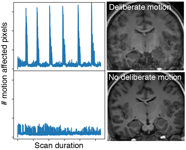
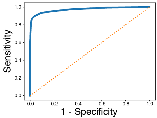
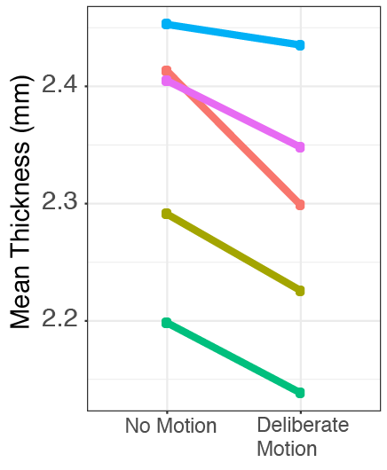
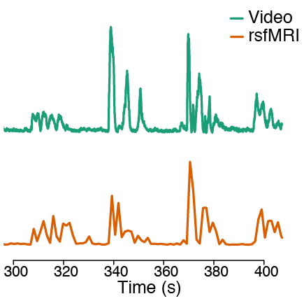
Five healthy control participants were imaged. Four whole brain T1-weighted MPRAGE acquisitions were obtained with voxel size = 1 mm³ on a Siemens 3T Prisma magnet. Two acquisitions were carried out with deliberate “no-no” head motion every 30 TRs, and two acquisitions were obtained without deliberate head motion. A 7 minute resting state EPI-BOLD acquisition was obtained (TR = 1.3 s). For this scan the first 2 minutes was obtained with the participant’s head held still, then the participant moved their head in series of deliberate rotational movements every 30s: (i) “no-no”; (ii) “yes-yes”; (iii) the head tilted with ears moving towards each shoulder. Video was recorded simultaneously using an in-scanner Eyelink 1000 Plus Eyetracker. Video data was processed using the OpenCV-Python software package. Motion affected pixels were detected in each frame by subtracting stationary background pixels identified using a mixture-of-gaussian background subtraction algorithm [2]. Frame-wise motion pixel counts were summed and ROC curves were calculated by comparing the number of suprathreshold pixels for a range of thresholds. True positive findings were defined as suprathreshold total pixel counts in frames occurring during deliberate motion epochs; false positive findings were defined as suprathreshold pixel counts in frames occurring outside deliberate motion epochs. Cortical thickness estimates were calculated for each volumetric scan using Freesurfer v6.0. Motion estimates were obtained in the resting-state fMRI acquisition using standard fMRI-based methods implemented in FSL [3].Results
Motion detection analysis of video data obtained from the in-scanner eyetracker during acquisition of MRI scans was able to clearly detect deliberate head motion in all five participants; an example plot of framewise motion pixel counts is shown in Figure 1. ROC analyses indicated that the technique was able to detect deliberate head movements with high sensitivity and specificity; mean AUC = 0.95 ± 0.03 SD (Figure 2). Mean whole brain cortical thickness in all participants was reduced in the motion-affected scans relative to motion-free scans (Figure 3), as reported in previous studies [1]. Comparison of the video-based motion assessment with standard image-based methods for motion correction in fMRI studies indicates high agreement between the two methods; an example subset timeframe is shown in Figure 4.Discussion
This study demostrates the feasibility of assessing head motion using computational analysis of in-scanner video. The method may be used to directly measure and therefore statistically control for head motion in quantitative studies of neuroanatomy. Although this study utilized video output from a commercial eye tracker, we believe that it would be straightforward to apply this methodology to any in-scanner camera system. The use of a video/optical signal allows for sampling at a significantly higher temporal frequency than existing fMRI-based methods; therefore the method may have utility for detecting head micro-movements that cannot be detected using standard fMRI-based methods. The technique may be readily applied to other MR acquisitions for which few (if any) motion assessment methods exist, such as diffusion MRI studies. Although our technique works well in healthy individuals undertaking deliberate head motion in the scanner, a primary future goal is to demonstrate the utility of the technique for detecting natural in-scanner head motion.Conclusions
We present a video-based technique for directly assessing in-scanner head motion during MRI acquisition. The method may find application as a quality assurance tool for quantitative studies of human neuroanatomy.Acknowledgements
This project was supported by the FACES foundation (Finding A Cure for Epilepsy and Seizures)References
1. Pardoe, H.R., R. Kucharsky Hiess, and R. Kuzniecky, Motion and morphometry in clinical and nonclinical populations. Neuroimage, 2016. 135: p. 177-85.
2. KaewTraKulPong, P. and R. Bowden, An Improved Adaptive Background Mixture Model for Real-time Tracking with Shadow Detection, in Video-Based Surveillance Systems: Computer Vision and Distributed Processing, P. Remagnino, et al., Editors. 2002, Springer US: Boston, MA. p. 135-144.
3. Power, J.D., et al., Spurious but systematic correlations in functional connectivity MRI networks arise from subject motion. Neuroimage, 2012. 59(3): p. 2142-54.
Figures