4436
A novel, coil-integrated camera for prospective optical motion correction of brain imaging at 7T1Stanford University, Stanford, CA, United States
Synopsis
The advancements in signal to noise ratio (SNR), contrast, and resolution enabled by high-field MR systems may visualize more nuanced brain anatomy and pathology. In order to translate these advancements to the discovery and clinical implementation of novel neuroimaging biomarkers, motion artifact resulting from requisite long scan times must be addressed. Here, we demonstrate a novel prospective optical motion tracking and correction system using a camera seamlessly integrated into the 7T Tx/Rx head coil. The integrated camera allows tracking of head motion by visualizing an optical marker on the forehead of human subjects in a 7T MR system.
Introduction
High-field (7T) MRI systems can produce images with higher SNR and spatial resolution than clinical MR systems (1.5, 3T). However, achieving both high SNR and high resolution requires long scan-times, often resulting in image artifacts due to subject motion. Addressing motion artifact is a pivotal step in translating 7T systems to identify novel imaging-based biomarkers of disease. Retrospective methods for motion correction are available1 but require longer post-processing time and may degrade images from overlaps and gaps in K-space.2 Prospective motion correction methods, which compensate for motion in real-time, often rely on optical monitoring using a camera to obtain the required tracking data. Several studies have successfully used MR-compatible cameras mounted inside the bore of the magnet for optical tracking at 7T. However, these studies have used mouthpieces to mount the tracking marker and/or large bore-mounted cameras,3-4 limiting line-of-sight on systems utilizing close-caged head coils. Our prior work implemented a proof-of-concept prospective motion correction system at 7T with a coil-mounted camera external to the coil pointing towards a marker attached to the subject’s cheek.5 The goal of the present study was to fully integrate the camera by mounting between Tx and Rx shells, allowing the optical marker to be placed on the subject’s forehead, providing a more rigid coupling between the subject’s head and the marker with much improved patient tolerance. This is expected to improve system performance and enable prospective tracking at 7T in patients, facilitating discovery of novel neuroimaging biomarkers.Methods
The prospective tracking system utilized here consisted of an MR‐compatible camera, a marker, and a tracking computer. Utilizing a CAD model of the transmit and receive portions of a Nova Medical 2 Tx/32 Rx head coil for a 7T GE MR950 scanner, a single MR-compatible camera was designed and built to be mounted between the Tx and Rx coils (Figure 1), enabling direct line-of-sight to the checkerboard-marker placed with adhesive on the subject’s left forehead. The location of the camera within the MRI bore was calibrated as in our prior work.6 Initial validation of was conducted using a ‘papaya phantom’, with deliberate rotational (rotating ~45-degrees to the left or right every 30 seconds) motion applied using a wooden rod. A T1-weighted whole-brain 3D-BRAVO sequence (0.94x0.94x1.5mm, TE=3.12, TR=9.4, 4min) was acquired twice, once without and once with motion correction. To test the system in vivo, the same 3D-BRAVO as well as 2D-GRE (6 slices at 0.31x0.31x1.2mm3, TE=20, TR=300, 2.5min) sequences were each acquired four times on a single subject: no motion with and without correction, with motion (discrete rotations ~45 degrees to the left or right every 30 seconds) with and without correction. To then demonstrate the utility of this system in enabling high-quality acquisitions of otherwise untenably long scans with ultra-high-resolution, a 2D GRE sequence (6 slices at 0.14x0.14x1.0mm3, TE=20, TR=300, 8NEX, 20min) was acquired with no deliberate motion and with correction.Results
The phantom validation study shows demonstrable improvement of motion by prospective motion-correction (Fig. 2B).
On a volunteer without deliberate rotational motion, the images are of similar quality without (Fig. 3A) or with (Fig. 3C) motion-correction. Deliberate motion without correction resulted in poor and almost unusable image quality (Fig. 3B, 4B). This was significantly improved with motion-correction (Fig. 3D, 4D), enabling visualization of both gray-white differentiation (Fig. 3D) and deep-gray and brainstem nuclei (Fig. 4D).
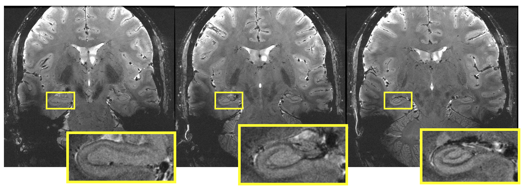
The ultra-high-resolution 2D GRE sequence with motion correction demonstrated exquisite quality images despite a long scan time and with high-resolution visualization of the hippocampus and remainder of visualized brain (Fig. 5).
Discussion
Motion-correction for both deliberate and involuntary motion was successfully demonstrated on human subjects, producing high-quality images, without introducing significant spurious artifacts. This study demonstrates the feasibility of a prospective optical motion-tracking system at 7T using a coil-integrated camera and marker attached to the subject’s forehead.Conclusion
Our motion-correction system demonstrates potential to overcome motion artifact at 7T, one of the biggest challenges precluding the utilization of ultra-high-field MR in both research and clinical settings. This practical and user-friendly design allows longer scan-times and finer image resolution, which may facilitate discovery of novel biomarkers of aging and disease in the brain.Acknowledgements
The authors would like to acknowledge research support by GE Healthcare, NIH P41 EB015891, NIH S10 RR026351-01A1, and ASNR Boerger Alzheimer’s Fund.References
1. Gallichan D, Marques JP, Gruetter R. Retrospective correction of involuntary microscopic head movement using highly accelerated fat image navigators (3D FatNavs) at 7T. Magn Reson Med. 2016; 75: 1030–1039. doi: 10.1002/mrm.25670 PMID: 25872755 2. Federau C, Gallichan D (2016) MotionCorrection Enabled Ultra-High Resolution In-Vivo 7TMRI of the Brain. PLoS ONE 11(5): e0154974. doi:10.1371/journal.pone.0154974 3. Stucht D, Danishad KA, Schulze P, Godenschweger F, Zaitsev M, Speck O. Highest Resolution In Vivo Human Brain MRI Using Prospective Motion Correction. PLoS One. 2015; 10: e0133921. doi: 10.1371/journal.pone.0133921 PMID: 26226146 4. Mattern, H. , Sciarra, A. , Godenschweger, F. , Stucht, D. , Lüsebrink, F. , Rose, G. and Speck, O. (2018), Prospective motion correction enables highest resolution time‐of‐flight angiography at 7T. Magn. Reson. Med., 80: 248-258. doi:10.1002/mrm.27033 5. DiGiacomo P, Maclaren J, Aksoy M, Burns B, Bammer R, Rutt B, Zeineh M. Optical prospective motion correction for brain imaging at 7T without a mouthpiece. Poster presentation at the International Society for Magnetic Resonance in Medicine, Paris, France, June 21, 2018. 6. Maclaren, J., Aksoy, M., Ooi, M. B., Zahneisen, B., & Bammer, R. (2018). Prospective motion correction using coil‐mounted cameras: Cross‐calibration considerations. Magnetic resonance in medicine, 79(4), 1911-1921.Figures