4434
Robust motion regression of resting-state data using a convolutional neural network modelZhengshi Yang1, Xiaowei Zhuang1, Karthik Sreenivasan1, Virendra Mishra1, and Dietmar Cordes1,2
1Cleveland Clinic Lou Ruvo Center for Brain Health, Las Vegas, NV, United States, 2University of Colorado, Boulder, CO, United States
Synopsis
The fluctuation introduced by head motion considerably confounds the interpretation of resting-state fMRI data. Specifying motion regressors without taking fMRI data itself into consideration may not be sufficient to model the impact of head motion. We proposed a robust and automated deep neural network (DNN) to derive motion regressors with both fMRI data and estimated realignment parameters considered. The results show that DNN-derived regressors outperform traditional regressors based on several quality control measurements.
Introduction
The fluctuation introduced by head motion considerably confounds the interpretation of resting-state functional magnetic resonance imaging (rs-fMRI) data [1]. Estimated rigid-body motion parameters and their variances were commonly used as nuisance regressors to reduce motion-related artifacts [2]. These regressors were generated without taking fMRI data itself into consideration and thus may not be sufficient to model the impact of head motion because of the complexity of motion artifacts. To better model how in-scanner motion affects rs-fMRI data, we have developed a robust and automated deep neural network model to derive motion regressors with both fMRI data and motion parameters considered.Methods
Subjects: The structural MRI and rs-fMRI data used in this study were downloaded from the publicly available ADNI database (http://adni.loni.usc.edu/). 76 subjects identified as normal controls by site investigators were used in this study. The rs-fMRI data were acquired from an echo-planar imaging sequence with parameters: 140 time points; TR/TE=3000/30 ms; flip angle=80 degrees; 48 slices; spatial resolution=3.3 mm x 3.3 mm x 3.3mm and imaging matrix=64 x 64. Before motion regression, slice timing correction, rigid-body head motion correction, co-registration, normalization and detrending were applied on fMRI data. cnn12 neural network architecture: As shown in Fig.1, the cnn12 network is constructed with two temporal convolutional layers in the sequential order. Both layers have filter size f=5, stride length s=1 and same padding so that the output has the same length as the original input. In these two convolutional layers, 32 temporal filters are specified for the first one, and 12 temporal filters are specified for the second one to match the number of traditional motion regressors used in this study. The 6 estimated rigid-body motion parameters R=[X Y Z yaw pitch roll] are replicated to generate samples matching the number of non-gray matter (non-GM) voxels. These duplicate samples become unique and meaningful when they are linked to different non-GM time series. The voxels are limited to non-GM voxels because white matter or ventricle voxels share similar motion-related artifacts as gray matter voxels but do not contain neural signal. The cnn12 network is trained for each subject separately and thus each subject has a unique set of model parameters. The correlation between non-GM time series and the 12 output regressors is defined as the loss function and maximized to train the cnn12 network. The optimal output regressors are applied on the same subject for reducing motion-related fluctuation. The neural network converges in less than 40 epochs for the fMRI data with 135 time points. The computational time for each subject is less than 2 minutes on a Tesla K40c GPU with 2,880 cores.Results
We have compared the fMRI data processed with only general preprocessing steps (raw), traditional motion regression with motion parameters and their derivatives (mot12) and cnn12-derived motion regression. Figure 2 shows the remaining variance (in %) of regional time series after motion regressing using cnn12 or mot12. The proposed cnn12 network significantly reduces more variance than mot12 (two-tailed ttest: t=129.8, p=0). 98.1% of mot12-regressed time series have higher remaining variance than the corresponding time series regressed by cnn12. The median percentages of variance retained for cnn12 and mot12 were 50.2% and 73.1%, respectively. For the raw fMRI data, the mean of the standard deviation across whole brain is significantly linearly correlated with quality control (QC) measurements including FD and rmsFD (see blue curve in Fig.3, p<10-8). The mot12-denoised fMRI data (red curve) has reduced the linear relationship with QC measurements but not significantly. In contrast, cnn12 (green curve) has significantly reduced the linear relationship with p<0.05. The fMRI data processed by different techniques from a single subject is shown in Fig.4, visually cnn12 has a better performance than mot12 in alleviating marked band effects introduced by head motion, as pointed out by blue arrows. The FD (red), sum of absolute translational parameters (blue) and sum of absolute rotational parameters (black) are presented in the top panel.Discussion and Conclusion
In this study, a deep neural network is proposed to derive motion-related artifacts using rigid-body motion parameters and fMRI data itself. With the same number of regressors, cnn12-derived regressors explain more variance than traditional regressors. The proposed cnn12 network but not mot12 significantly reduces the linear trend between mean whole-brain standard deviation and QC measurements, indicating improved data quality. To the best of our knowledge, this is the first study where a deep neural network is designed for denoising resting-state functional MRI data.Acknowledgements
This research project was supported by the NIH (grant 1R01EB014284 and COBRE grant 5P20GM109025) and a private grant from Peter and Angela Dal Pezzo. Data collection and sharing for this project was funded by the Alzheimer's Disease Neuroimaging Initiative (ADNI) (National Institutes of Health Grant U01 AG024904) and DOD ADNI (Department of Defense award number W81XWH-12-2-0012).References
[1]. Caballero-Gaudes, C., Reynolds, R.C., 2017. Methods for cleaning the BOLD fMRI signal. Neuroimage 154, 128-149. [2]. Friston, K.J., Williams, S., Howard, R., Frackowiak, R.S., Turner, R., 1996. Movement‐related effects in fMRI time‐series. Magnetic resonance in medicine 35, 346-355.Figures
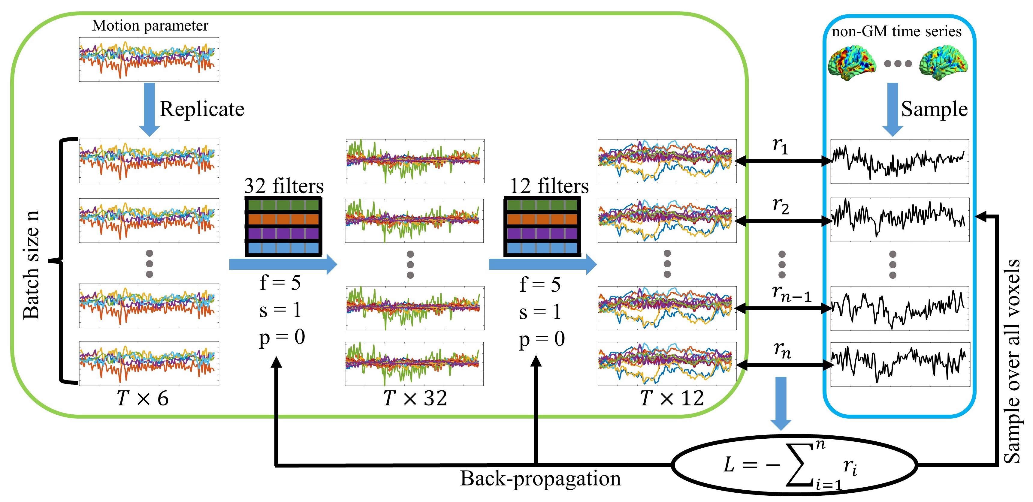
Figure 1. Schematic diagram of the denoising neural
network.
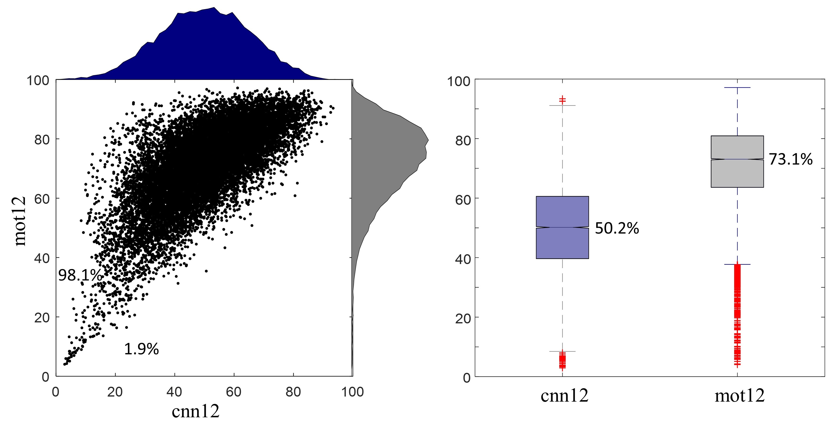
Figure 2. Remaining variance of regional time series after motion regression.
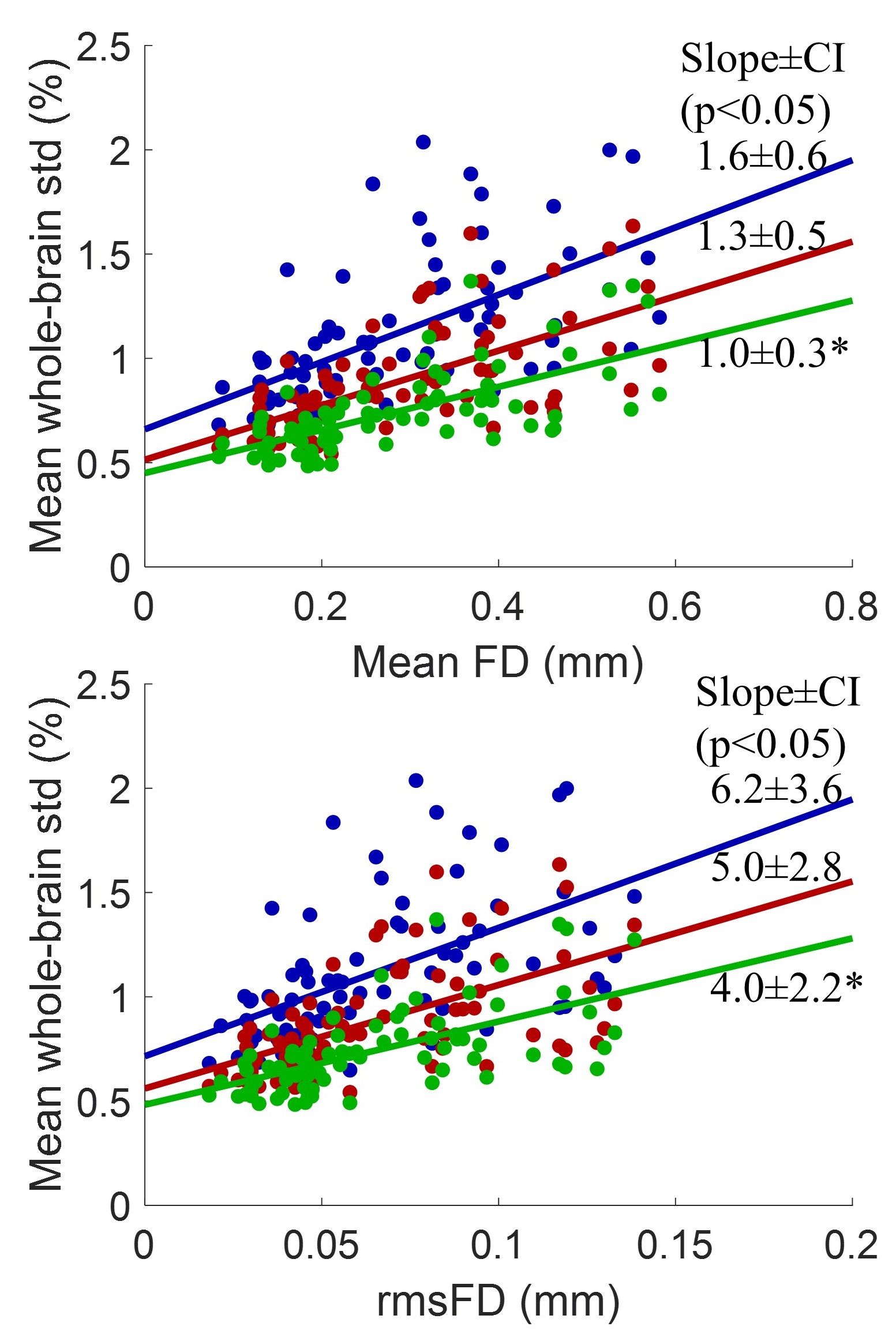
Figure 3. Plots of mean whole-brain standard deviation versus quality control measurements for raw (blue), mot12 (red) and cnn12 (green) fMRI data.
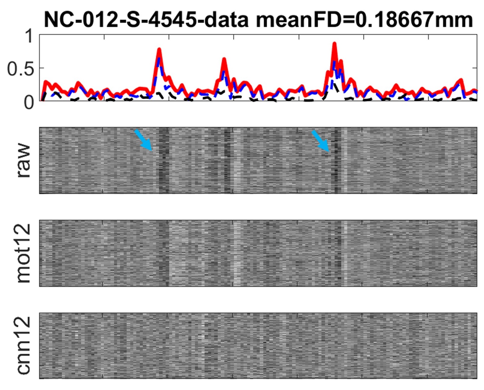
Figure 4. Different processed time series from an example subject.