4423
Silent 3D Parameter Mapping using Variable Flip Angle Looping Star1GE Healthcare, Munich, Germany, 2Department of Neuroimaging, Institute of Psychiatry, Psychology and Neuroscience, King's College London, London, United Kingdom, 3GE Healthcare, London, United Kingdom
Synopsis
This abstract presents a new method for silent and 3D parameter mapping, including proton density (PD) , T1, T2*, and quantitative susceptibility mapping (QSM), by combining Looping Star 3D silent T2*-weighted imaging with the concept of variable flip angle (VFA) PD and T1 mapping.
Introduction
MRI allows detailed investigation and depiction of numerous tissue properties, including proton density (PD), MR relaxation times (T1, T2, T2*), diffusion and magnetic susceptibility. While providing excellent soft-tissue contrast weighting, the non-quantitative nature of standard MRI, together with its loud acoustic noise are still considered notorious pain-points and are often complained about by researchers, clinicians and patients.
Recently, quantitative MR parameter mapping and synthetic MR contrast generation have (re-)gained significant interest and advancement with methods such as MR fingerprinting and synthetic MR (1–3). In parallel, MR pulse sequences have been presented allowing virtually silent 3D MR imaging (4–6).
Here we present a novel method for silent and 3D parameter mapping, including PD, T1, T2*, and quantitative susceptibility mapping (QSM), by combining Looping Star 3D silent T2* imaging (6) with variable flip angle (VFA) PD and T1 mapping (7,8).
Methods
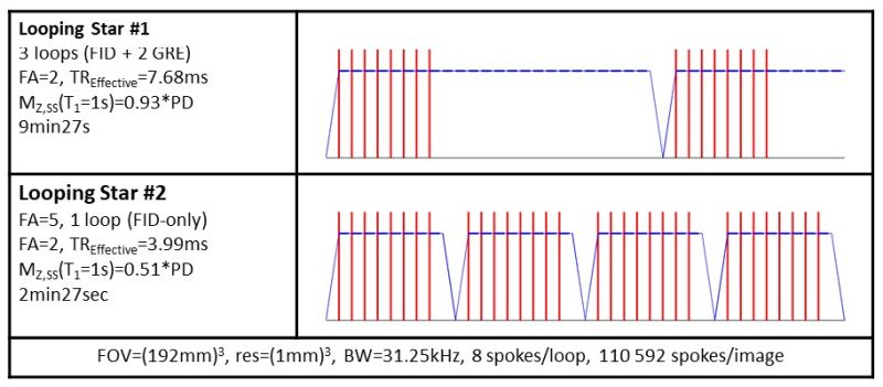
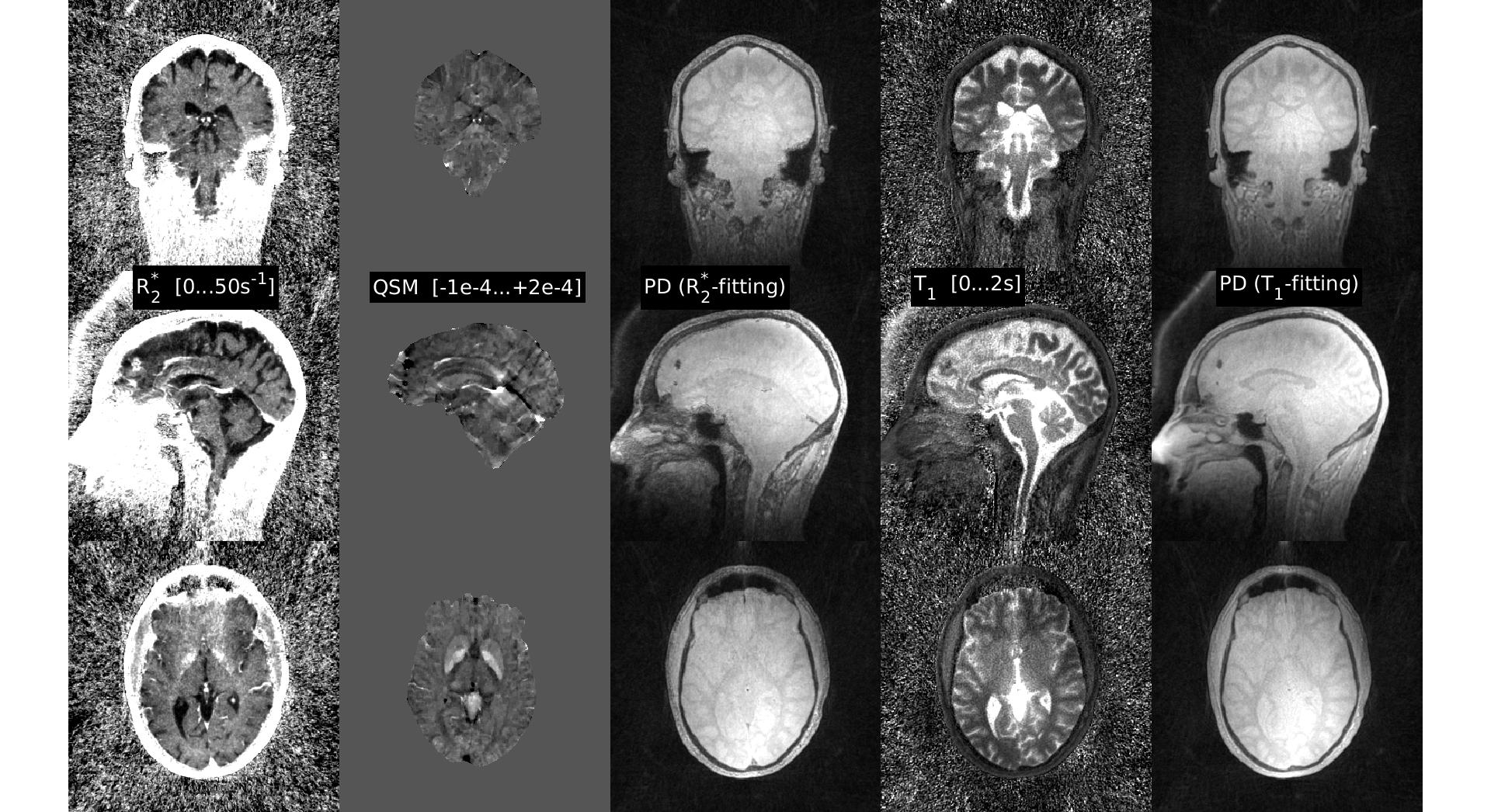
The Looping Star pulse sequence is based on the 3D radial Rotating Ultra‐Fast Imaging Sequence (RUFIS) extended by a time‐multiplexed gradient‐refocusing mechanism. First, multiple magnetic coherences are excited, which are subsequently gradient‐refocused in form of a looping k‐space trajectory. Looping Star captures an initial free induction decay (FID) image followed by gradient echo (GRE) images at equidistant echo times (cf. Fig. 1). Like in the RUFIS method, the 3D radial readout gradients are not ramped down in between repetitions, thereby eliminating most of the gradient switching (except minor directional gradient updates), resulting in virtually silent scanning. The multi-gradient echo nature of Looping Star provides T2* and magnetic susceptibility information. For QSM the images were processed using background field removal, projection onto dipole fields, L1 regularization (λ=200) and SMV correction (radius=5mm).
Repeating Looping Star with a different flip angle and using the VFA concept provides additional PD and T1 information. More specifically, for flip angle α<<1rad and repetition time TR<<T1, the Looping Star longitudinal steady-state magnetization (MZ,LS) can be approximated as:
$$M_{Z,LS}=\frac{PD*(1-e^{-\frac{TR_{Effective}}{T1}})}{1-e^{-\frac{TR_{Effective}}{T1}}*\cos(\alpha)} \approx \frac{PD}{1+\frac{T1}{TR_{Effective}}\frac{\alpha^2}{2}} \space [1] $$
allowing linear fitting of PD and T1 maps using two Looping Star acquisitions acquired with different α and/or TREffective (the average duration between two RF excitations).
Looping Star was implemented and tested on a GE 3T MR750w scanner (GE Healthcare, Chicago, IL). Phantom and volunteer experiments were conducted in the head using an 8ch receive brain coil. Two sequential silent 3D Looping Star scans were performed; the first one using α=2° and 3 loops (i.e. 1 FID + 2 gradient echoes), the second one using a α=5° and only a single loop (i.e. FID-only). Other scan parameters are listed in Fig. 1. Gridding image reconstruction and parameter fitting was performed off-line in MATLAB (MathWorks, Natick, MA).
Results
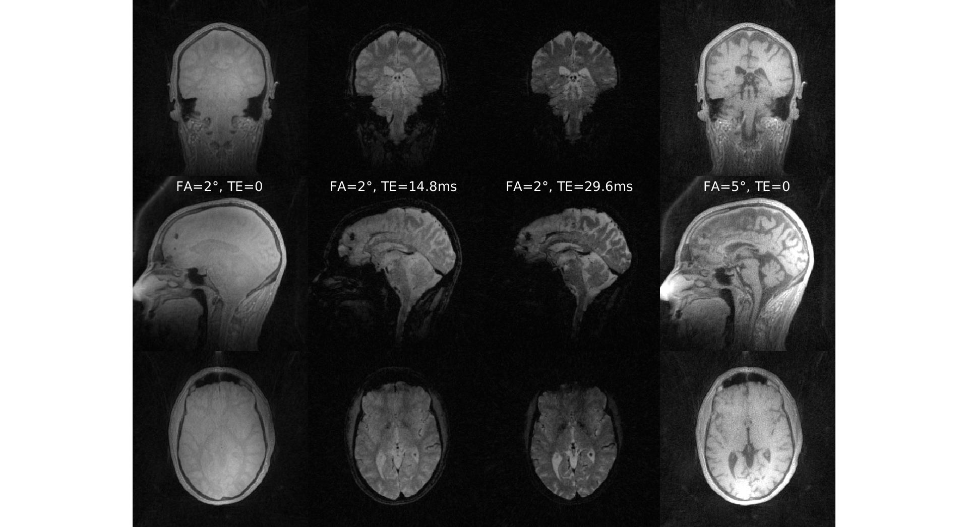
Figure 2 shows native head images obtained from VFA Looping Star. The first Looping Star experiment (i.e. α=2°, 3 loops) shows the expected PD-weighting and T2* decay for the later gradient echoes (columns 1-3). The higher α and shorter TREffective of the second Looping Star experiment (α=5°, 1 loops) provides extra T1-weighting (right column). Parameter maps are illustrated in Figure 3, showing R2* relaxivity (1/T2*), QSM and PD maps (columns 1-3) obtained from the first Looping Star experiment and T1 and PD via VFA processing of the two FID images. A benign calcification in the prefrontal cortex is well depicted in all four parameter maps. The acoustic noise was measured to be 68.4dB(A) which is only 4.2dB(A) above the ambient noise level inside the scanner.
Discussion
Conventional multi-GRE imaging requires extensive gradient switching and, hence, ranks among the loudest pulse sequences used in diagnostic MRI. In contrast, Looping Star generates multiple coherences and carefully refocuses them in a soft looping manner, enabling silent 3D radial GRE imaging. Combining Looping Star with the VFA concept, provides 3D silent parameter mapping of PD, T1, T2*, and QSM.
Typical Looping Star scan parameter (i.e. α, TREffective) results in a steady-state signal response close to the SNR-optimal maximum (i.e. Ernst angle). The additional high scan efficiency (90% of the scan time is used for sampling) renders this method fast and efficient.
The parameter fitting nicely separates and is well-conditioned. Obtained initial results in the head are promising in terms scan efficiency and patient comfort but need further validation. The accuracy of parameter mapping is expected to improve by acquiring more loops and incorporation of B1 correction.
Acknowledgements
No acknowledgement found.References
1. Warntjes et al. Rapid magnetic resonance quantification on the brain. MRM 60: 320-329 (2008).
2. Ma et al. Magnetic resonance fingerprinting. Nature 495: 187-192 (2013).
3. Tanenbaum et al. Synthetic MRI for Clinical Neuroimaging. AJR: 38: 1103-110 (2017).
4. Madio et al. Ultra-fast imaging using low flip angles and fids. MRM 34: 525-529 (1995).
5. Alibek et al. Acoustic noise reduction in MRI using Silent Scan. DIR 20: 360-363 (2014).
6. Wiesinger et al. Looping Star. MRM DOI: 10.1002/mrm.27440 (2018).
7. Cheng et al. Rapid high-resolutionT1 mapping by variable flip angles. MRM 55: 566-574 (2006).
8. Ljungberg et al. Silent T1-Mapping Using the Variable Flip Angle Method with Zero Echo Time. ISMRM Paris: p270 (2018).
Figures