4422
Six-direction diffusion tensor MRI using a convolutional neural network1Athinoula A. Martinos Center for Biomedical Imaging, Department of Radiology, Massachusetts General Hospital, Harvard Medical School, Charlestown, MA, United States, 2Harvard-MIT Division of Health Sciences and Technology, Massachusetts Institute of Technology, Cambridge, MA, United States, 3Radiological Sciences Laboratory, Department of Radiology, Stanford University, Stanford, CA, United States
Synopsis
Diffusion tensor imaging (DTI) is widely used for clinical neuroimaging and neuroscientific research but has traditionally suffered from relatively length acquisition. Here, we propose a new approach to obtain both scalar and orientational DTI metrics from six diffusion-weighted images with optimal directional encoding. Through the careful choice of diffusion directions, we compute initial tensor results that are then denoised using a convolutional neural network. Our results provide comparable scalar and orientational DTI metric maps to those acquired with 90 directions.
Target Audience
Introduction
Diffusion tensor MRI (DTI) is a neuroimaging method for probing the microstructure of the in vivo human brain. It has been widely adopted in neuroscientific research and clinical neuroimaging for measuring white matter integrity and tracing the major white matter bundles. Nonetheless, DTI acquisition is relatively lengthy since many repetition times (TRs) are needed to encode the water diffusion along different directions. Mathematically, six diffusion-weighted images (DWIs) and one non-DWI image are required to fit the DT model. In practice, however, at least 30 DWIs from unique and uniformly distributed encoding directions are required for a rotationally invariant estimation of fractional anisotropy (FA), tensor orientation and mean diffusivity (MD)1. For low signal-to-noise (SNR) data (e.g. high-resolution data), even more DWIs are needed for the robustness to noise. Even though the recent advances of the simultaneous multislice imaging have dramatically shortened the TR for acquiring each DWI, the minimum number of DWIs for DTI has remained unchanged. Previous works successfully reduced the q-space sampling requirement for estimating scaler metrics from DTI (e.g. FA, MD), diffusion kurtosis imaging and neurite orientation dispersion and density imaging using deep learning2,3. Nonetheless, the fiber orientations can not be resolved in these studies. In this work, we obtained both scalar and orientational DTI metrics from six DWIs that were comparable to those from 90 DWIs. By choosing optimized six encoding directions, we computed initial tensor results, which were then denoised using a convolutional neural network (CNN)-based denoising approach.Methods
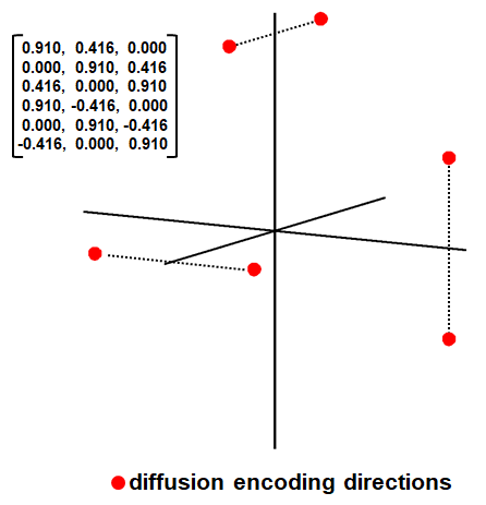
Data. Pre-processed diffusion MRI data of 20 subjects (19 for training, 1 for evaluation) from the Human Connectome Project (HCP) WU-Minn-Oxford Consortium were used (https://www.humanconnectome.org/). Diffusion data were acquired at 1.25-mm isotropic resolution using a b-values of 1000 s/mm2, 90 uniform directions. The diffusion tensor model was fitted using all 90 directions using FSL’s “dtifit” function to derive tensor, FA, MD, primary eigenvector (V1). One b=0 image and DWIs along six chosen directions (Fig.1) that minimize the condition number of the diffusion tensor transformation matrix4 were selected from each subject for a tensor model fit to derive tensor, FA, MD, and V1. FreeSurfer volumetric segmentation of the T1w data was resampled to diffusion space to obtain binary masks of cerebrospinal fluid (CSF).
Data Formatting. The six-element diffusion tensor from six DWIs served as the input of the CNN while the tensor from 90 DWIs served as the target of the CNN. Blocks of 64×64×64 voxel size were extracted (stride of 32) and normalized by subtracting the mean value of block voxels and dividing by their standard deviation prior to training.
Network. A 3D U-Net5 was used to learn the mapping from the diffusion tensor from 6 DWIs to that from 90 DWIs. The U-Net was implemented using the Keras API (https://keras.io/) with a Tensorflow (https://www.tensorflow.org/) backend, L2 loss, leaky ReLU activation, 3×3×3 kernels, 5 levels and 64 kernels at the highest level, ×3 kernels at each lower level. The training was performed on 19 subjects (~1,000 samples) and tested on 3 (~180 samples) using an Nvidia V100 GPU for 20 epochs (~3 hours).
Results
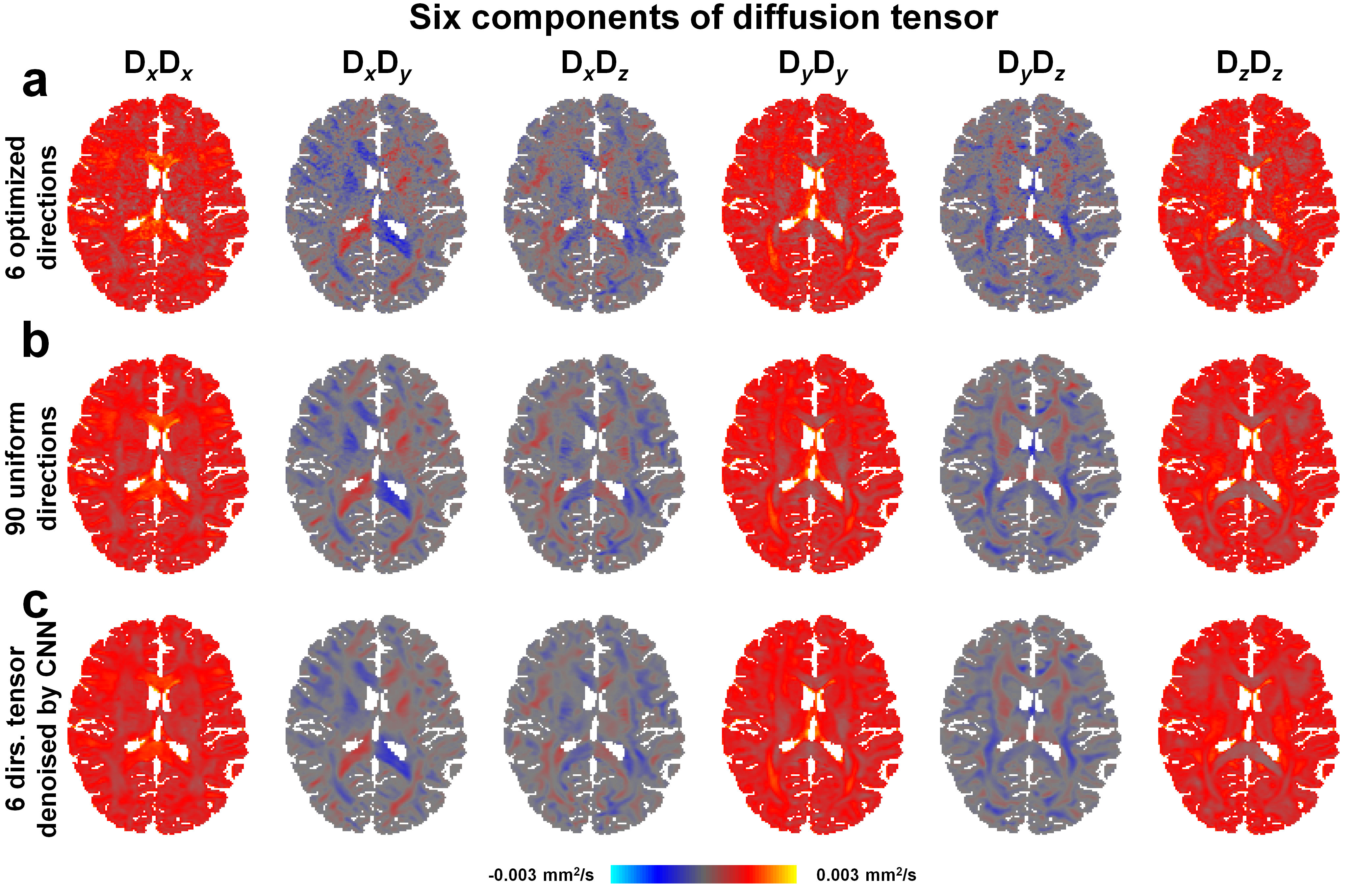
Figure 2 shows the diffusion tensor can be estimated from six chosen diffusion encoding directions but is noisy due to the low number of DWIs. The U-Net prediction results have higher signal-to-noise ratio and is comparable to the diffusion tensor estimated from 90 DWIs.
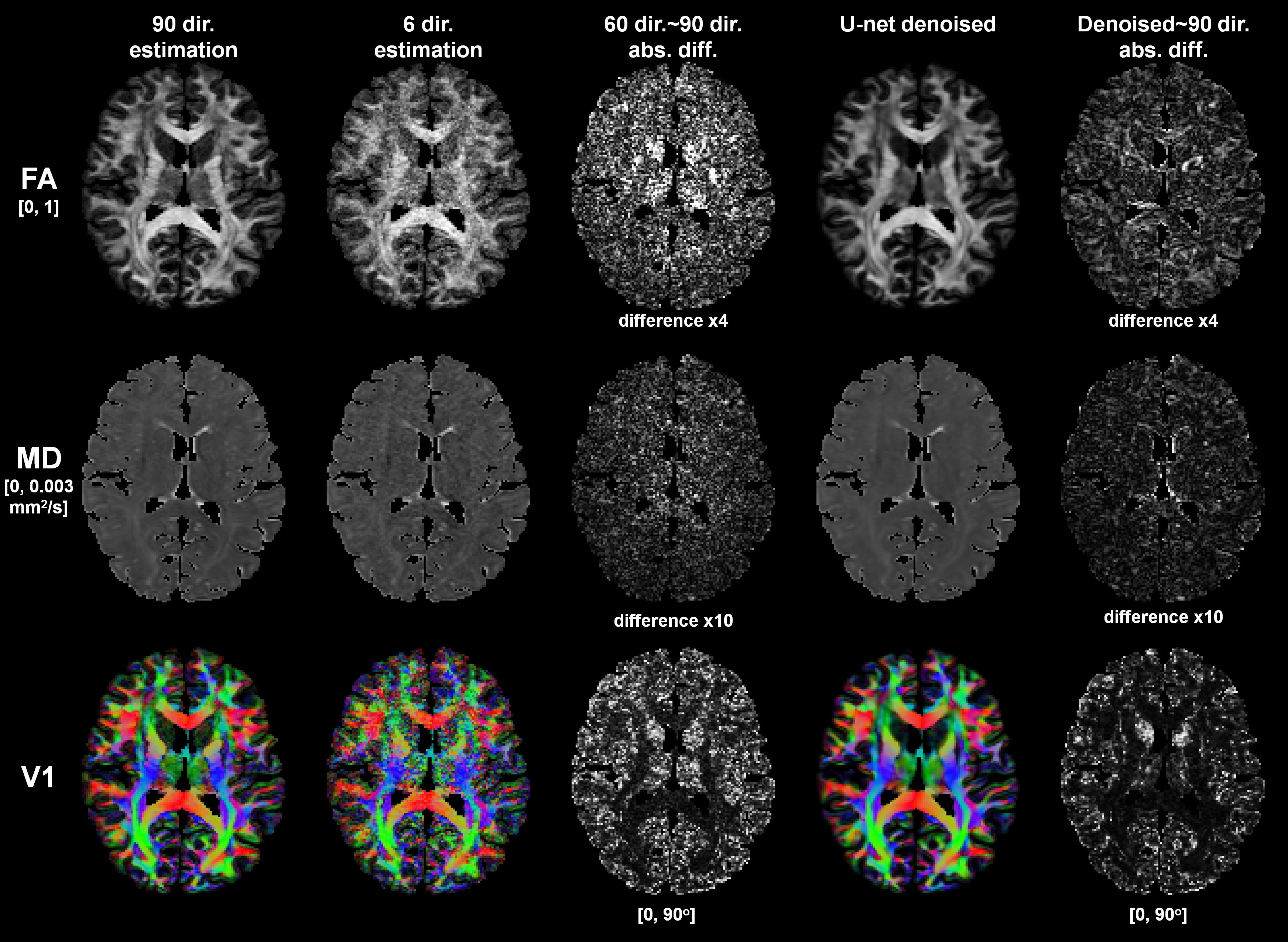
Figure 3 demonstrates the scalar (FA, MD) and orientational metrics (V1) derived from the U-Net results are more similar to those derived from 90 DWIs compared to those from 6 DWIs. However, the U-Net results are slightly blurry compare to the results from 90 DWIs.
Discussion
We demonstrate the application of CNN in estimating both the scalar and orientational DTI metrics from only six DWIs. We formulated the problem as a denoising framework by using optimized 6 diffusion encoding directions, which is unique from other studies.Acknowledgements
Funding was provided by the NIH: P41-EB015896, S10-RR019307, K23-NS096056, R01-MH111419, and an MGH Claflin Distinguished Scholar Award.References
[1] Jones DK. The effect of gradient sampling schemes on measures derived from diffusion tensor MRI: a Monte Carlo study. Magnetic Resonance in Medicine: An Official Journal of the International Society for Magnetic Resonance in Medicine. 2004;51(4):807-15.
[2] Golkov V, Dosovitskiy A, Sperl JI, Menzel MI, Czisch M, Samann P, Brox T, Cremers D. q-Space Deep Learning: Twelve-Fold Shorter and Model-Free Diffusion MRI Scans. IEEE transactions on medical imaging. 2016;35(5):1344-51.
[3] Li H, Zhang C, Liang Z, et al. Deep learning diffusion tensor imaging with accelerated q-space acquisition. 2018; ISMRM workshop on machine learning (Part II).
[4] Skare S, Hedehus M, Moseley ME, Li T-Q. Condition number as a measure of noise performance of diffusion tensor data acquisition schemes with MRI. Journal of Magnetic Resonance. 2000;147(2):340-352.
[5] Ronneberger O, Fischer P, Brox T. U-net: Convolutional networks for biomedical image segmentation. Paper presented at: International Conference on Medical image computing and computer-assisted intervention 2015.
Figures