4414
To Evaluate Effect of SENSE and CSENSE on Quantitative T1 and T2 mapping of Human Brain1Indian Institute of Technology Delhi, New Delhi, India, 2AIIMS, New Delhi, India, 3Philips India Limited, Gurugram, India, 4University of Pennsylvania, Philadelphia, PA, United States, 5Fortis Memorial Research Institute, Gurugram, India, 6Philips Health Tech Asia Pacific, Tokyo, Japan
Synopsis
Parallel-imaging and compressed-sensing based approaches are playing crucial role in accelerating MRI data acquisition. Objective of the study was to accelerate the data acquisition of T1, T2 and PD-weighted TSE images and to evaluate the accuracy of T1 and T2 mapping in the human brain. Data was acquired using SENSE parallel-imaging and Compressed-SENSE technique for different factors as well as without any acceleration. T1 and T2 values obtained using data with SENSE (upto factor of 3) and CSENSE (upto factor of 6) were comparable to those acquired without any acceleration. Errors in T1 and T2 increased with increase in acceleration factor.
Introduction
Spin lattice relaxation time constant (T1) and spin-spin relaxation time constant (T2) are two important parameters required for quantitative MRI and several methods have been developed for estimating these parameters individually1,2,3. To estimate these parameters together, a novel method was previously reported4 that require three independent T1, T2 and PD-weighted(W) images acquired using SE/TSE sequence, and is less sensitive to the effect of B1 field inhomogeneity on the relaxivity maps. Conventional images such as T1, T2 and PD-W, based upon TSE sequence, are routinely acquired during human brain exam; however, takes a long scan time depending upon resolution. Several methods including parallel imaging6, Compressed Sensing (CS)7 and Compressed Sense (CSENSE)5 are being developed to accelerate data acquisition. However, the effect of these acceleration techniques on the accuracy of T1 and T2 maps needs to be evaluated. In this study, we have evaluated the effect of SENSE parallel imaging and a novel scan acceleration method by combining compressed sensing technology with the SENSE infrastructure called Compressed-SENSE (CSENSE) on T1, T2 and PD-W images as well as on T1 and T2 maps.Methods
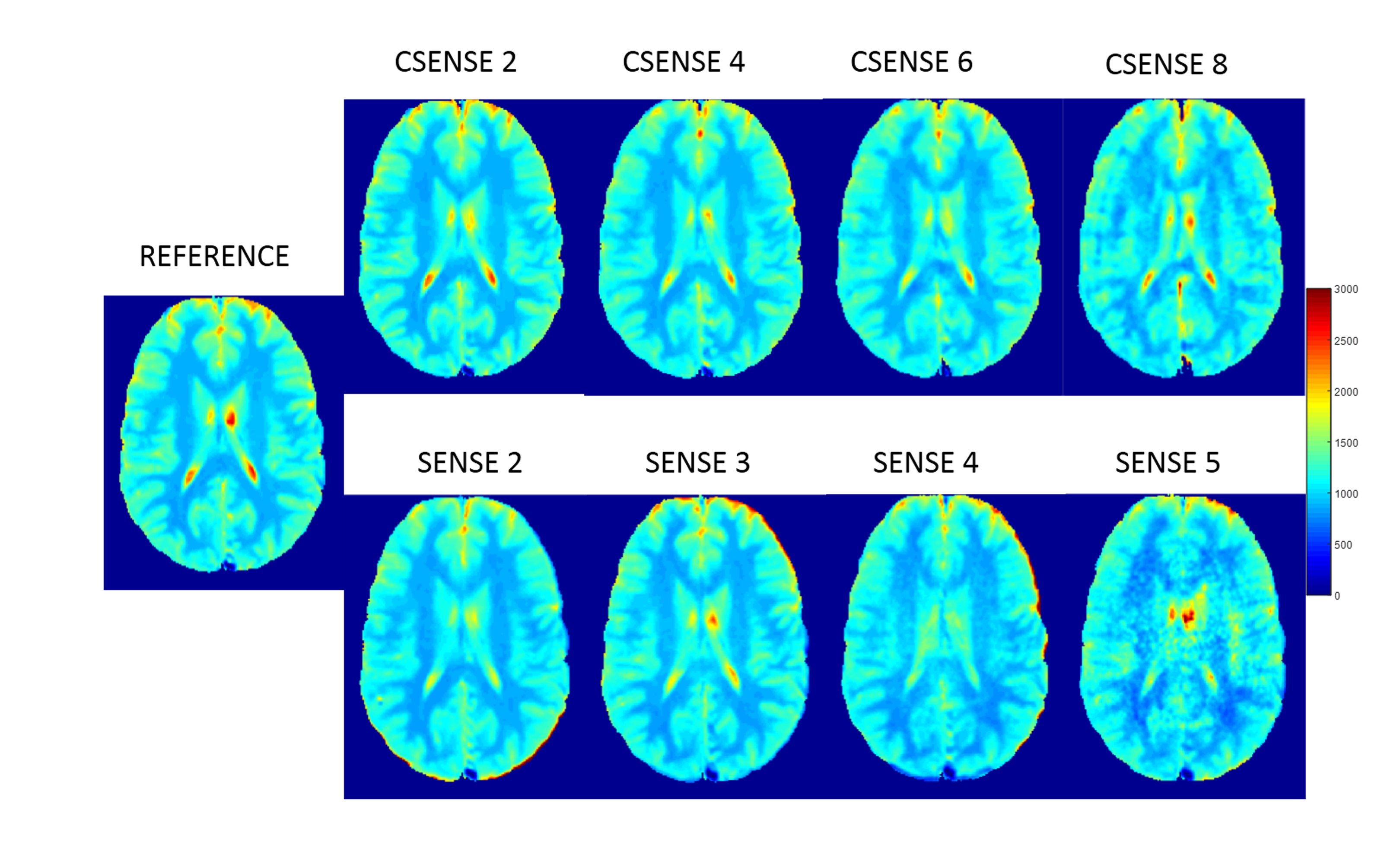
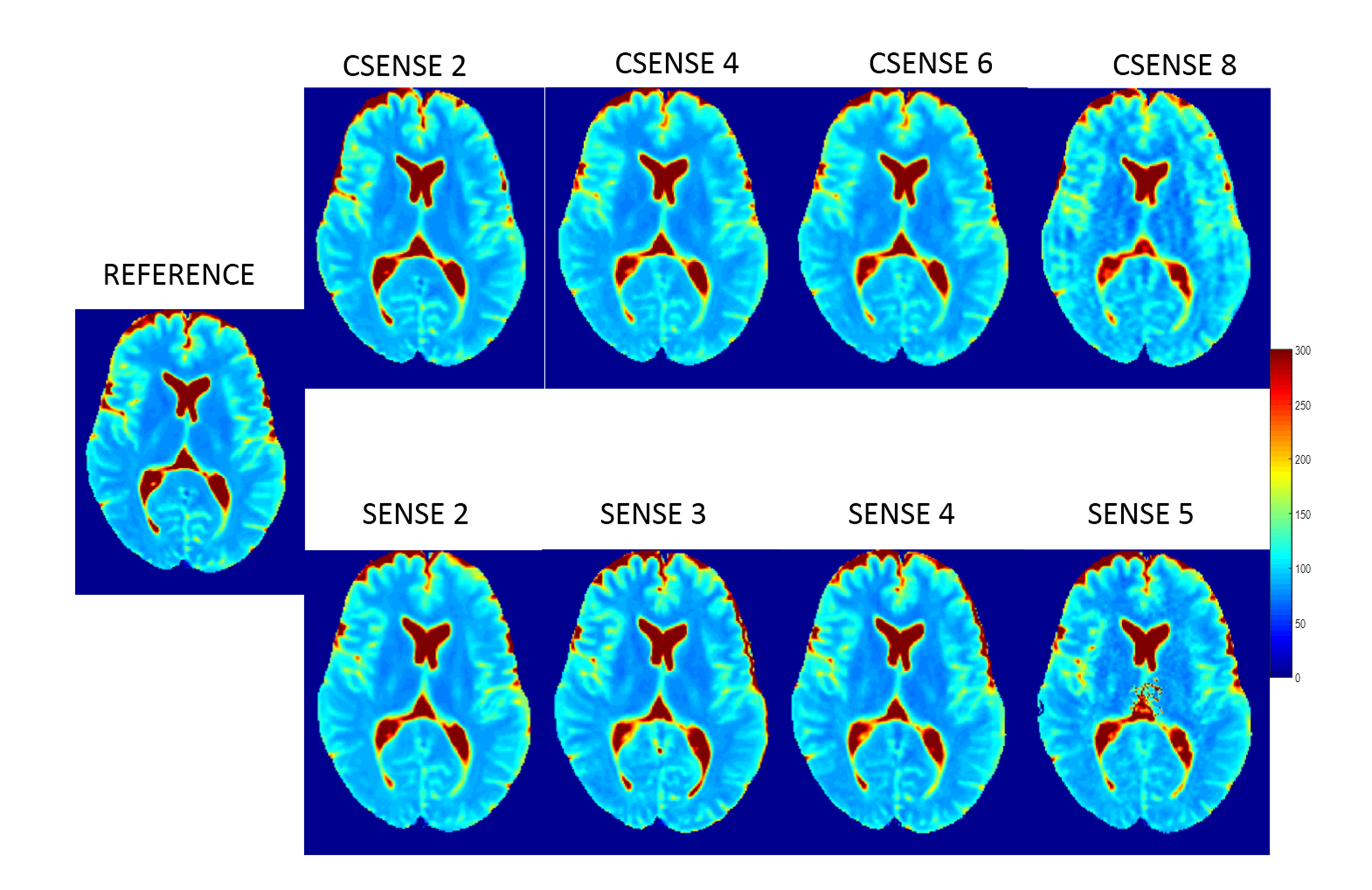
MRI data of nine human subjects were acquired at 3.0 T (Ingenia, Philips, The Netherlands) with a 15 channel head coil. Nine sets of T1 and dual PD-T2 W images were acquired for each subject using TSE sequence. First data set was acquired without any SENSE and CSENSE (considered as reference for comparison). Four data sets were acquired with SENSE factors 2,3,4,5 and the other four were acquired with CSENSE factors 2,4,6,8 respectively. Other MRI parameters were: FOV = 240×240 mm2, number of slices= 20, slice thickness=5mm and acquisition matrix= 240×240. TE/TR values for dual PD-T2 and T1 W images were 7.2ms/90ms/3500ms and 10ms/360ms respectively. Three subjects were scanned twice to check for reproducibility. Data was processed using in-house developed programs in MATLAB. T1 and T2 maps were generated using previously described procedure4 and the white matter (WM) part was segmented out. Normalized mean squared error (NMSE) was calculated in WM ROI for each combinations to analyze the nature of error propagation.Results
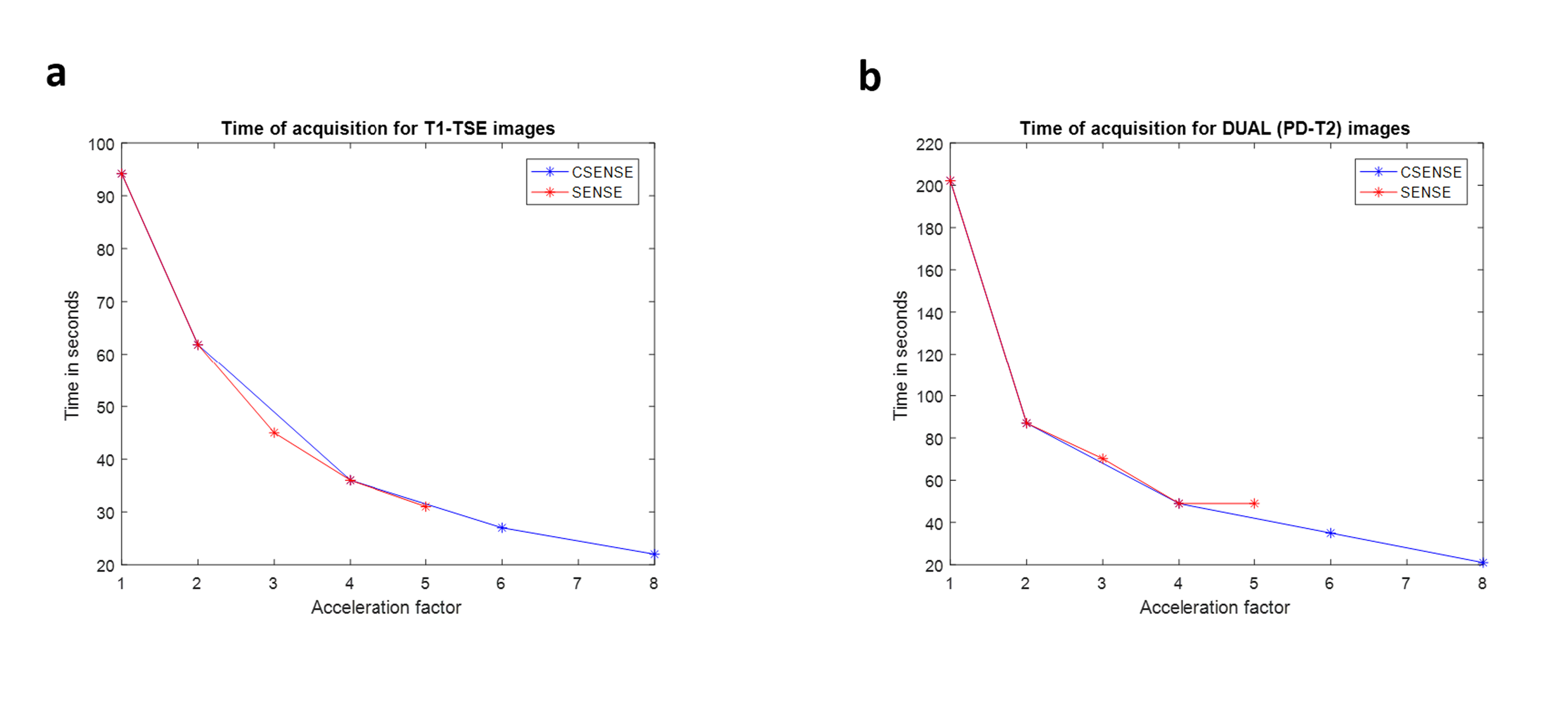
Data acquisitions of both T1 -W and PD- T2 W with both SENSE and CSENSE gives similar amount of acceleration in time, which follows a reciprocal like pattern as shown in Figure-1. T1 and T2 maps obtained using data from SENSE (acceleration factor upto 3) as well as from CSENSE (acceleration factor upto 6) looked similar to those obtained using conventional approach as shown in Figure-2 and Figure-3. While going for higher factors, SENSE factor introduces more noise compared to CSENSE. Visually T1 and T2 maps looks stable till CSENSE factor 6, while noise breakthrough is clearly visible for maps with SENSE factor 3. Figure-4 shows the mean and standard deviation of NMER calculated for WM ROI for each subjects. The error propagation in both T1 and T2 maps for different SENSE and CSENSE factor follow an exponential pattern. On increasing the CSENSE factor, the error stay below 1% for both T1 and T2 map until an acceleration factor 6. While for SENSE acquisition, the error stay below 1% till an acceleration factor 3. The error propagation is observed to be more for SENSE acquisition when compared to CSENSE. Results were also reproducible.Discussion
Use of SENSE and CSENSE approaches in data acquisition results in a substantial reduction in data acquisition time. Results of current study show that T1 and T2 maps of human brain obtained using SENSE factor upto 3 and CSENSE factor upto 6 are comparable to those obtained using conventional data acquisition approach. Moreover, maps corresponding to SENSE or CSENSE approaches are also reproducible. Therefore, acceleration gained in data acquisition using these factors can be used either to reduce data acquisition time or to further increase spatial resolution at same scan time. In the current study, T1 values were comparable to reported literature values; however, T2 values show slightly higher values compared to reported T2 values using other methods. However, both T1 and T2 values were reproducible. From the analysis, error propagation is more for SENSE based data acquisition and more acceleration factor is observed to be stable for CSENSE acquisition.Conclusion
In conclusion, quality of T1, T2 and PD W TSE images as well as accuracy of T1 and T2 maps derived from these images is not affected by the use of SENSE(upto factor of 3) and CSENSE approach(upto a factor of 6). Therefore, data required for T1 and T2 mapping can be accelerated significantly using CSENSE approach without loss of accuracy compared to conventional approach.Acknowledgements
The Authors acknowledge technical support of Philips India Limited and Fortis Memorial Research Institute Gurugram for MRI data acquisition. This work was supported by Science and Engineering Research Board (IN) (YSS/2014/000092).References
[1] Crawley, Adrian P., and R. Mark Henkelman. "A comparison of one‐shot and recovery methods in T1 imaging." Magnetic resonance in medicine 7.1 (1988): 23-34.
[2] Evans, Avery J., et al. "Evaluation of steady and pulsatile flow with dynamic MRI using limited flip angles and gradient refocused echoes." Magnetic resonance imaging 5.6 (1987): 475-482.
[3] Kay, I., and R. M. Henkelman. "Practical Implementation and Optimization of One‐shot T1 imaging." Magnetic resonance in medicine 22.2 (1991): 414-424.
[4] Singh A, Singh A, Haris M, Rathore D, Purwar A, Sarma M, Bayu G, Husain N, Rathore RK, Gupta RK. Quantification of physiological and hemodynamic indices using T(1) dynamic contrast-enhanced MRI in intracranial mass lesions. J Magn Reson Imaging. 2007; 26: 871-880.
[5] Geerts-Ossevoort, L., de Weerdt, E., Duijndam, A., van IJperen, G., Peeters, H., Doneva, M., Nijenhuis, M. and Huang, A., Speed done right. Every time. Philips Healthcare; 2018 Jan. Report No: 4522 991 31821. https://www.philips.de/content/dam/b2bhc/de/resourcecatalog/landingpages/ingeniaelition/White_Paper_Compressed_SENSE-opt.pdf
[6] Pruessmann, K. P., Weiger, M., Scheidegger, M. B., & Boesiger, P. (1999). SENSE: sensitivity encoding for fast MRI. Magnetic resonance in medicine, 42(5), 952-962.
[7] Michael Lustig, David Donoho, and John M. Pauly. Sparse MRI: The Application of Compressed Sensing for Rapid MR Imaging. Magnetic Resonance in Medicine. 2017; 58:1182–1195 (2007)
[8] Heidemann, R.M., Özsarlak, Ö., Parizel, P.M., Michiels, J., Kiefer, B., Jellus, V., Müller, M., Breuer, F., Blaimer, M., Griswold, M.A. and Jakob, P.M., 2003. A brief review of parallel magnetic resonance imaging. European radiology, 13(10), pp.2323-2337
[9]David S. Smith, E. Brian Welch, Xia Li, Lori R. Arlinghaus, Mary E. Loveless, Tatsuki Koyama, John C. Gore and Thomas E. Yankeelov. Quantitative effects of using compressed sensing in dynamic contrast enhanced MRI. Phys Med Biol., 2011; 56(15): 4933–4946
[10] SoHyun Hana, Jeffrey L. Paulsenb, Gang Zhuc, Youngkyu Songd, SongI Chund, Gyunggoo, Chod, Ellen Ackerstaffe, Jason A. Koutchere, and HyungJoon Choa. Temporal/spatial resolution improvement of in vivo DCE-MRI with compressed sensing-optimized FLASH. Magn Reson Imaging, 2012; 30(6): 741–752
Figures