4413
Improving T2 and B1 parametric estimation in the brain with multi spin-echo MR and fusion bootstrap moves solver (FBMS)1ISR-Lisboa/LARSyS and Department of Bioengineering, Instituto Superior Técnico – Universidade de Lisboa, Lisbon, Portugal, 2Centre for the Developing Brain, King's College London, London, United Kingdom
Synopsis
Multi spin-echo (MSE) sequences have been prescribed for efficient T2 mapping. This can be further improved by matching to pre-computed echo-modulation curves (EMC). Previous use of this method to estimate T2 and B1 resulted in bias in the latter. We investigated the possibility to improve B1 by taking advantage of its spatial smoothness, using a fusion bootstrap moves solver (FBMS). The two methodologies were compared using a numerical phantom and in-vivo brain data. While T2 estimation was accurate and equivalent, B1 accuracy was improved using the FBMS. Future work is required to tune the regularization parameters of the FBMS algorithm.
Purpose/Introduction
Quantitative MR provides valuable clinical information, based on parametric properties of the tissues. Multi spin-echo (MSE) sequences have been suggested as an efficient T2 mapping technique, allowing for shorter scan times than conventional single SE. Additionally, T2 accuracy with MSE has been shown to be further improved using a pre-computed dictionary of Echo Modulation Curves (EMC) and the MR fingerprinting concept1. Previous simulation work revealed that voxelwise B1 estimation often led to bimodal distributions, even though the main peak coincided with the ground truth value2. In the present work, we used the fact that B1 maps are spatially smooth, taking advantage of the information provided by neighbouring pixels to improve the final B1 map estimation. This was undertaken by adapting an iterative parametric estimation methodology, previously prescribed for stabilizing fits to the intravoxel incoherent motion model in diffusion MR, known as the fusion bootstrap moves solver (FBMS)3.Methodology
A numerical brain phantom was generated considering three distinct tissues corresponding to the cerebrospinal fluid (CSF), white matter and grey matter in the brain, with corresponding T1, T2 and proton densities at 3T. Estimation of T2 and B1 parametric maps were obtained by dictionary matching to pre-computed EMC. Simulation of the EMC dictionary was implemented using the Extended Phase Graph (EPG) formalism4 and considering a T2 range of 20:5:400 ms and 400:100:2000 ms, B1 range of 60:2:140% and a fixed T1 = 1000 ms, as previously suggested1. The simulated dictionary also took into account the same acquisition parameters as the in-vivo protocol regarding echo-train length (ETL), flip angle and RF waveforms. Furthermore, the FBMS methodology3,5 was implemented with Matlab 2015b (Mathworks, USA). The FBMS solver iteratively updates parametric estimations (such as T2 and B1) by applying a binary graph-cut solver allowing to fuse previous parametric estimates with the new proposal of parameters that better fits the observed data. Two combinations of smoothing factors were tested (αB1 = 5, αT2 = 2) and (of αB1 = 10, αT2 = 3) and the FBMS loop was set to a maximum number of iterations of 10. In-vivo brain data with 6 axial slices were acquired on a Philips Achieva 3T scanner with a MSE sequence: 32 ETL (dTE = 10 ms), spatial resolution 1.6✕1.6✕4.0 mm3, FOV 250✕250 mm2 and TR = 10 ms. T2 maps obtained from a monoexponential fit tof the even echoes only and B1 maps obtained with the Actual Flip Angle (AFI) method were used as in-vivo reference2.Results
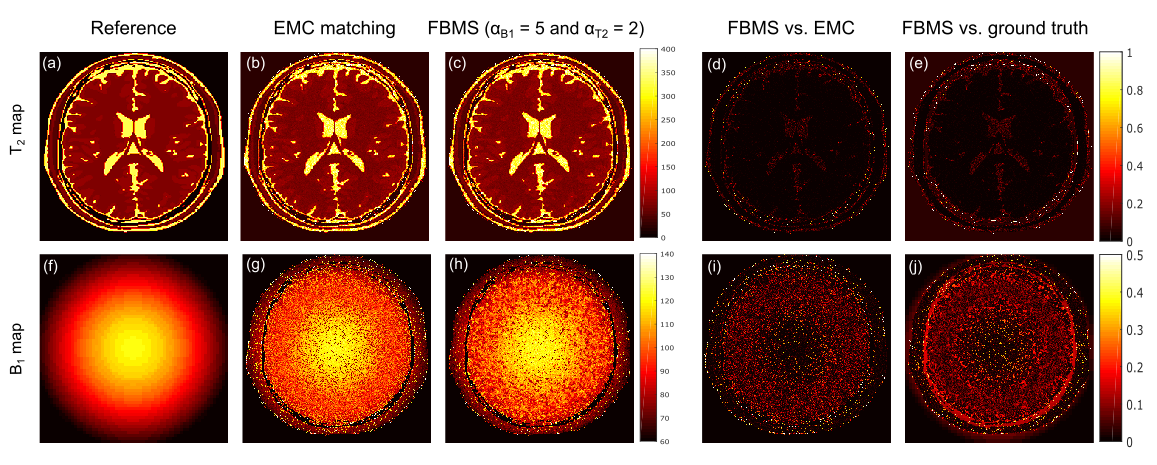
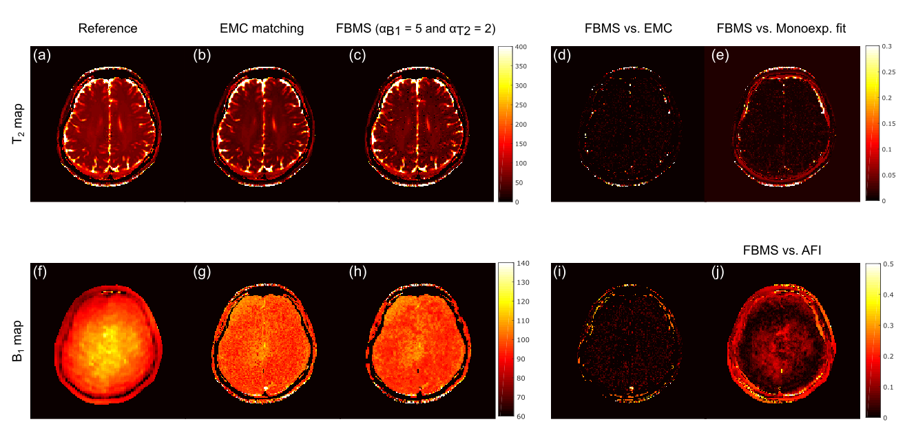
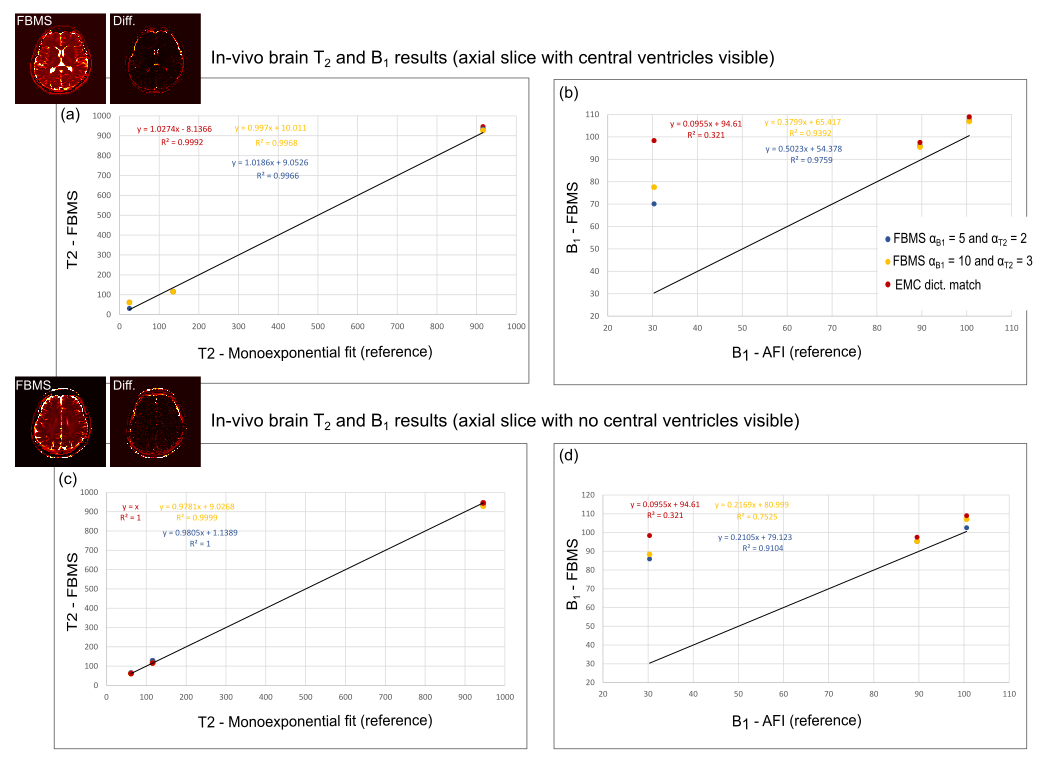
Figure 1 shows the numerical phantom simulation results. Noticeable differences in T2 estimation (Figure 1 d) between the two methods are present at the edges of the brain and in the centre, encompassing the ventricles. Figure 2 presents T2 and B1 maps from in-vivo brain data (axial slice above the ventricles) using EMC and FBMS. Similarly to the simulation results, most differences between the two methods are visible in regions corresponding to high T2 values (possibly due to CSF). Quantitative T2 and B1 measurements for three selected regions of the brain (CSF: ~1400 px and WM and GW: ~6000 px) are shown in Figure 3. Although no noticeable difference was found for T2 estimation between EMC and FBMS (Figure 3 a,c), some improvement in B1 accuracy of the mean values in the selected regions was achievable using the FBMS compared to EMC approach (b,d).Discussion and conclusions
Accurate T2 estimation (even for very long T2 values) was possible with both the EMC and FBMS method. Improved B1 estimation was achieved when using the FBMS solver compared to the EMC matching only (Figure 3). Use of a lower smoothing factor for the B1 parameter resulted in improved B1 accuracy in both in-vivo data sets. Future work is required to optimize FBMS regularization and fully understand its effect in T2 and B1 accuracy. In conclusion, the FBMS is a promising tool to further improve B1 estimation from MSE sequences in the brain, enabling to estimate both parameters (T2 and B1) from a single scan. It would also be desirable to investigate strategies for limiting the impact on neighbouring pixels of poor B1 estimates in regions with very high T2 values for which the imaging protocol may not have been optimal (e.g. CSF).Acknowledgements
Portuguese Foundation for Science and Technology (FCT - IF/00364/2013, UID/EEA/50009/2013, SFRH/BD/120006/2016)References
1 Ben-Eliezer N, Sodickson D K, Block K T. Rapid and accurate T2 mapping from multi-spin-echo data using Bloch-simulation-based reconstruction. Magn Reson Med, 2015; 73: 809-817.
2 Santos N, Teixeira R, Hajnal J and Nunes RG. Assessing the accuracy of T2 and B1 maps estimated from multi-echo spin echo MRI sequences using extended phase graph signal predictions. In the proceedings of 24th ISMRM, Singapore, 2016.
3 Freiman M, Perez-Rossello J M, Callahan M J, Voss S D, Ecklund K, Mulkern R V, Warfield S K. Reliable estimation of incoherent motion parametric maps from diffusion-weighted MRI using fusion bootstrap moves. Med Image Anal, 2013; 17: 325-336.
4 Weigel M. Extended phase graphs: dephasing, RF pulses and echoes - pure and simple. J Magn Reson Imaging 2015; 41: 266-295.
5 While P. A comparative simulation study of bayesian fitting approaches to intravoxel incoherent motion modeling in diffusion-weighted MRI. Magn Reson Med, 2017; 78: 2373-2387.
Figures