4408
Fast multiparametric imaging in the brain using a stationary balanced steady state cartesian approach1Institute for Biomedical Engineering, University and ETH Zurich, Zurich, Switzerland, 2AMT Lab, Department of Biomedical Engineering, University of Basel, Allschwil, Switzerland, 3MIAC AG, Basel, Switzerland
Synopsis
We propose a multiparametric balanced steady-state 3D Cartesian sequence that exploits model based and pattern matching reconstruction strategies for a series of 20 flip angles and repetition times, allowing for the simultaneous quantification of B0, B1+, T1, T2, and proton density. Time-varying signal patterns at the steady state are reached that allow for the acquisition of unique signal patterns in each image voxel for any acquisition scheme. We show the feasibility of our technique in-vivo in the human brain in 11 minutes, here with Cartesian acquisition and no acceleration strategies.
Introduction
The basis of Magnetic Resonance Fingerprinting (MRF) relies on the simultaneous quantification of tissue parameters exploiting spatio-temporal signal correlations from pseudo-randomized series of hundreds of flip-angles (FAs) and repetition times (TRs) acquired in highly undersampled spiral readouts1. In this work, we propose to use a dictionary based reconstruction similar to the MRF framework, however using a 3D fully sampled balanced steady-state Cartesian sequence that is being optimized2,3 with only 20 time-points sequentially repeated to reach a steady-state4 of time varying signal patterns.Methods
Our sequence uses a series of 20 different FAs and TRs. Since the sequence parameter space is greatly reduced, optimization of trajectories can be performed to achieve better discrimination of the encoded parameters T1, T2, B0, B1+, and proton density2,3. Discriminatory power of a FA/TR trajectory can be evaluated by calculating the dot product matrix H of a simulated dictionary D2,5:
H = D†D
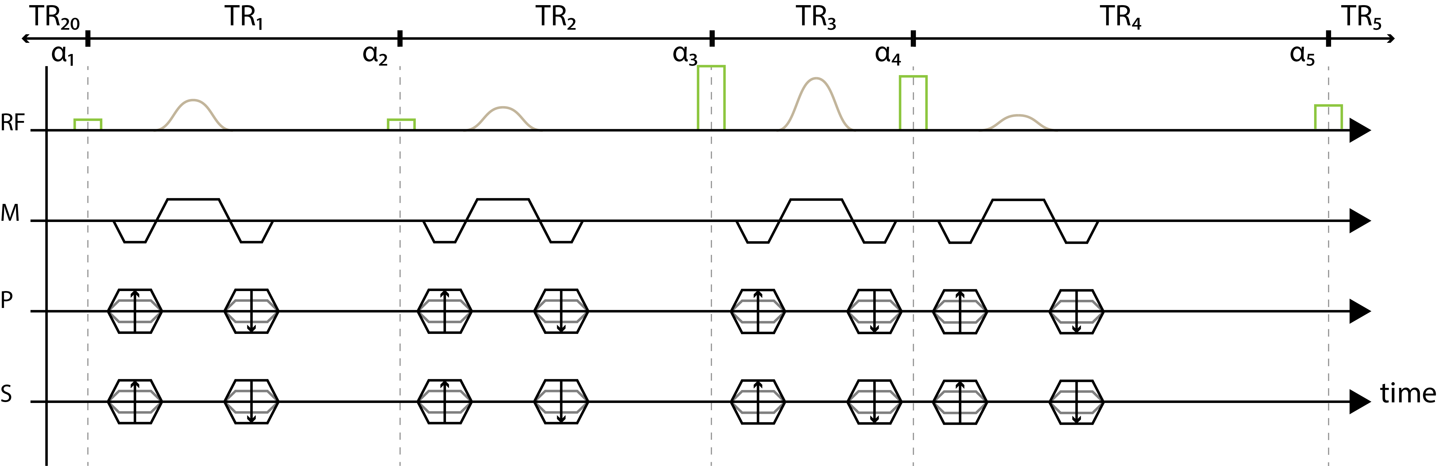
The FA and TR train is chosen using stochastic optimization within a predefined set, selecting candidates presenting the minimum averaged correlation coefficient. The optimized sequence presented uses FAs between 10° and 170° and TRs between 11 ms to 41 ms, leading to a total sequence duration of 500 ms per k-line. This sequence allows for classical Cartesian acquisition through establishing a stationary steady-state, where the whole sequence is repeated without delay but different imaging gradients4. The pulse sequence diagram of the proposed sequence is shown in Figure 1.
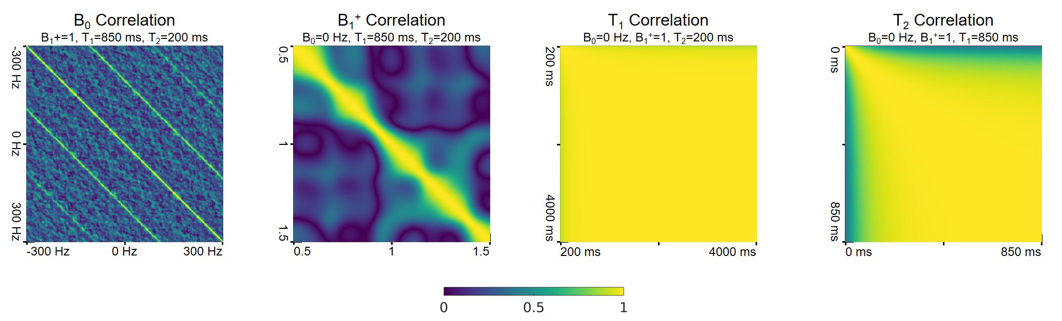
The bSSFP-nature of the presented sequence together with the variation of repetition times lead to high discriminatory power with respect to B0 and B1+ (correlation < .995), but lower ability to discriminate T1 and T2 (correlation >.999) (see Figure 2). Thus, in order to correctly resolve T1 and T2 variations, fine resolution dictionaries for B0 and B1+ need to be simulated. The dictionary is generated in a two-step process. First, a subspace spanned by B0 and B1+ is simulated, while T1 and T2 are fixed. This dictionary is grouped into subgroups using the greedy grouping approach6,7. In a second step, these sub-dictionaries are extended by simulating the respective T1 and T2 dimensions. In this way, efficient grouping can be realized without the need to perform full-dictionary comparisons.
The sequence was implemented on a 1.5 T Philips Achieva system and data was acquired using an 8-channel head coil. Full head coverage was achieved using non-selective excitation and phase encoding along both phase and slice directions with an elliptical shutter and 1.3-fold slice oversampling. The FOV was 240x225x150 mm3, with an acquisition matrix size of 80x75x25 and 3x3x6 mm3 resolution and no parallel imaging (scan duration: 11 min). Reconstruction was performed within 11 minutes using MATLAB on a workstation with twelve cores (2.4 GHz) and a dictionary of 250M entries.
Results
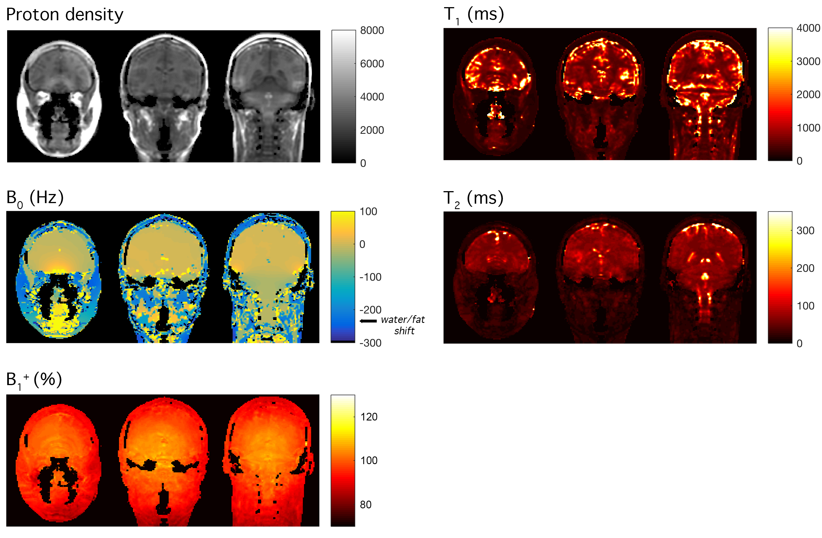
In Figure 3, in-vivo results are shown for three selected coronal slices. Both T1 and T2 values are within the expected range, and grey matter can be well separated from white matter and CSF. Variations in the static magnetic field B0 can be appreciated, revealing strong gradients at the air-tissue interface close to the ear canals and at the vicinity of the sinuses. Homogeneous off-resonance areas in the skin and cheeks are in accordance with the typical 3.5ppm chemical shift of water/fat (220 Hz at 1.5 T). Spatial homogeneity of the transmit coil is also well represented in the corresponding B1+ map. Residual Gibbs ringing can be seen in the quantitative maps that originates from the sharp signal cut-off at the edge of the low-resolution k-space.Discussion
Added with time-varying signal patterns reaching the steady state, we obtain simple and robust implementation of quantitative MRI with 3D Cartesian schemes. We maintain a balanced sequence to keep B0 as a parametric map and envision future T2* characterization when most MRF and related work currently make use of spoiled steady state sequences8 insensitive to off-resonance effects. A limitation of our approach though is that it can result in very large dictionaries finely resolving B0 and B1+ parameters needed for good matching, ultimately impacting computational time and accuracy.Conclusions
We have demonstrated that an optimized multiparametric sequence of length=20 TRs can be used for rapid quantitative parametric imaging with Cartesian sampling, providing five fully-quantified maps in 3D in the living human brain in 11 minutes using full sampling of k-space. From our Cartesian scheme, performance can be easily enhanced using conventional acceleration strategies widely available in the clinics, e.g. parallel imaging. Our approach also benefits from simple and robust Fourier transform reconstruction that may result in more straightforward adoption over non-Cartesian acquisition schemes.Acknowledgements
This project has received funding from the European Union’s Horizon 2020 research and innovation programme under grant agreement No 668039.References
1. Ma D, Gulani V, Seiberlich N, et al. Magnetic resonance fingerprinting. Nature. 2013;495(7440):187-192.
2. Cohen O, Rosen MS. Algorithm comparison for schedule optimization in MR fingerprinting. Magn Res Imaging. 2017;41:15-21.
3. Cohen O, Sarracanie M, Ackerman JL, Rosen MS. Magnetic resonance fingerprinting trajectory optimization. Proc Int Soc Magn Reson Med. 2014;p27, Milan, Italy
4. Amthor T, Doneva M, Koken P, Sommer K, Meineke J, Börnert P. Magnetic Resonance Fingerprinting with short relaxation intervals. Magn Reson Imaging. 2017;41:22-28.
5. Sommer K, Amthor T, Doneva M, Koken P, Meineke J, Börnert P. Towards predicting the encoding capability of MR fingerprinting sequences. Magn Reson Imaging. 2017;41:7-14
6. Cauley SF, Setsompop K, Ma D, et al. Fast group matching for MR fingerprinting reconstruction. Magn Reson Med. 2015;74(2):523-528
7. Buonincontri G, Sawiak SJ. MR fingerprinting with simultaneous B1 estimation. Magn Reson Med. 2016;76(4):1127-1135.
8. Jiang Y, Ma D, Seiberlich N, Gulani V, Griswold M a. MR fingerprinting using fast imaging with steady state precession (FISP) with spiral readout. Magn Reson Med. 2015;74(6):1621-1631.
Figures