4390
Development of a 3D UTE MP2RAGE sequence for mouse pulmonary T1 mapping at 7T1CNRS - Univ. Bordeaux, CRMSB UMR 5536, Bordeaux Cedex, France, 2INSERM, LAMC INSERM U1029, Pessac, France, 3CNRS - Univ. Bordeaux, IMB UMR5251, Talence, France
Synopsis
The 3D Magnetization Prepared 2 Gradient Echo (MP2RAGE) sequence is very useful to obtain high contrasts between brain tissues and between metastases and the surrounding healthy brain at high clinical magnetic fields (≥3T). In order to apply this sequence for the detection and T1 mapping of lung metastases in mice at 7T, major modifications were done. We developed an ultra-short echo time (UTE) MP2RAGE sequence by replacing the Cartesian encoding by a radial one. This encoding enables (i) to shorten echo time to less than 0.1ms and consequently obtain lung T1 maps; and (ii) to track respiration motion through a self-gating strategy to evaluate the displacements of the metastases due to breathing.
INTRODUCTION
The 3D Magnetization Prepared 2 Gradient Echo (MP2RAGE) sequence is very useful to obtain high contrasts between brain tissues at high clinical magnetic field (≥3T). It has been also used to detect and measure the T1 of brain metastases in mice at 7T1. Metastases growing in the lungs are commonly found in many types of cancer. In order to apply the latter sequence on the lungs, major modifications of this sequence are necessary. To decrease the sensitivity to respiration motion, radial encodings are the most suitable. Nevertheless, due to extremely short T2* value of the pulmonary tissue, a shortening of the echo time of the sequence is also necessary. One advantage of these encodings is the ability to track respiration motion through a self-gating strategy. This would enable to evaluate the displacement of pulmonary metastases, and then either delete respiration-corrupted projections or correct the T1 maps. Consequently, we developed an ultra-short echo time (UTE) MP2RAGE sequence by replacing the Cartesian encoding by a radial one. This new sequence was applied on the mouse lungs at 7T to obtain T1 maps and to track pulmonary metastases during respiration.METHODS
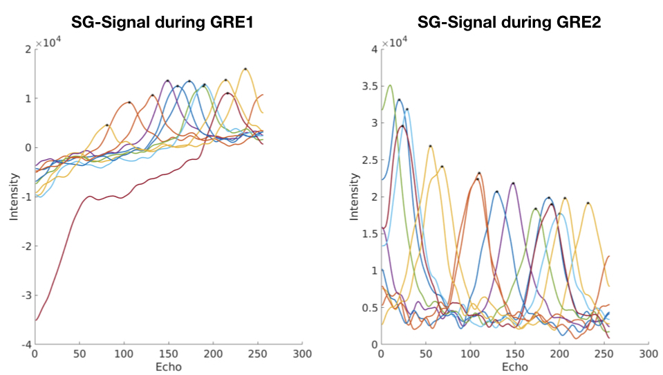
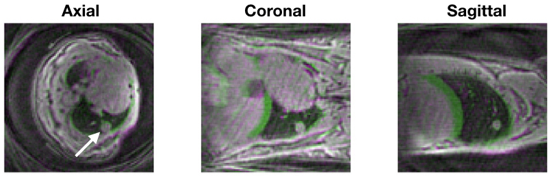
The Cartesian encoding of the 3D MP2RAGE sequence was replaced by a radial one, with an in-out configuration and a pseudo golden-angle distribution of the spokes. Echo time was thus reduced to <0.1ms. Mice (healthy or injected intravenously with RENCA cells) were imaged using the following parameters : FOV: 203; matrix: 963; spatial resolution: 208μm3; 128 echoes per train; TE/TR:0.06/3ms; Ti1-Ti2-MP2RAGETR: 1000/3300/8250ms; 5 points of self-gating signal; 128 or 512 inversions; total amount of spokes: 16384 or 65536, respectively; acquisition time: 17min or 1h, respectively; respiration : 80-100beats/min. The 3D T1 maps were reconstructed without SG correction using the 17min acquisitions. In parallel, the SG signal from each projection of a GRE block (from the 1h scans) was reconstructed over time2. The mean signal was then measured from the 512 inversions. Then, each signal was compared one-by-one to the mean. If superior to 3 times the Standard Deviation, it was considered to be acquired during breathing. The respiration peaks were identified. Reconstruction of 10 images per respiration cycle was performed, each containing 6553 spokes. Then, two Ti2 images were extracted : one at the peak of the respiration and one during the end of expiration phase in order to create a displacement map3.RESULTS
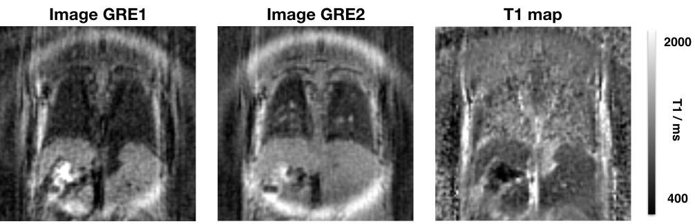
The new 3D UTE MP2RAGE sequence enabled to measure the T1 of the lung parenchyma at 1300±122ms at 7T (Figure 1). This was obtained in only 17 min, during free-breathing of the animal. T1 of this pulmonary metastasis model was 1739±94ms. The SG signal varied intensively during respiration motion, enabling to detect respiration cycles in a non-steady state, i.e. during magnetization relaxation, at both GRE blocks (Figure 2). During breathing, the displacement of the lung metastases was different depending on their positions within the lungs (Figure 3). Indeed, a metastasis located in the middle of the lungs moved of 0.47mm (i.e. 2 pixels approximatively) and 0.27mm in the z and y directions, respectively; whereas a metastasis located at the bottom of the lungs moved of 0.43mm and 0.93mm (i.e. more than 4 pixels approximatively) in the z and y, respectively.DISCUSSION
Due to the UTE encoding, the new MP2RAGE sequence enabled to measure the T1 of the lungs in 3D, with high spatial resolution and in only 17min. The T1 of the lung parenchyma measured here is similar to the ones found in literature4. The T1 of pulmonary metastases were also measured, without respiration-triggering. Due to the SG technique, projections affected by respiration motion were sorted. This is the first study that demonstrate that a respiration signal can be extracted from the MP2RAGE sequence. The motion of the metastases during breathing can be as intense as 1mm. This information can be used either to correct T1 maps from motion or to measure the displacement of pulmonary metastases during breathing. This development extends the application of the MP2RAGE sequence to thorax mapping.CONCLUSION
This new sequence would be interesting to apply when breathing oxygen in order to evaluate ventilation, which is a biomarker of lung function. In addition, it would enable to characterize the intensity of displacement of lung metastases or tumors before applying radiotherapy.Acknowledgements
No acknowledgement found.References
[1] Trotier AJ, Rapacchi S, Faller TL et al.Compressed-Sensing MP2RAGE sequence: application to the detection of brain metastases in mice at 7T. Magn Reson Med 2018. doi: 10.1002/mrm.27438
[2] Winter P, Kampf T, Helluy X et al. Self-Navigation Under Non-Steady-State Conditions: Cardiac and Respiratory Self-Gating of Inversion Recovery Snapshot FLASH Acquisitions in Mice. Magn Reson Med 76:1887–1894, 2016.
[3] Zachiu C, Papadakis N, Ries M et al. An improved optical flow tracking technique for real-time MR-guided beam therapies in moving organs. Phys Med Biol. 2015 Dec 7;60(23):9003-29.
[4] Guo J, Cao X, Cleveland ZI et al.Murine Pulmonary Imaging at 7T: T2* and T1 With Anisotropic UTE. Magn Reson Med. 2018;79(4):2254-2264.
Figures