4389
Efficient Quantitative Susceptibility Mapping of Popliteal Artery Wall1Meinig School of Biomedical Engineering, Cornell University, new york, NY, United States, 2Radiology, Weill Cornell Medicine, New York, NY, United States
Synopsis
The objective of this study was to develop and optimize the pulse sequence and post-processing for an efficient and high quality QSM of the popliteal artery wall. We showed that high quality QSM could be achieved in 4 minutes without the need for cardiac gating.
INTRODUCTION
To date much of the imaging of the popliteal artery are focused on measuring luminal stenosis but not on the site of pathology1. Quantitative susceptibility mapping (QSM) is a sensitive imaging technique for detecting and quantifying calcifications and hemorrhages2 that are often presented in popliteal artery atherosclerosis. The quantitative insights provided by QSM could potentially characterize high-risk plaque, which can useful for risk stratification in patients. QSM has been attempted for arterial wall imaging3, but its utility is limited by long scan time and artifacts associated with blood flow, chemical shift, and imperfect background field removal. Our objective was to develop and optimize a robust and efficient QSM acquisition and reconstruction methods for popliteal wall imaging in healthy volunteers.METHODS
Popliteal wall MRI typically requires cardiac gating to synchronize the data acquisition with the pulsatile blood flow pattern in the lower extremities to eliminate flow artifact, leading to long scan time. We proposed to shorten acquisition time of popliteal QSM by developing a full 3D flow-compensated multi-echo pulse sequence without cardiac gating. To further minimize artifact caused by residual flow, QSM maps were reconstructed with the preconditioned total field inversion methods (pQSM)4 with 2 additional regularization terms added to the inversion, one for the artery and one for the vein, to restrict the susceptibility variations within the blood vessels (pQSM+0)5:
$$y^*=argmin_y\frac{1}{2}{\parallel}w(f-d{\otimes}Py){\parallel}^2_2+{\lambda\parallel}M_G{\triangledown}Py{\parallel}_1+{\lambda_{artery}\parallel}M_{artery}P(y-\overline{y}^{artery}){\parallel}^2_2+{\lambda_{vein}\parallel}M_{vein}P(y-\overline{y}^{vein}){\parallel}^2_2$$
The first two terms are respectively the data fidelity term and structure consistency regularization terms for pQSM. The last two terms constrain the susceptibility variation the artery and the vein blood pools, where $$$\lambda_{artery}$$$ and $$$\lambda_{vein}$$$ are regularization parameters, $$$M_{artery}$$$ and $$$M_{vein}$$$ are the mask for artery and vein obtained through manual segmentation on the GRE images, and $$$\overline{y}^{artery}$$$ and $$$\overline{y}^{vein}$$$ are the average susceptibility over the artery and vein blood pools, respectively. The QSM map, $$$\chi$$$, is then $$$\chi=Py^*$$$. The susceptibility difference between artery and vein in QSM was converted to oxygenation difference as described in our previous work6.
The popliteal arteries of 5 healthy volunteers were scanned with the developed QSM sequence on a GE MR750 scanner using an 8-channel transmit/receive knee coil. The scan parameters were: 1st TE/TE/#TE/TR=3.7ms/4.7ms/6/30ms, acquisition resolution=.667x.625x2mm3, FOV=200mm2, number of slices=32, ky interleaved sequential view order, 20 views per segment, and trigger delay=350ms for the cardiac gated scan. The scan time for the gated scan was approximately 10 minutes for heart rate of 60 bpm, and the scan time for the ungated scan was 6 minutes. The echo spacing of 4.7ms, which corresponds to a 4 inter-echo phase evolution of fat relative to water at 3T, was chosen to minimize the effect of fat chemical shift, allowing for the estimation of field map without the need for water/fat separation.
To study the effect of the number of echo times (which is proprotional to scan time) on QSM quality, popliteal QSM maps were also reconstructed using the first 2 and 4 echoes and compared with the QSM maps reconstructed using all 6 echoes.
RESULTS
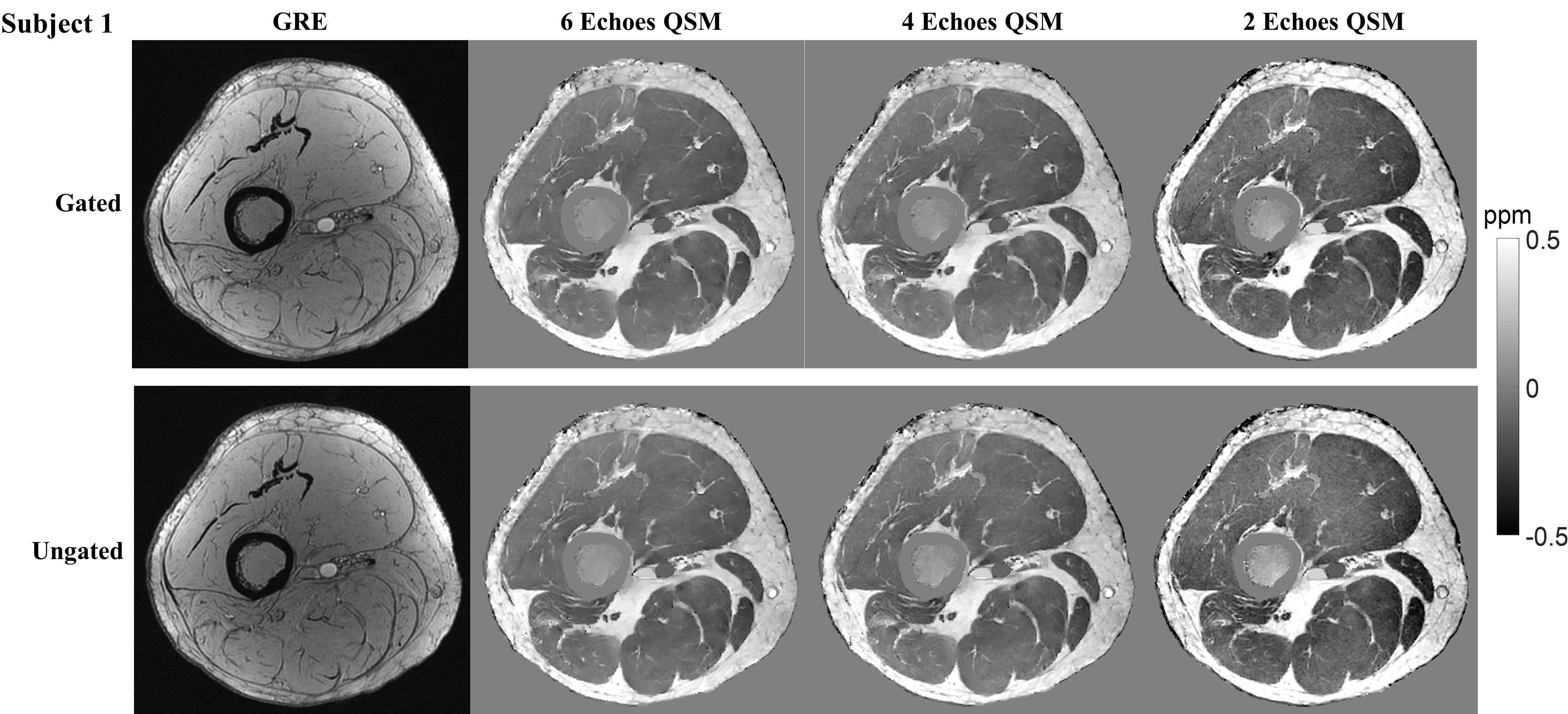
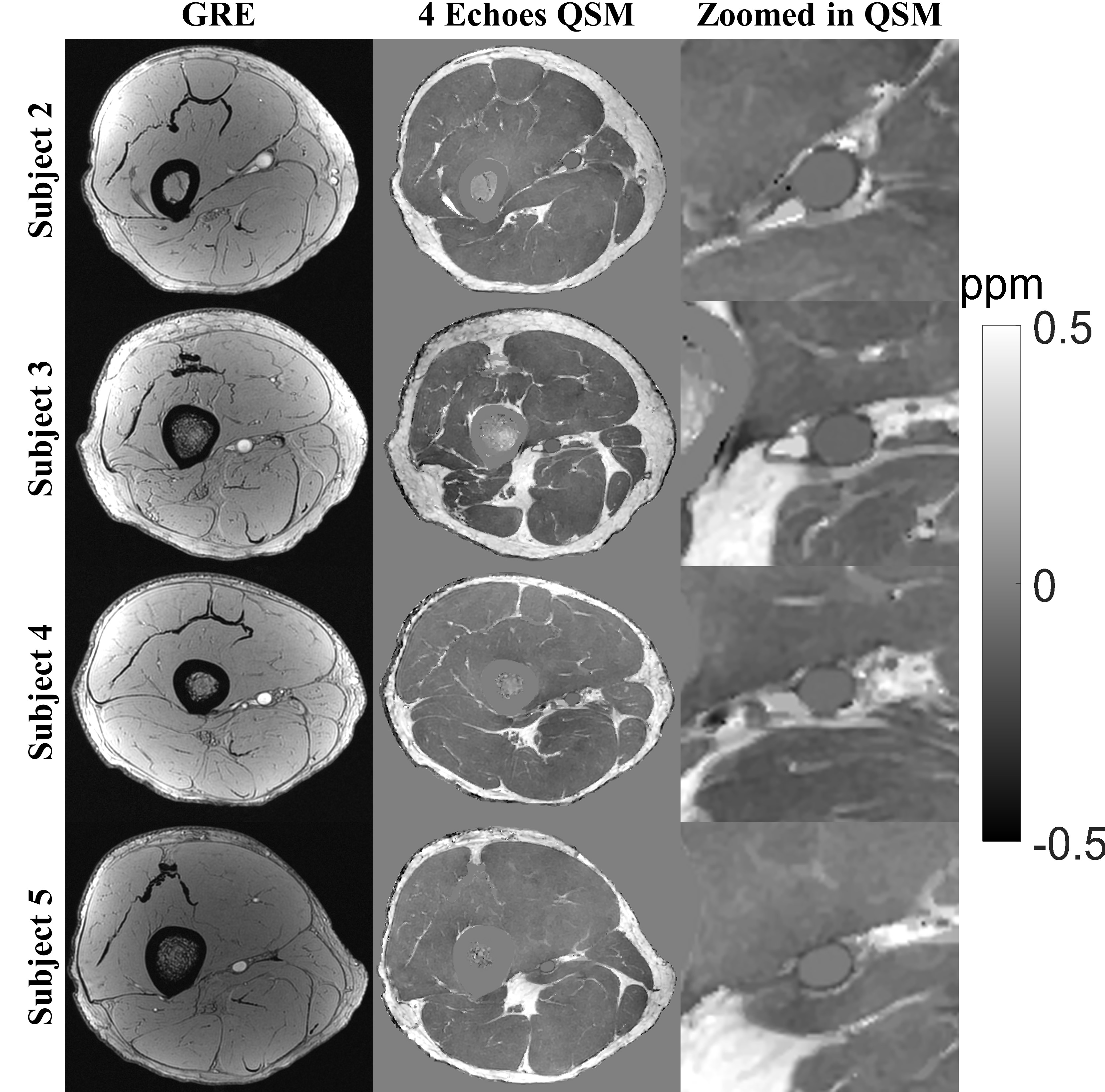
Figure 1 shows comparison of QSM maps reconstructed using 6, 4, and 2 echoes in both gated and ungated scans obtained from one subject. In both scans, QSM maps reconstructed using 6 echoes and 4 echoes have similar high quality, and both are superior to QSM map reconstructed using only 2 echoes. The difference in QSM image quality and contrast between gated and ungated acquisitions was found to be minimal. The average oxygenation difference between artery and vein measured from the gated and ungated scans were similar (24.3±3.0% vs. 24.2±5.5%, p=.99). The 4 echoes and 6 echoes reconstructions also produced similar average oxygenation difference (24.3±4.3% vs. 26.0±3.8%, p=.48). High quality QSM maps of the popliteal vessel wall were also obtained in the other 4 volunteers using 4 echoes (Figure 2).DISCUSSION
This is the first study demonstrating the feasibility of obtaining high quality QSM of the popliteal artery wall in reasonable scan time. The developed ungated 3D flow-compensated QSM sequence provided similar quality to the cardiac gated sequence while reducing scan time. Four echo times are sufficient to achieve high quality QSM, which corresponds to 4 min scan time.Acknowledgements
This work was supported in part from NIH grant R01NS072370, R01NS090464, and R01NS095562.References
1. Wright LB, Matchett WJ, Cruz CP, James CA, Culp WC, Eidt JF, McCowan TC. Popliteal Artery Disease: Diagnosis and Treatment. RadioGraphics 2004;24(2):467-479.
2. Wang Y, Liu T. Quantitative susceptibility mapping (QSM): Decoding MRI data for a tissue magnetic biomarker. Magnetic Resonance in Medicine 2015;73(1):82-101.
3. Wang, C., S. Liu, S. Buch, H.S. Choi, E.-J. Hwang, Z. Fan, et al. Quantitative Susceptibility Mapping of Atherosclerosis in Carotid Arteries. in Proceedings of the 24th scientific meeting, International Society for Magnetic Resonance in Medicine, Singapore. 2016.
4. Liu, Z., et al., Preconditioned total field inversion (TFI) method for quantitative susceptibility mapping. Magnetic Resonance in Medicine, 2016: p. doi:10.1002/mrm.26331.
5. Liu, Z., et al., MEDI+0: Morphology enabled dipole inversion with automatic uniform cerebrospinal fluid zero reference for quantitative susceptibility mapping. Magnetic Resonance in Medicine: 79: 2795-2803. doi:10.1002/mrm.26946.
6. Wen, Y., et al., Cardiac quantitative susceptibility mapping (QSM) for heart chamber oxygenation. Magnetic Resonance in Medicine.
Figures