4388
Free-running 3D Radial Myocardial T1 Mapping using Self-Calibrating GRAPPA Operator Gridding for Accelerated Iterative Reconstruction1School of Biomedical Engineering and Imaging Sciences, King's College London, London, United Kingdom
Synopsis
Free-running 3D radial (kooshball) sampling is suitable for fast and self-navigated whole-heart cardiovascular imaging. However, iterative undersampled 3D radial reconstruction requires computational demanding gridding/regridding steps in each iteration, which leads to long reconstruction time and may limit the applications of this imaging strategy. In this work, we investigate the feasibility of accelerating iterative reconstruction for a free-running 3D myocardial T1 mapping sequence using GRAPPA Operator Gridding (GROG)-based pre-reconstruction interpolation. Image quality and T1 estimation accuracy of the accelerated GROG-based reconstruction were compared with conventional non-uniform FFT (NUFFT)-based reconstruction in a standardized phantom and five healthy subjects.
Introduction
Application of 3D radial (kooshball) sampling to cardiovascular MR imaging has become very popular, due to its efficient 3D sampling, potential for respiratory or cardiac self-navigation (1), and flexible retrospective reconstruction for quantitative mapping (2). However, iterative reconstruction of undersampled 3D radial data using sparsity or low-rank regularization needs computationally demanding gridding/regridding (3) steps in each iteration. Recent studies (4, 5) have demonstrated the feasibility of accelerating 2D radial undersampled reconstruction by first interpolating the data onto a Cartesian grid and then performing the iterative reconstruction in the Cartesian space. Self-calibrating GRAPPA Operator Gridding (GROG) (6) is suitable for 3D gridding, as it is effective, requiring low memory and has shown good performance in maintaining image quality (7). In this work, we investigate the feasibility of accelerating iterative reconstruction for a free-running 3D myocardial T1 mapping sequence using GROG-based pre-reconstruction interpolation.Methods
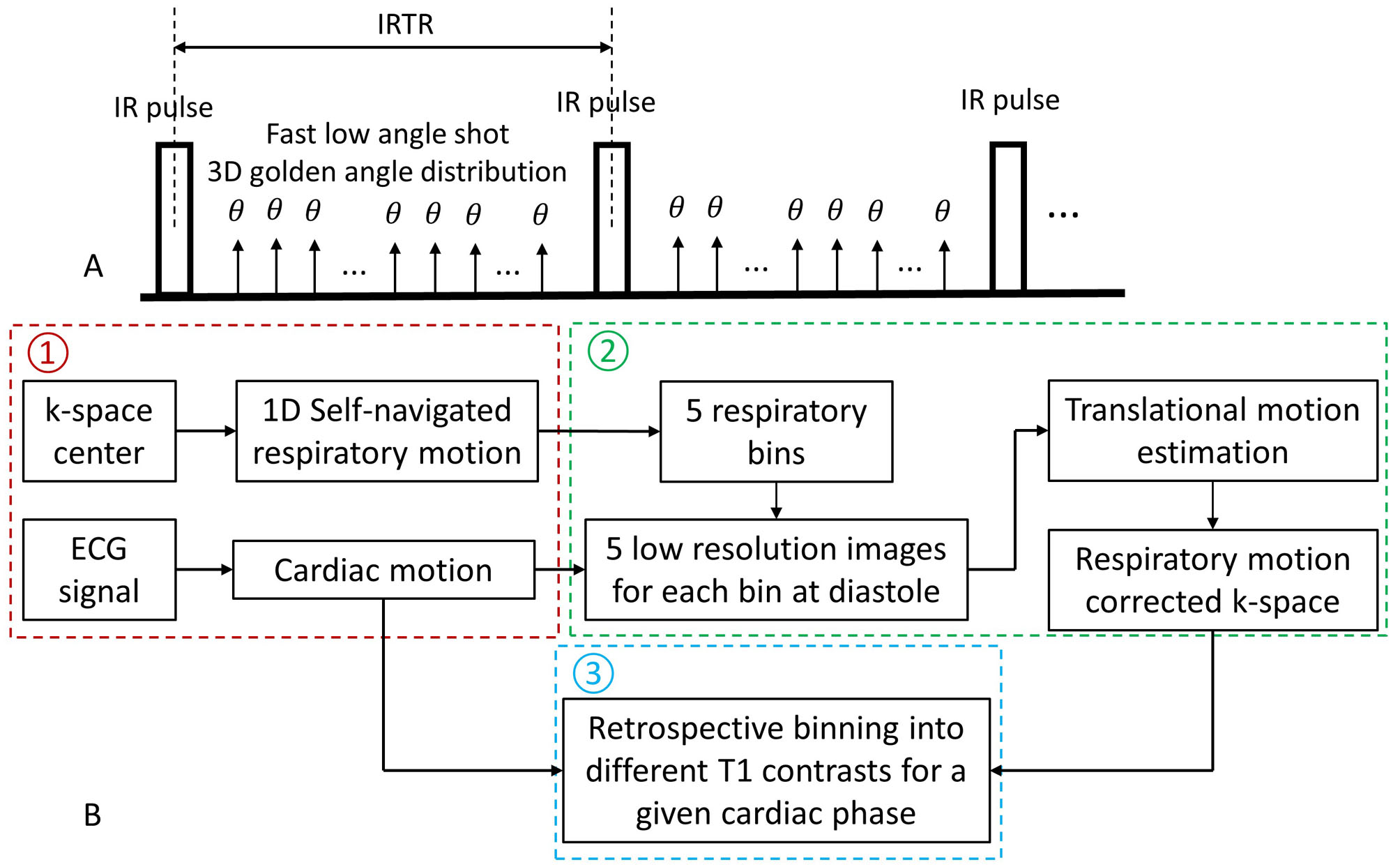
Pre-reconstruction Interpolation using GROG: The adopted free-running 3D myocardial T1 mapping sequence is shown in Fig. 1A. For retrospective reconstruction of T1 image series for a specific cardiac phase, the data sorting process is shown in Fig. 1B. For a given cardiac phase, the highly undersampled 3D radial data is reconstructed over multiple T1 contrasts by combining a dictionary-based low-rank reconstruction with a recently proposed parallel imaging 3D patch-based reconstruction, exploiting local, non-local and contrast redundancies (8). The proposed reconstruction is formulated in the following unconstrained Lagrangian:$$L\left(I,\alpha,Y\right)=argmin\parallel EI-\sqrt{D}K \parallel_2^2+\lambda\parallel \alpha \parallel_0+\mu\parallel I-P\alpha-Y \parallel_2^2$$where $$$E$$$ is the encoding operator $$$E=\sqrt{D}AU_{r}FS$$$, including the non-Cartesian density compensation function $$$D$$$, sensitivity maps $$$S$$$, Fourier Transform $$$F$$$, low rank operator $$$U_{r}$$$, obtained by truncating the singular value decomposition of the simulated dictionary, and the convolutional gridding operator $$$A$$$, transforming Cartesian data back to 3D radial; $$$K$$$ is the undersampled 3D radial data; $$$P$$$ is the patch grouping operator and $$$\alpha$$$ are the associated sparse coefficients; $$$Y$$$ is the Lagrangian multiplier; $$$\lambda$$$ is the sparsity regularization parameter and $$$\mu$$$ is the penalty parameter. The above equation can be solved by operator-splitting via alternating direction method of multipliers (ADMM) (8). For the iterative minimization of the data consistency term $$$\parallel EI-\sqrt{D}K \parallel_2^2$$$, $$$E$$$ and its Hermitian transpose $$$E^{H}$$$ involving convolutional gridding needs to be performed in each iteration. Introducing GROG by pre-interpolating $$$K$$$ into corresponding undersampled Cartesian k-space $$$K_{c}$$$, the data consistency term is modified to be $$$\parallel E_{GROG}I-WK_{c} \parallel_2^2$$$. The GROG-based encoding operator is $$$E_{GROG}=WMU_{r}FS$$$, where $$$M$$$ is the sampling mask of $$$K_{c}$$$, $$$W$$$ is the GROG weighting factor introduced to resolve the blurring caused by the variable-density data distribution after GROG (5) and is given by:$$W=\sqrt{D_{GROG,n}/D_{GROG,act}}$$where $$$D_{GROG,n}$$$ and $$$D_{GROG,act}$$$ are the density filters given by the GROG algorithm using the actual number of spokes and $$$n$$$ times the actual number of spokes, separately. After reconstruction, T1 maps were generated by a dot product matching between the reconstructed singular images and the dictionary.
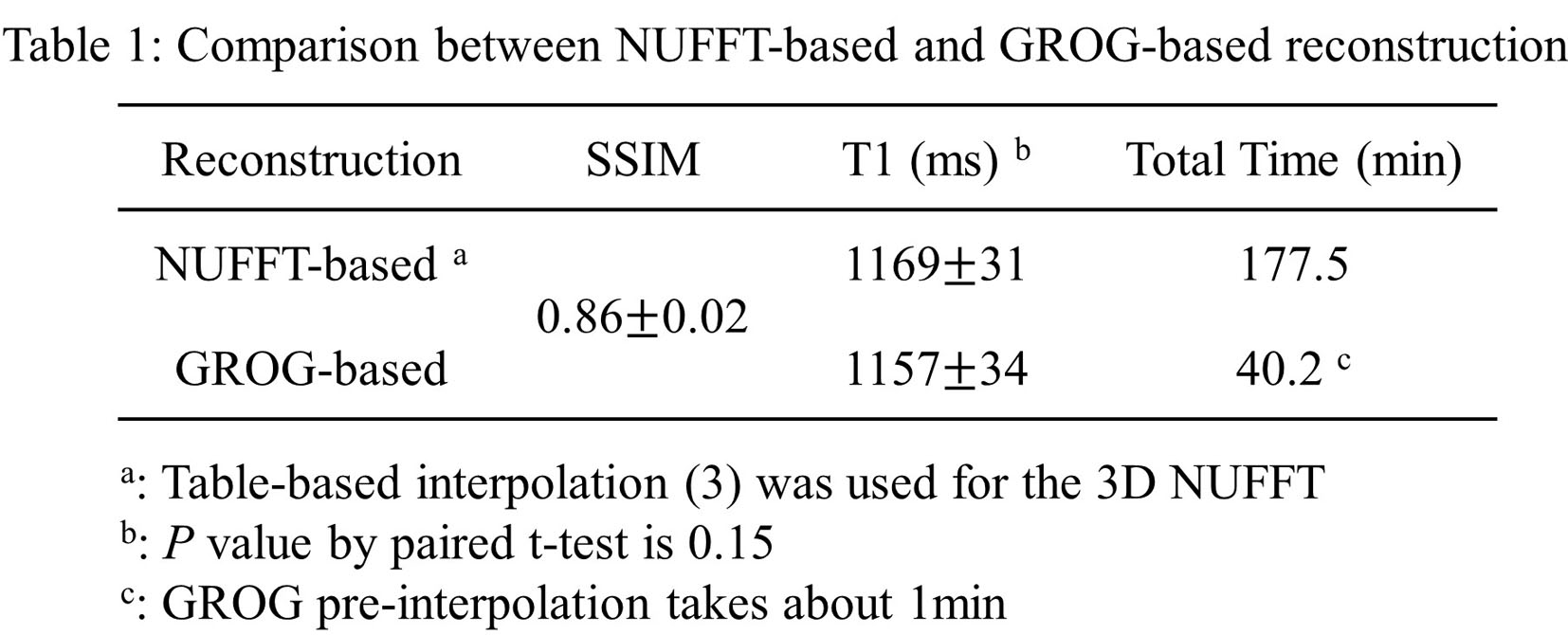
Data Acquisition and Analysis: Imaging was performed in a standardized T1 phantom and five heathy subjects on a 1.5T scanner (Ingenia, Philips Healthcare). Imaging parameters of the free-running 3D radial sequence were: FOV=200mm3, spatial resolution=1.5mm3, TR/TE/flip angle=11.6ms/5.1ms/6°, scan time=9.5min. Phantom T1 mapping accuracy by non-uniform FFT (NUFFT)-based and GROG-based reconstruction was compared against gold standard 2D inversion recovery spin echo (IR-SE) acquisition. For in vivo analysis, T1 images were reconstructed at systolic cardiac phase. The structural similarity index (SSIM) of the singular images reconstructed by the two methods was measured. Three short-axis slices were selected from each subject, and mean myocardium T1 was calculated for each slice and compared between the two reconstruction methods by paired t-test. Five ADMM iterations were performed for NUFFT-based and GROG-based reconstructions, with other reconstruction parameters optimized separately for each method. All reconstructions were performed on a server with dual 16-core CPUs, 256GB RAM.
Results
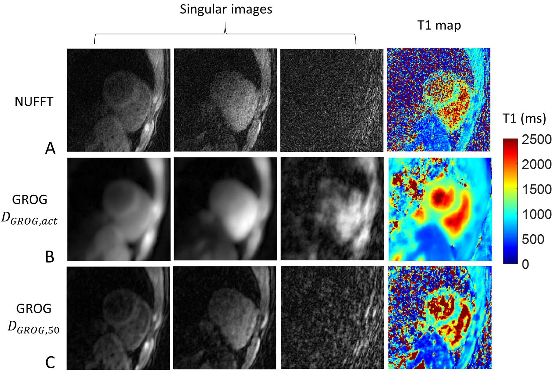
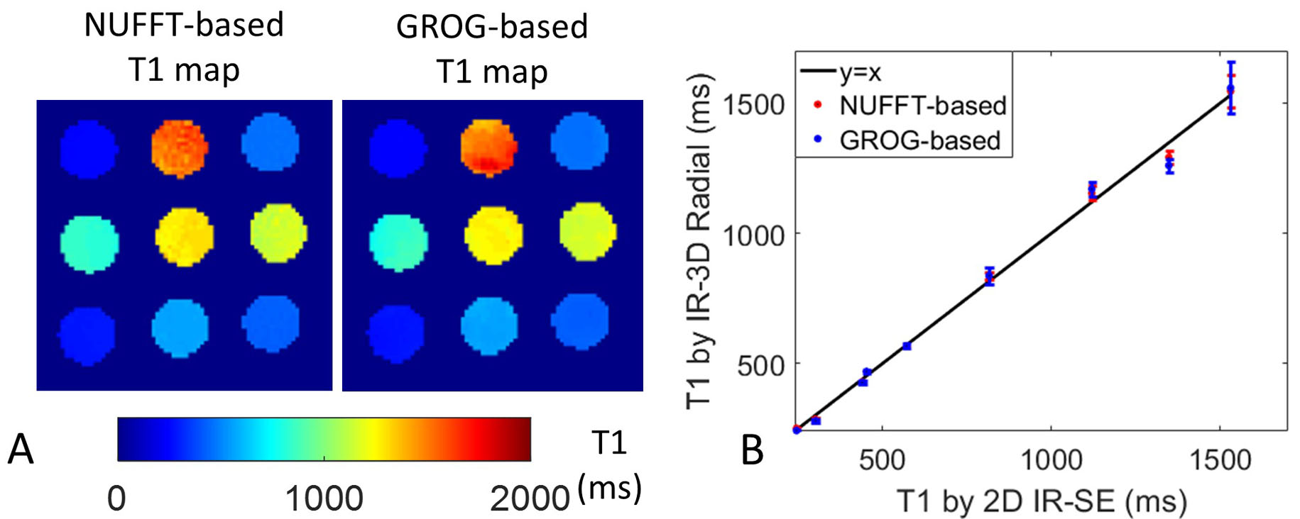
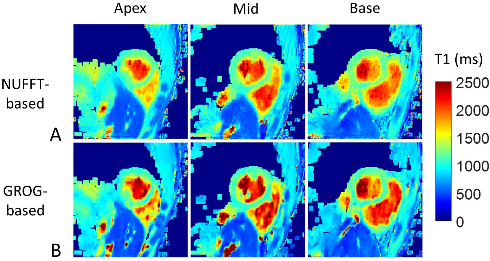
As shown in Fig. 2, compared with NUFFT, GROG performed with $$$D_{GROG,50}$$$ instead of $$$D_{GROG,act}$$$ can resolve the blurring, so $$$D_{GROG,50}$$$ was used in the following GROG-based iterative reconstructions. Phantom T1 mapping by NUFFT-based and GROG-based reconstructions have similar performance (Fig. 3A), and both agree well with the gold standard 2D IR-SE (Fig. 3B). Representative myocardial T1 maps by the two reconstruction approaches are compared in Fig. 4, where no blurred and even sharper maps were observed for GROG-based reconstruction, which can be justified by the fact that pseudo-uniform distributions of radial spokes may cause blurring in NUFFT-based reconstruction. GROG-based reconstruction had similar image quality and T1 measurements to NUFFT-based reconstruction while achieving 4.4-fold reduction of reconstruction time (Table 1).Conclusions & Discussion
Pre-reconstruction interpolation using GROG can substantially accelerate iterative reconstruction of undersampled 3D radial data without sacrificing image quality. This may broaden the applications of 3D radial trajectory for fast and self-navigated acquisitions.Acknowledgements
This work was supported by the following grants: EPSRC EP/P032311/1, EP/P001009/1 and EP/P007619/1.References
1. Pang JN, Sharif B, Fan ZY, Bi XM, Arsanjani R, Berman DS, Li DB. ECG and Navigator-Free Four-Dimensional Whole-Heart Coronary MRA for Simultaneous Visualization of Cardiac Anatomy and Function. Magnetic resonance in medicine 2014;72(5):1208-1217.
2. Qi H, Sun J, Qiao H, Chen S, Zhou Z, Pan X, Wang Y, Zhao X, Li R, Yuan C, Chen H. Carotid Intraplaque Hemorrhage Imaging with Quantitative Vessel Wall T1 Mapping: Technical Development and Initial Experience. Radiology 2018;287(1):276-284.
3. Fessler JA, Sutton BP. Nonuniform fast Fourier transforms using min-max interpolation. Ieee T Signal Proces 2003;51(2):560-574.
4. Tran-Gia J, Stab D, Wech T, Hahn D, Kostler H. Model-Based Acceleration of Parameter Mapping (MAP) for Saturation Prepared Radially Acquired Data. Magnetic resonance in medicine 2013;70(6):1524-1534.
5. Benkert T, Tian Y, Huang CC, DiBella EVR, Chandarana H, Feng L. Optimization and validation of accelerated golden-angle radial sparse MRI reconstruction with self-calibrating GRAPPA operator gridding. Magnetic resonance in medicine 2018;80(1):286-293.
6. Seiberlich N, Breuer F, Blaimer M, Jakob P, Griswold M. Self-calibrating GRAPPA operator gridding for radial and spiral trajectories. Magnetic resonance in medicine 2008;59(4):930-935.
7. Tian Y, Erb KC, Adluru G, Likhite D, Pedgaonkar A, Blatt M, Iyer SK, Roberts J, DiBella E. Technical Note: Evaluation of pre-reconstruction interpolation methods for iterative reconstruction of radial k-space data. Med Phys 2017;44(8):4025-4034.
8. Bustin A, Ginami G, Cruz G, Correia T, Ismail TF, Rashid I, Neji R, Botnar RM, Prieto C. Five-minute whole-heart coronary MRA with sub-millimeter isotropic resolution, 100% respiratory scan efficiency, and 3D-PROST reconstruction. Magnetic resonance in medicine 2018.
Figures