4381
Field Map Estimation from Magnitude-Based Water-Fat Separation1Perspectum Diagnostics Ltd, Oxford, United Kingdom
Synopsis
Complex-based MRI chemical-shift encoded water-fat separation depends on accurate field map convergence, which is often mitigated with spatial regularization. This is prone to error propagation and over-smoothing of fat-fraction maps. Magnitude-based separation circumvents field mapping but is reportedly limited in fat-fraction range (0-50%). We have recently presented MAGO, a magnitude-based method that resolves this water-fat ambiguity. In this study, we compare MAGO to state-of-the-art fat-fraction quantification on N=150 volunteers, and we expand the method for field map calculation using previously estimated water and fat images. MAGO is comparable to regularized hybrid-based decomposition and shows promise in higher field inhomogeneity regimes.
INTRODUCTION
Chemical-shift encoded (CSE) water-fat separation MRI methods have emerged as non-invasive tools for proton density fat fraction (PDFF) quantification in the liver. Most advanced CSE methods (e.g. IDEAL1) are complex-based, in that they need both magnitude and phase images, and estimate PDFF indirectly through iterative optimization of the “field map”. However, erroneous field map convergence leads to fat-water swap artefacts in the PDFF map. Spatial regularization is often used but smoothness assumptions and sensitivity to initial seed pixels may lead to over-smoothed or incorrect PDFF values2. CSE methods that use only magnitude images have also been proposed3. These do not require field map estimation and optimize PDFF directly, but remain prone to water-fat ambiguity and are reportedly limited to a dynamic range of 0 to 50% PDFF. We have recently developed MAGO, a magnitude-based method that estimates PDFF over the entire range and has shown excellent accuracy and reproducibility in phantoms across manufacturers and clinical field strengths4. Although not required for MAGO, field map estimation can be useful to assess image quality. In this study, we extend MAGO to estimate the field map from complex data after magnitude-based PDFF calculation. We compare PDFF and field map values to those calculated using implementations of the Hybrid IDEAL algorithm5.METHODS
The MAGO algorithm uses the phase-constrained signal model where $$$\phi_W(x)=\phi_F(x)=\phi(x)$$$6:
$$|s[t_i]|=\left|\left(\rho_W+\rho_F\cdot\sum_{p=1}^{P}\alpha_p\cdot e^{j2\pi f_pt_i}\right)\cdot e^{j(2\pi \psi t_i+\phi)}\cdot e^{-R_2^*t_i} \right|=\left|\rho_W+\rho_F\cdot\sum_{p=1}^{P}\alpha_p\cdot e^{j2\pi f_pt_i}\right|\cdot e^{-R_2^*t_i}$$
Water $$$w(x)$$$, fat $$$f(x)$$$ and $$$R_2^*(x)$$$ are estimated directly at each pixel $$$x$$$ from magnitude images using multi-peak fat modelling and multipoint search coupled to non-linear optimization (ITK LevenbergMarquardtOptimizer). Two separate runs of the algorithm at each voxel suffice to ensure correct convergence (initial conditions $$$\left\{\rho_W,\rho_F,R_2^*\right\}_1=\left\{1000,0,50\right\}$$$ and $$$\left\{\rho_W,\rho_F,R_2^*\right\}_2=\left\{0,1000,50\right\}$$$). At each pixel independently, the converged parameters with lower associated residual sum of squares are chosen. Field map $$$\psi(x)$$$ and phase offset $$$\phi(x)$$$ can then be estimated given $$$w(x)$$$, $$$f(x)$$$ and $$$R_2^*(x)$$$ using the full complex-valued data (Matlab lsqcurvefit, $$$\psi_0=\phi_0=0$$$ for all pixels) and the reduced expression
$$FW_i\equiv\left(\rho_W+\rho_F\cdot\sum_{p=1}^{P}\alpha_p\cdot e^{j2\pi f_pt_i}\right),\,\,\,R_i\equiv e^{-R_2^*t_i},\,\,\,s[t_i]/(FW_iR_i)=e^{j(2\pi \psi t_i+\phi)}$$
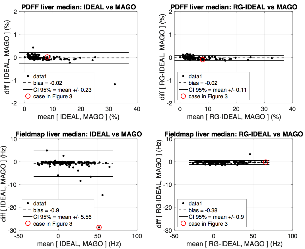
PDFF and field maps were calculated on N=150 UK Biobank7 volunteers (Siemens 1.5T, single-slice six-echo 2D spoiled gradient echo protocol, $$$\text{TE}_1=1.2$$$ ms, $$$\Delta\text{TE}\approx2$$$ ms, 5° flip angle) with MAGO and with two implementations of the Hybrid IDEAL algorithm5, one pixel-independent (“IDEAL”, $$$\psi_0=0$$$ for all pixels), the other with spatial regularization (“RG-IDEAL”, includes an initial region growing step from Yu et al., 20052). One case was discarded due to poor positioning. Median hepatic PDFF and field map values from all three methods were extracted using automatic liver segmentation masks8 and compared for absolute agreement with Bland-Altman analyses (mean ± 95% CI).
RESULTS
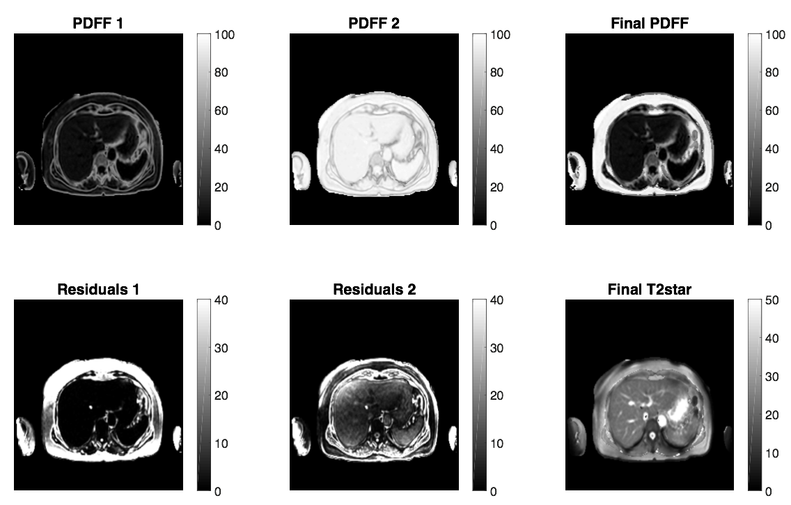
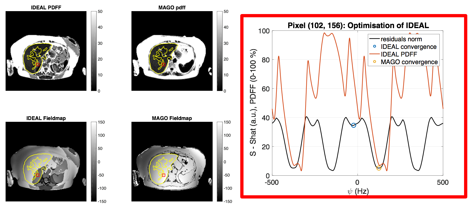
Figure 1 illustrates the intermediate results of the MAGO algorithm, showing how it finds the correct solution at each pixel independently. Bland-Altman comparisons in Figure 2 show good PDFF agreement between MAGO and pixel-independent IDEAL ($$$-0.02\pm0.23$$$) %, but poorer agreement for median field map values ($$$-0.90\pm5.56$$$) Hz. Excellent agreement was observed between MAGO and RG-IDEAL both in median PDFF ($$$-0.02\pm0.11$$$) % and median field map values ($$$-0.38\pm0.90$$$) Hz. Figure 3 illustrates the convergence of the pixel-independent implementations in regions of high field inhomogeneity. For field map values outside the liver and with an associated PDFF>60%, a mapping was empirically found between MAGO and IDEAL/RG-IDEAL ($$$\psi(x)_{IDEAL}=\psi(x)_{MAGO}-0.59*\text{PDFF}(x)_{MAGO}$$$ in those pixels), attributed to differences between the IDEAL formulation1 and the phase constrained model6.DISCUSSION
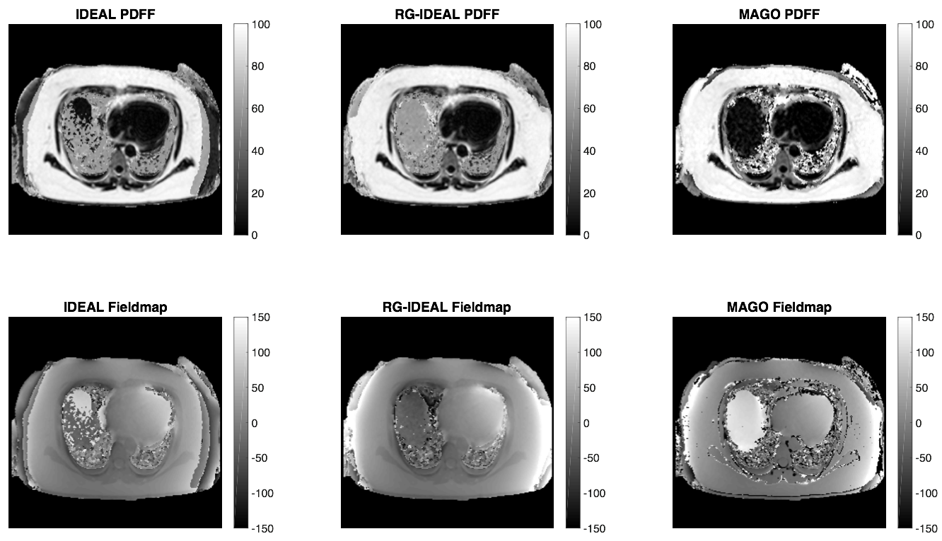
Pixel-independent MAGO PDFF and field map calculation showed comparable performance to a regularized state-of-the-art method on UK Biobank data, which generally consists of well-shimmed single-slice acquisitions. Figure 4 includes a comparison on a peripheral slice from another study (Siemens 1.5T) where Hybrid IDEAL suffered from a full-liver swap; this shows the potential of the MAGO approach in more challenging cases with higher field variation. Future work will aim to validate this approach in 3T acquisitions, notably under bipolar readouts, and explore field map regularization, e.g.
$$\widehat{\psi(x)}=\text{arg min}\sum_x|s(x)-\hat{s}(x)|^2+\lambda\cdot\text{reg}(\psi)$$
where $$$\hat{s}(x)$$$ is the estimated signal, $$$\lambda$$$ a Lagrange multiplier and $$$\text{reg}(\cdot)$$$ a suitable regularizer. We may initially follow a region growing approach for comparison, but regularizers that respect tissue boundaries are of particular interest, including anisotropic diffusion (total variation) or Markov measure fields.
CONCLUSION
Our results suggest that MAGO can robustly estimate PDFF from magnitude data and then use it to estimate a field map from complex data. This approach for water-fat separation may be more robust and widely applicable than complex- and hybrid-based approaches, which estimate the field map first and therefore rely on the availability and reliability of phase images and assumptions that propagate errors to the PDFF map.Acknowledgements
This research has been conducted using the UK Biobank Resource under application 9914 and access to these data was facilitated by Steve Garratt, UK Biobank, Stockport, UK and Jimmy Bell, University of Westminster, London, UK.References
- Reeder SB, Wen Z, Yu H, Pineda AR, Gold GE, Markl M, Pelc NJ. Multicoil Dixon chemical species separation with an iterative least-squares estimation method. Magn Reson Med. 2004 Jan;51(1):35-45. DOI: 10.1002/mrm.10675.
- Yu H, Reeder SB, Shimakawa A, Brittain JH, Pelc NJ. Field map estimation with a region growing scheme for iterative 3-point water-fat decomposition. Magn Reson Med. 2005 Oct;54(4):1032-9. DOI: 10.1002/mrm.20654.
- Bydder M, Yokoo T, Hamilton G, Middleton MS, Chavez AD, Schwimmer JB, Lavine JE, Sirlin CB. Relaxation effects in the quantification of fat using gradient echo imaging. Magn Reson Imaging. 2008 Apr;26(3):347-59. DOI: 10.1016/j.mri.2007.08.012.
- Bagur A, Hutton C, Irving B, Gyngell ML, Robson MD, Brady M. Magnitude-Intrinsic Water-Fat Ambiguity can be Resolved with Multi-Peak Fat Modelling and a Multipoint Search Method. [Submitted manuscript, 2018].
- Yu H, Shimakawa A, Hines CD, McKenzie CA, Hamilton G, Sirlin CB, Brittain JH, Reeder SB. Combination of complex-based and magnitude-based multiecho water-fat separation for accurate quantification of fat-fraction. Magn Reson Med. 2011. DOI: 10.1002/mrm.22840.
- Yu H, Reeder SB, McKenzie CA, Brau AC, Shimakawa A, Brittain JH, Pelc NJ. Single acquisition water-fat separation: feasibility study for dynamic imaging. Magn Reson Med. 2006 Feb;55(2):413-22. DOI: 10.1002/mrm.20771.
- Sudlow C, Gallacher J, Allen N, Beral V, Burton P, Danesh J, Downey P, Elliott P, Green J, Landray M, Liu B, Matthews P, Ong G, Pell J, Silman A, Young A, Sprosen T, Peakman T, Collins R. UK biobank: an open access resource for identifying the causes of a wide range of complex diseases of middle and old age. PLoS Med. 2015 Mar 31;12(3):e1001779. DOI: 10.1371/journal.pmed.1001779.
- Irving B, Hutton C, Dennis A, Vikal S, Mavar M, Kelly M, Brady, JM. Deep Quantitative Liver Segmentation and Vessel Exclusion to Assist in Liver Assessment. MIUA 2017. DOI: 10.1007/978-3-319-60964-5_58.
Figures