4373
Dynamic Imaging of Vitamin C Pharmacokinetics by 13C Chemical Shift Imaging without Hyperpolarization1National Cancer Institute, Bethesda, MD, United States
Synopsis
Intravenously injected vitamin C is re-emerging as a potential chemotherapeutic agent but questions remain about its bioavailability and metabolic fate. Using newly developed methods for signal denoising, we show that is possible to image vitamin C pharmacokinetics by non-hyperpolarized 13C MRI with significant sensitivity and time resolution that the accumulation of vitamin C into a tumor and its metabolism into downstream metabolites can be visualized. Since the method does not rely on hyperpolarization and therefore does not have restrictions on T1 relaxation, it can be potentially extended to other highly tolerated drugs.
Purpose
The discovery that intravenous injection can overwhelm the tight regulation on oral doses of vitamin C has sparked renewed clinical investigation into its use as a potential chemotherapy agent.1 Since cancer cells likely differ greatly in their ability to import and metabolize vitamin C due to differences in the tumor microenvironment, an in vivo imaging technique able to trace intravenously injected vitamin C would greatly aid in the development of vitamin C as a clinical technique. Two techniques currently exist for in vivo imaging of vitamin C. Luminescence imaging is limited to preclinical studies and subcutaneous tumors by poor tissue penetration.2 The short T1 relaxation of vitamin C limits its use as a tracer in hyperpolarized MRI. The oxidized form, dehydroascorbic acid, can be used instead3-5 but its substantial pancreatic toxicity restricts its use to preclinical studies. We propose a new method of imaging vitamin C distribution and metabolism using 13C MRI without hyperpolarization. In trial studies, we are able to show the selective uptake of vitamin C into pancreatic adenocarcinoma (PDAC) tumors and its further downstream metabolism.Methods
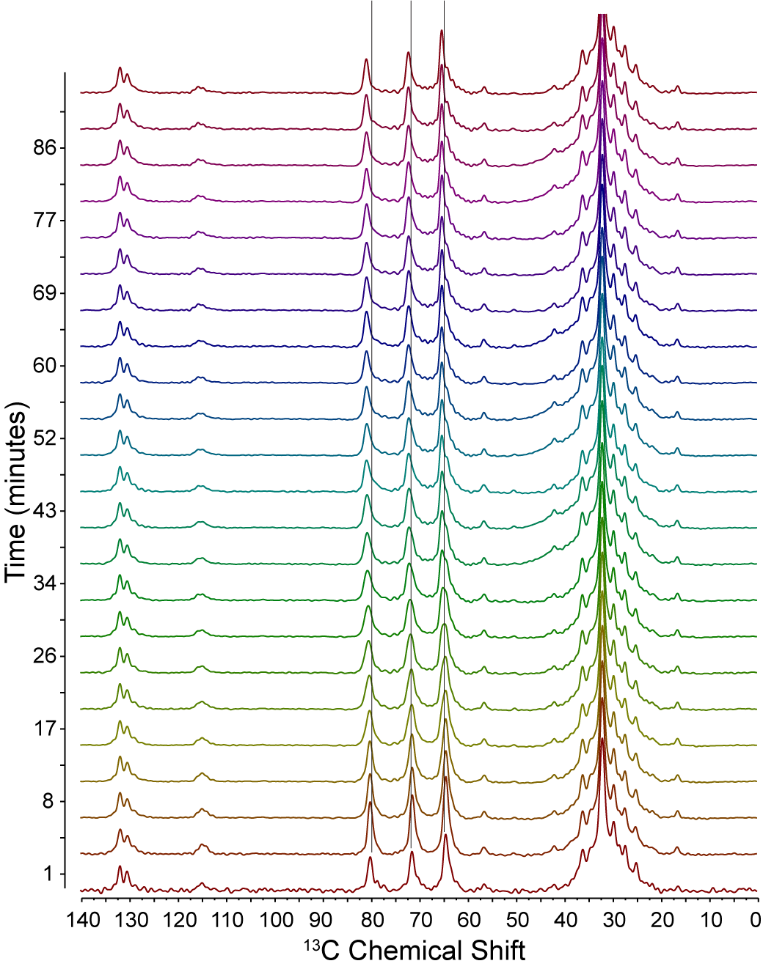
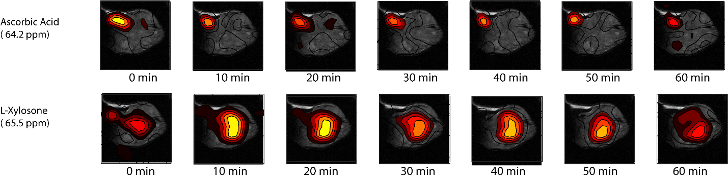
Mice bearing a Hs766t leg xenograft were anesthetized by 1.5–2.0% isoflurane to keep respiration and body temperature within a normal physiological range of 35-37º C and 60-90 breaths per min. For the non-localized experiments, spectra were acquired without hyperpolarization following an IP injection of 25 mg uniformly 13C labeled vitamin C at 9.4 T using a 50 ms repetition time, Ernst Angle excitation of 12º, 512 FID points, a sweep width of 198.6 ppm, 16 averages per scan, and 6750 scans for a total acquisition time of 1.5 hours. MLEV16 decoupling6, 7 was applied during acquisition using -20 dB of decoupling power and a 0.2 ms decoupling element centered on the main proton lipid resonance at 1.3 ppm. Due to the strong decoupling used, the isolation of the proton channel from the carbon channel was key for the acquisition of a strong signal. To achieve this isolation, the leg coil was designed as a quasi-Helmholtz pair with a solenoidal inner 13C coil and an outer 1H coil consisting of two saddle loops with 120° arcs arranged coaxially to the solenoidal coil to generate orthogonally oriented 13C and 1H RF fields. Denoising by singular value decomposition rank reduction (rank 5) was used to enhance the signal to detectable levels.8 Chemical shift imaging experiments were performed similarly except an 8x8 image with 0.15 cm x 0.15 cm x 1.6 cm voxels was acquired every 48 seconds for 90 minutes. Tensor decomposition was then used to enhance the signal to detectable levels (rank 5x6x6x5).8Results
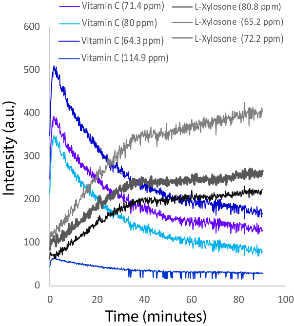
A representative set of spectra obtained after IP injection of 25 mg uniformly 13C labeled vitamin C into a mouse bearing a HS766t PDAC xenograft is shown in Fig. 1. After singular value decomposition to reduce noise to bring the signal to a detectable level, three distinct sets of peaks with different kinetics are detected. The first set is constant in time and corresponds to the natural abundance lipid signals. The second set of three peaks of comparable intensity at 71.4, 64.2, and 80 ppm and a less intense, broader peak at 114.9 ppm spikes within the first two minutes of acquisition before decaying. These peaks can be assigned to unmodified vitamin C by the peak at 114.9 pm, which is not found in dehydroascorbic acid or any other downstream metabolites. The final set can be detected as three peaks ~ 0.8 ppm downfield from the peaks 71.4, 64.2, and 80 ppm (grey lines in Fig. 1) which increase in intensity steadily throughout the experiment. The growth of the second set of peaks is tightly correlated with the disappearance of the vitamin C (Fig. 2), implying they are a metabolic byproduct of vitamin C. These peaks can be tentatively assigned to L-xylosone, a known byproduct of vitamin C metabolism under non-oxidizing conditions,9 based on the observed chemical shifts. Chemical shift imaging showed Vitamin C rapidly built up in the bladder, indicating most of the dose failed was removed from circulation before being transported into the cells (Fig. 3). While vitamin C is not detected in either normal tissue outside the bladder or in a normal leg without a xenograft, it is imported into the tumor and retained, most likely by the GLUT1 transporter in the form of dehydroascorbic acid. The downstream metabolite L-xylosone is confined to the tumor as it is highly polar and unable to cross the cell membrane.Conclusion
Non-hyperpolarized 13C imaging may emerge as a viable technique for determining vitamin C in vivo pharmacokinetics.Acknowledgements
The authors would like to thank Helmutt Merkle of NINDS for coil construction and Helmutt Merkle and Jeeva Munasinghe technical assistanceReferences
[1] Shenoy, N., Creagan, E., Witzig, T., and Levine, M. (2018) Ascorbic Acid in Cancer Treatment: Let the Phoenix Fly, Cancer Cell. DOI: 10.1016/j.ccell.2018.07.014
[2] Song, B., Ye, Z., Yang, Y., Ma, H., Zheng, X., Jin, D., and Yuan, J. (2015) Background-free in-vivo Imaging of Vitamin C using Time-gateable Responsive Probe, Sci Rep 5, 14194.
[3] Keshari, K. R., Sai, V., Wang, Z. J., Vanbrocklin, H. F., Kurhanewicz, J., and Wilson, D. M. (2013) Hyperpolarized [1-13C]dehydroascorbate MR spectroscopy in a murine model of prostate cancer: comparison with 18F-FDG PET, J Nucl Med 54, 922-928.
[4] Bohndiek, S. E., Kettunen, M. I., Hu, D. E., Kennedy, B. W., Boren, J., Gallagher, F. A., and Brindle, K. M. (2011) Hyperpolarized [1-13C]-ascorbic and dehydroascorbic acid: vitamin C as a probe for imaging redox status in vivo, J Am Chem Soc 133, 11795-11801.
[5] Keshari, K. R., Kurhanewicz, J., Bok, R., Larson, P. E., Vigneron, D. B., and Wilson, D. M. (2011) Hyperpolarized 13C dehydroascorbate as an endogenous redox sensor for in vivo metabolic imaging, Proc Natl Acad Sci U S A 108, 18606-18611.
[6] Levitt, M. H., Freeman, R., and Frenkiel, T. (1983) Broad-Band Decoupling in High-Resolution Nuclear Magnetic-Resonance Spectroscopy, Adv Magn Reson 11, 47-110.
[7] Levitt, M. H., Freeman, R., and Frenkiel, T. (1982) Supercycles for Broad-Band Heteronuclear Decoupling, J Magn Reson 50, 157-160. [8] Kang, S. O., Sapper, H., and Lohmann, W. (1982) The Oxidative-Degradation of L-Ascorbic-Acid Via an Alpha-Ketoaldehyde, Z Naturforsch C 37, 1064-1069.
[8] Brender, J. R., Kishimoto, S., Merkle, H., Reed, G., Hurd, R. E., Chen, A. P., Ardenkjaer-Larsen, J. H., Munasinghe, J., Saito, K., Seki, T., Oshima, N., Yamamoto, K., Choyke, P. L., Mitchell, J., and Krishna, M. C. (2018) PET by MRI: Glucose Imaging by 13C-MRS without Dynamic Nuclear Polarization by Noise Suppression through Tensor Decomposition Rank Reduction, bioRxiv 265793.
[9] Kang, S. O., Sapper, H., and Lohmann, W. (1982) The Oxidative-Degradation of L-Ascorbic-Acid Via an Alpha-Ketoaldehyde, Z Naturforsch C 37, 1064-1069.
Figures