4372
Metabolic differences between hyperpolarized 13C pyruvate MRI and 18F-FDG PET imaging in a rat model of spontaneous brain cancer1Department of Radiology, Stanford University, Stanford, CA, United States, 2Department of Neurology, Stanford University, Stanford, CA, United States
Synopsis
Hyperpolarized [1-13C]pyruvate MRI and 18F-FDG PET imaging signals were compared in a rat model of spontaneous slow growing glioma, which mimics the growth pattern of some human gliomas. No enhancement of 18F-FDG was observed in the tumor regions relative to normal appearing brain. Hyperpolarized 13C MRI however did show metabolic differences in all animals. The present study suggests that hyperpolarized 13C MRS potentially may be used to identify and characterize gliomas, which are 18F-FDG negative.
Introduction
Hyperpolarized 13C pyruvate MRI and 18F-FDG PET imaging are two distinctive imaging techniques that report on complementary aspects of intracellular glucose metabolism. Combining the two techniques may provide better insight in the metabolic processes that drives diseases such as cancer. In this study we compared hyperpolarized [1-13C]pyruvate MRI and 18F-FDG PET Imaging signals in a rat model of spontaneous slow growing glioma 1.Methods
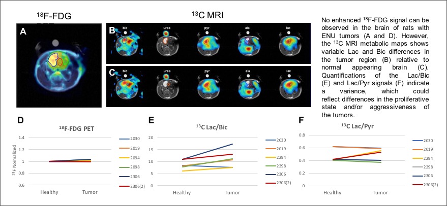
N-ethyl-N-nitrosourea (ENU) a carcinogen chemical, was injected into female rats during pregnancy, which result in birth of rats that later spontaneous developed tumors in neuronal tissue including the brain (gliomas) 2. Rats (n = 6, 450-550g, 110-120 days old) with suspected ENU gliomas were anesthetized with Isoflurane and scanned in a GE 3T Signa scanner with hyperpolarized [1-13C]pyruvate (125mM, 7s bolus injection). 13C MRI signals was acquired using a 13C surface coil (diam: 15mm) and a 16x16 phase encoded FID-CSI sequence (FA: 100, TR=75ms, FOV: 64mm, special resolution: 4x4x5mm). The 13C MRI was followed by a gadolinium enhanced MRI scan (T1-weighted spin echo). 1-2 days later the rats were imaged in a PET scanner (Siemens Inveon D-PET, microPET scanner). 18F-FDG (1mCi in saline) was injected iv. followed by 1 hour of dynamic PET acquisition. All rats were fasted for 18-20 hours prior to 18F-FDG injection. The Gd-enhanced T1-weighted MRIs were collected for co-registration of MR and PET images and for identifying tumor regions of interest (ROIs). Visualization of 18F radio tracer uptake, ROI identification and analysis of signal differences were performed using PMOD software (Version 3.7, PMOD Technologies, Zürich, Switzerland). Normalized uptake of 18F-FDG at 60 min was used for comparison between groups and in the tumor ROIs in contrast to the normal-appearing brain ROIs (figure 1). Hyperpolarized 13C metabolic maps of pyruvate, lactate and bicarbonate and ROI analysis, were performed in MATLAB by integrating the area under the curve of the 13C metabolic peaks.Results
The Gd-enhanced T1-weighted MRIs revealed tumors in 5 out of 6 animals and 2 animals had more than 1 tumor. PET imaging showed no enhancement of 18F-FDG signal in the tumor regions compared to the contralateral normal appearing brain regions (FDG negative). Hyperpolarized 13C MRI however did show metabolic differences in all animals (Figure 1).Discussion
Low grade human gliomas are known to be 18F-FDG negative and much focus have recently been on developing new radio tracers that can identify these types of tumors. The ENU rat model is a spontaneous and slow growing glioma model that mimics the growth pattern of many human gliomas. The present study suggests that hyperpolarized 13C MRS potentially may be used to identify and characterize 18F-FDG negative gliomas similar to the ENU tumors. The results from the metabolic imaging will be compared with histologic data to established if the metabolic differences correlate with the proliferative state of the tumor.Acknowledgements
NIH grants R01CA176836, R01EB019018, P41EB01589, S10OD012283
S10 grant S10OD018130-01 (PET scanner)
GE Healthcare
Lucas Foundation
References
1. Druckrey H, Ivankovic S, Preussmann R: Teratogenic and carcinogenic effects in the offspring after a single injection of ethyl-nitrosourea to pregnant rats. Nature 210:1378–1379, 1966
2. Jang T, Sathy B, Hsu YH, Merchant M, Recht B, Chang C, Recht L. A distinct phenotypic change in gliomas at the time of magnetic resonance imaging detection. J Neurosurg 108:782-90, 2008
Figures