4371
Overcoming short T2 issue with the use of 3D UTE for imaging of 19F-loaded theranostic nanocapsules1Department of Magnetic Resonance Imaging, Institute of Nuclear Physics Polish Academy of Sciences, Kraków, Poland, 2Jerzy Haber Institute of Catalysis and Surface Chemistry, Polish Academy of Sciences, Kraków, Poland
Synopsis
The development of heteronuclear magnetic resonance imaging is one of the strategies in a control of a biodistribution of therapeutic agents. Molecules containing 19F atoms are particularly attractive for use as MRI markers due to favorable physical properties of fluorine nucleus. Advances in MRI instrumentation and ultrafast pulse sequence development expanded the range of fluorine compounds available as new MRI markers. Optimization of imaging parameters of such sequences enables the visualization of 19F compounds with the less preferable characteristic. In this paper, we report the application of the 3D UTE sequence at a high field for imaging of compounds with a complex 19FNMR spectrum and very short T2.
INTRODUCTION
One of the strategies in a control of biodistribution of therapeutic agents is the development of visualization methodology based on heteronuclear magnetic resonance imaging. Molecules containing 19F atoms are particularly attractive for use as MRI markers. The close to zero natural concentration of 19F nuclei in the human body makes fluorine atoms a perfect marker without any natural background signal. This creates the opportunity to locate and identify only exogenous fluorinated compounds with an excellent contrast-to-noise ratio and specificity of the signal.1 Furthermore, a 19F nucleus has a 100% natural abundance, its gyromagnetic ratio is close to a 1H value (40.08 vs 42.58 MHz/T), and it has a spin of ½. Also, the sensitivity of 19F MRI is very high, equal to 83% of 1H MRI.2 It is desirable for the tracer to be characterized by high fluorine content, simple 19FNMR spectrum, with a single, sharp, and intense peak, short T1 and long T2.3 Nevertheless, due to advances in MRI instrumentation, and ultrashort echo pulse sequence development, the range of fluorine compounds available as a new MRI marker has expanded. Optimization of imaging parameters of such sequences allows the visualization of 19F compounds with the less preferable characteristic. In this paper, we report the application of the 3DUTE pulse sequence at a high field for imaging of compounds with a complex 19FNMR spectrum and T2 values in the range of single milliseconds.MATERIALS AND METHODS
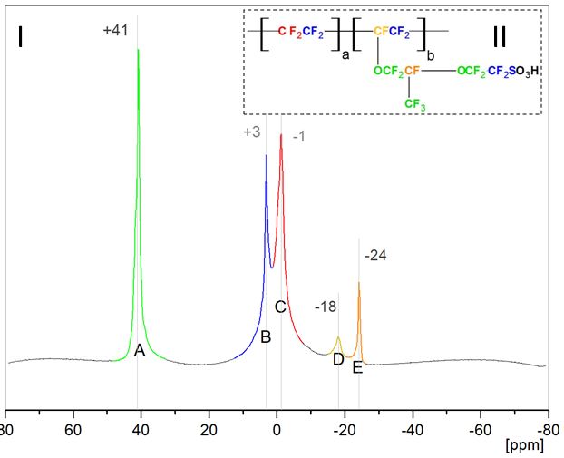
As an example of a 19F containing compound with complex spectrum, the Nafion polymer was chosen, as a compound relatively easy to incorporate into shell of theranostic nanocapsules. Its spectrum exhibits five broad peaks arising from equivalent fluorine nuclei groups4 (Fig.1.I). Furthermore, the structure of a Nafion unit (Fig.1.II) illustrates the variability of the material (unspecified length of a and b chains) which results in differences in peaks area from one synthesis to another. Both MR spectroscopy and 3DUTE imaging were performed at the 9.4 T Bruker Biospec 94/20 research MRI scanner with an in-house built transmit-receive volume coil which can be tuned either to 1H or 19F resonant frequency. The 3DUTE sequence was first applied to Nafion solutions of a different concentration. Imaging parameters were as follow: TR: 8 ms, TE: 0.16 ms, FA: 5°, RF pulse BW: 4.27 kHz, FOV: 2.0 or 2.5 cm, MTX: 32x32x32, acquisition time: 27 min. Prior to imaging, an MR spectroscopy (with a 17 μs 90° pulse and SW: 59.52 kHz) and T1 and T2 measurements were performed in order to choose the peaks with the largest area and consequently highest NMR signal and to set imaging sequence parameters to values enabling an optimal visualization of Nafion molecules distribution.RESULTS
Spectrum with five visible resonances at +41, +3, -1, -18 and -24 ppm with reference to 376.63 MHz was obtained. Measured longitudinal relaxation times varied from ∼700 to ∼900 ms for different resonances, while transverse were estimated to be in a range of single milliseconds. Due to the largest peak area, peaks at +3 and -1 ppm were chosen for imaging. SNR was calculated as a quotient of the signal intensity of the area of interest and the standard deviation of the signal intensity in the area surrounding an object. Imaging with acquisition time ∼27 min gave images with sufficient value of SNR (∼2.3) for a specimen of concentration as low as 0.44 g/l (Fig.2). Preliminary imaging of a phantom containing a 19F loaded core-shell nanocapsules was performed in order to check the possibility of visualizing the distribution of complete theranostic carriers in samples (Fig.2).DISCUSSION
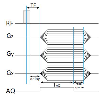
Conventional MRI sequences generate relatively long echo times (>1ms). Therefore the majority of the signal from short transverse relaxation time components decays to near zero prior to echo formation or even during RF pulse excitation. The application of 3DUTE sequence allows enables imaging of very short T2 compounds. Due to non-selective RF excitation, the minimum TE is defined only by RF pulse duration and the time necessary to change between RF excitation and acquisition mode (Fig.3).5 NMR signal is proportional to a field strength, therefore high field systems provide higher SNR than typical clinical scanners. This advantage allows to carry out experiments at very low concentrations of 19F nuclei giving a sufficient amount of signal from a specimen, necessary for further analysis. Achieving a full MR spectrum of a fluorine compound enables one to choose suitable imaging sequence and to set its parameters to values that yield good quality images with possibly least apparent artifacts. We showed that advances in MRI instrumentation and UTE pulse sequence development may expand the range of fluorine compound available as new MRI markers.Acknowledgements
This work was supported by InterDokMed project no. POWR.03.02.00-00-I013/16References
1. Colotti R, et al, MRM 2017; 77:2263–2271.
2. Tirotta I, et al, Chem. Rev. 2015; 115: 1106−1129.
3. Bartusik D, et al, Biomedicine & Pharmacotherapy 2014; 68: 813–817.
4. Bas C, et al, JAPS 2010; 117: 2121–2132.
5. Schmieder AH, et al, Engineering 2015; 1(4): 475–489.
Figures