4370
Temperature Sensitive 19F-Substituted Molecules for Combined Proton-/Fluorine-Imaging in a 7 T Whole-Body MRI System1Institute for Biometrics and Medical Informatics, Otto-von-Guericke University, Magdeburg, Germany, 2Department of Biomicrosystem Technology and Korea Artificial Organ Center, Korea University, Seoul, Korea, Republic of
Synopsis
19F MR spectroscopy and imaging represent important tools for the development of new MR contrast agents and pharmaceutics. Until now, there are only a few published data that described the influence of temperature changes to the 19F chemical shifts in aqueous solutions. Temperature coefficients up to ~ 8.7Hz/K were determined. Thermoresponsive agents are of high interest in, e.g. hyperthermia studies. Changes in signal intensity and chemical shift give information of the local temperature. Here, we present novel MR spectroscopic and imaging data, which describe the 19F MR signal temperature dependency of different fluorinated organic substrates in isotonic saline solution and their temperature calculation methods.
Introduction
An increasing number of fluorinated substrates with pharmaceutical applications were under investigation in the last years1. Important topics are the development of new MR contrast agents and the detection of fluorine nuclei in living organisms, e.g. in metabolisms studies2-4. The huge chemical shift range and the high MR sensitivity of fluorine predestines the determination of changes e.g. in temperature, which opens up novel opportunities2. Until now, there are only a few published data that described the influence of temperature changes on the 19F chemical shifts in aqueous solution. The very low natural abundance of 19F in living organisms leads to background-free 19F images.
These facts result to the idea of this study to develop new types of temperature sensitive contrast agent. Substances with temperature sensitive signals have a high relevance in the temperature control of medical applications like, e.g. laser-induced thermal therapy, hyperthermia treatment or RF-ablation5. Furthermore, contrast agents with background-free images are of interest in fine structures with low contrast.
In this study we present a MR imaging and spectroscopy study of temperature sensitive molecules using a recently developed transmit-receive system for 7T whole-body MRI6.
Methods
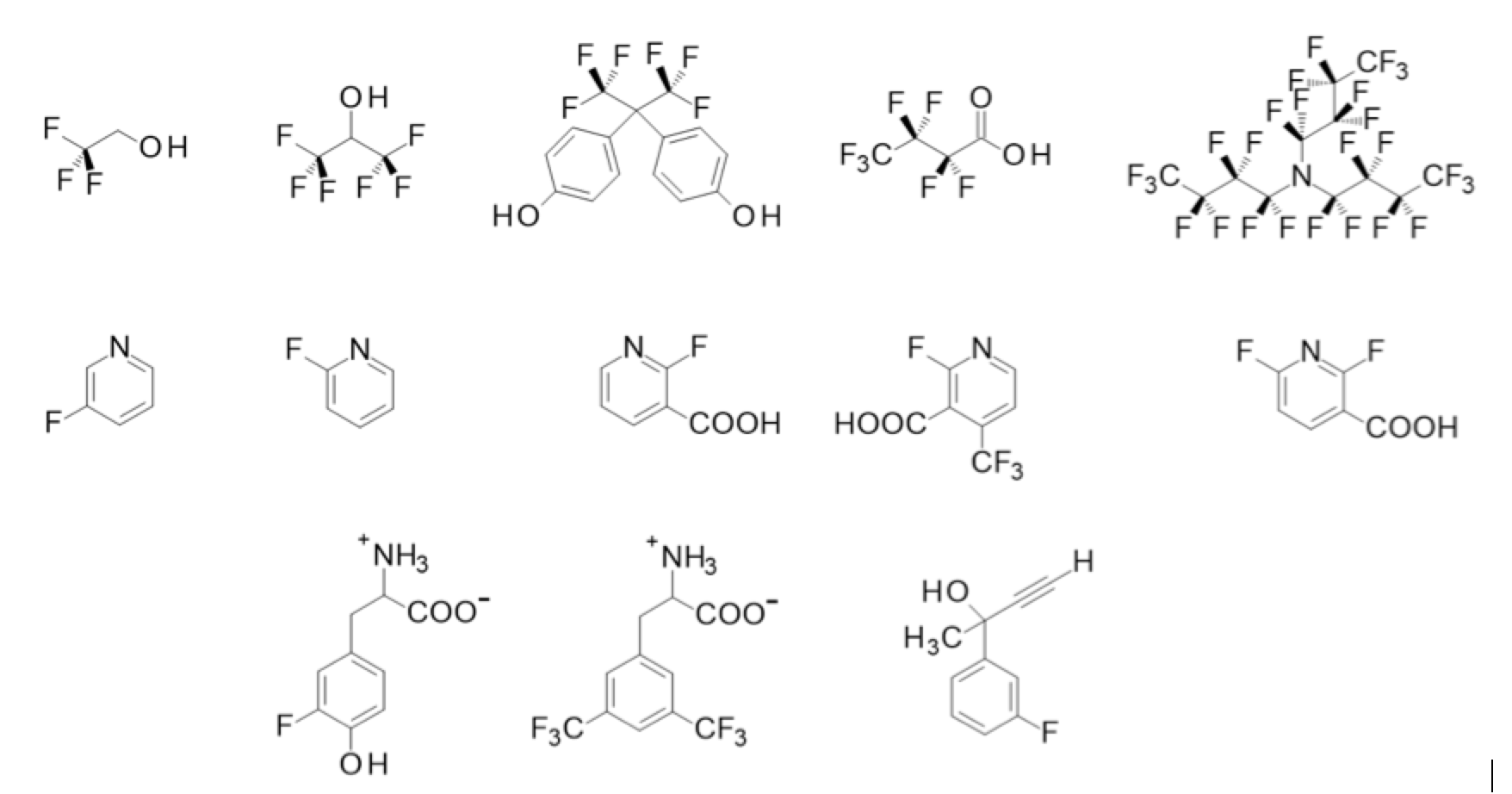
Trifluoroethanol, 1,1,1,3,3,3-hexafluoro-2-propanol, 4,4’-(hexafluoroisopropylidene)diphenol, heptafluorobutyric acid, heptacosafluorotributylamine, 3-fluoropyridine, 2-fluoropyridine, 2-fluoro-3-pyridinecarboxylic acid, 2-fluoro-4(trifluoromethyl)-pyridine-3-carboxylic acid, 2,6-difluoronicotinic acid, 3-fluoro-D, L-tyrosine, 3,5-bis(trifluoromethyl)-D,L-phenylalanine and 2-(3-fluorophenyl)-3-butyne-2-ol (Figure 1) are chosen as model compounds for temperature dependence 19F NMR measurements. Temperatures between 300K and 330K were adjusted on the used Bruker WB-300 NMR spectrometer and the spectra for each substance at different temperatures in 5 K-steps were measured.
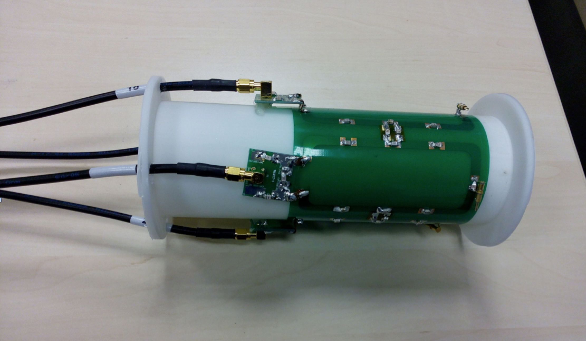
Furthermore, the same solutions were measured in a human whole-body 7T MRI (Siemens, Erlangen, Germany) system. For these measurements, a 4-elements transmit-/receive coil was developed. The RF-coil was simulated and optimized by CST Microwave Studio (Dassault Systèmes, Vélizy-Villacoublay, France) and afterwards with capacitive decoupling constructed (Figure 2). Additionally a transmit-/receive box based on Wilkinson power dividers and transmit-/receive switch were constructed in-house. Both, the RF-coil and the transmit-/receive box were broad banded optimized to the 19F MR signal6. This aims a sufficiently high gain of the 1H MR signal for imaging (in the final version the anatomic imaging) and shimming. The samples in the 7T whole-body scanner were heated up and 19F MR spectra and images were obtained during the following cooling process inside the scanner.
Results
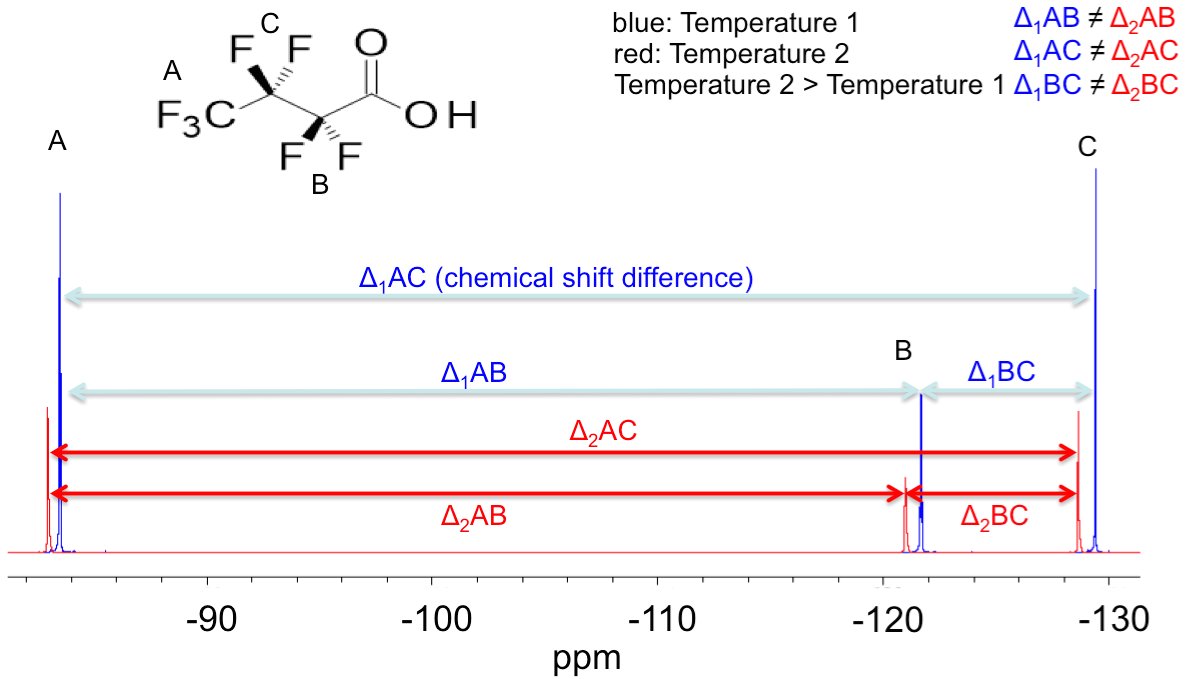
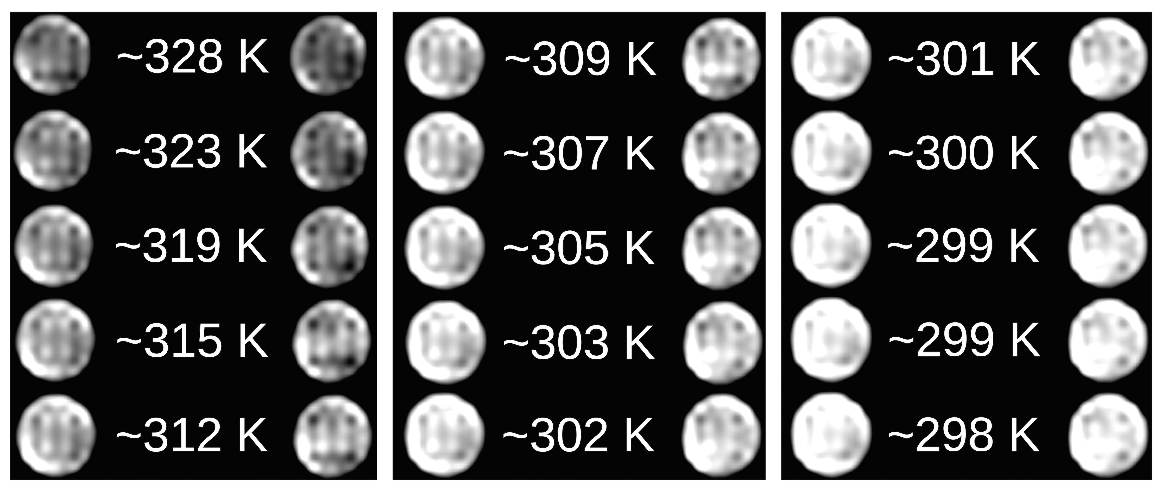
An overlay of 19F NMR spectra demonstrates in Figure 3 that all signals shifted to lower field and their intensities decrease upon heating the sample (because of temperature dependent Boltzmann distribution, different T1 times, etc.). The chemical shift differences vary between 1.4Hz/K (3-fluoropyridine) and 8.7Hz/K (2-fluoro-4-(trifluoromethyl)-pyridine-3-carboxylic acid, -CF3 group). Because the temperature dependent chemical shift changes differ of each group, each temperature has a characteristic difference between the peaks. This allows to calculate a calibration curve of the peak-difference and the temperature. The MRI experiments were measured with a resolution of 0.3x0.3x1.00mm3 and concentrations beneath 1.9mM. A decrease of signal intensity like in the NMR experiments was detected for higher temperatures, leading to lower contrasts (Figure 4).Discussion
Our preliminary results indicate, fluorinated substances may be successfully used to measure the temperature of the sample (and its surroundings) as well in MRS as in MRI by using chemical shift differences and contrast changes, respectively. The results of these measurements show that the 19F signals of CF groups and fluorine next to carboxy groups can be shifted strongly by temperature variation. Especially in case of carboxylic acids, the presence of Na+ ions in aqueous solution can increase this influence. Further substrates are in investigation. Important aspects for new molecules are no toxicity, acceptable water solubility and different fluorinated groups. A further idea is to use Gadolinium to reduce T times. This could help to further reduce the acquisition times. First experiments for heptafluorobutyric acid with a concentration of 1.766mM with Gadolinium (0.56 mM GdCl3·6H2O) in water showed up a reduction of T to 300 to 800 ms for the different 19F atoms in contrast to heptafluorobutyric acid without Gadolinium with T between 1s and 2s.Conclusion
These first comparable data concerning the relation of molecular structure and 19F chemical shifts are important for future studies in the field of development of temperature sensitive contrast agents.Acknowledgements
This work was supported by the BMBF Project 01DR12111: "EDUHF-LAB MRI“.References
1. Wang J, et al. Fluorine in pharmaceutical industry: fluorine-containing drugs introduced to the market in the last decade (2001-2011). Chem Rev, 2014;114(4):2432−2506.
2. Ojima I. Fluorine in Medical Chemistry and Chemical Biology, 1. Ed., Wiley-Blackwell: Chichester, 2009. 3. Ruiz-Cabello J, et al. Fluorine F MRS and MRI in biomedicine. NMR Biomed., 2011;24(2):114-129.
4. Jiang Z-X et al. Symmetry-guided Design and Fluorous Synthesis of A Stable and Rapidly Excreted Imaging Tracer for F MRI. Angew. Chem. Int. Ed., 2009;48: 4755–4758.
5. Pinakin R. Jethwa, et al. Magnetic Resonance Thermometry-Guided Laser-Induced Thermal Therapy for Intracranial Neoplasms: Initial Experience. Op. Neurosurg, 2012;71(1):133-145.
6. Bruns C. et al. Unified proton and fluorine imaging of small and low spin density samples at a human whole-body 7 T MRI: ISMRM 2017
Figures