4365
Innate immune cell tracking in the glioma microenvironment by correlated magnetic resonance imaging and multi photon microscopy (MR-MPM)1Neuroradiology, Neuroradiology Department, University Hospital Heidelberg, Heidelberg, Germany, 2Clinical Cooperation Unit Neurooncology, German Cancer Consortium (DKTK) within the German Cancer Research Center (DKFZ), Heidelberg, Germany, 3Clinical Cooperation Unit Neuroimmunology and Brain Tumor Immunology, German Cancer Research Center (DKFZ), Heidelberg, Germany, 4Neurology Clinic and National Center for Tumor Diseases, University Hospital Heidelberg, Heidelberg, Germany, 5Department of Neurology, University Medical Center Mannheim, Heidelberg University, Mannheim, Germany
Synopsis
The tumor microenvironment (TME) plays a key role for tumor biology. Composition of the TME correlates with overall survival and governs therapy response. Non invasive assessment of the TME has been notoriously difficult. We have designed an imaging strategy to non invasively visualize innate immune cell dynamics in the TME by correlated MRI and multiphoton microscopy. This approach allowed us to visualize the single steps of nanoparticle uptake by blood born monocytes that give rise to tumor macrophages in an experimental glioma model. We further found that nanoparticle uptake also occurs via the disrupted blood-brain barrier into the brain parenchyma where NP are taken up by tumor associated microglia.
Introduction
The tumor microenvironment (TME) is composed of various stroma and immune cells that interact within tumor. The composition of the TME is a predictive marker for survival and therapy response. Thus, the TME serves as a main target for novel therapies, including immunotherapies and antiangiogenic treatments (1,2). Glioma are highly malignant brain tumors with limited prognosis (3). Glioma is characterized by an immunosuppressive microenvironment that shows large infiltrates of M2-like macrophages / microglia (4). Recently, a number of novel immunotherapies have been developed for glioma that module the tumor environment and exploit various immunotherapeutic strategies (5). Innate immune cells are actively modulated by the tumor towards an anti-inflammatory phenotype, thus mediating tumor immune escape. Monitoring anti-tumor immune responses is a major challenge in clinical practice (6). Imaging is the main modality to monitor brain tumors but so far functional methods to visualize cellular and molecular changes in the TME have been limited. Iron oxide nanoparticles that can be detected by MRI have been shown to accumulate in phagocyte subsets and thus allow monitoring of immune responses (7). We have previously established iron oxide nanoparticle (NP) imaging using cross linked iron oxide nanoparticles conjugated with fluorescent dyes (8). We combine this strategy with multiphoton microscopy (MPM) to visualize the cellular and subcellular dynamics of nanoparticle uptake and sequestration in the same animal. To achieve this goal of dual modality imaging by MRI and MPM we developed a new holding system for cranial windows used in MPM to reduce metal artifacts.Methods
CX3CR1-GFP+/- mice with fluorescently labeled macrophages/microglia were used to assess NP uptake. To avoid metal artifacts we replaced the conventional titanium ring with a newly constructed, custom-made ring of polytetrafluorethylene (Teflon®). Fluorescently-labeled, cross-linked iron oxide NP (USPIO, 30nm in size) were used as a contrast agent (7). 2 weeks after cranial window implantation, 50.000 tumour cells (GFP-labeled Gl261) were stereotactically injected into the mouse brain at a depth of about 500 μm. MR imaging was performed on a 9.4 Tesla animal NMR scanner, including a RARE T2-w, a T1-w post-Gd-contrast sequence, to monitor tumor volume, a 3D T2-w sequence and a customized T2*-weighted gradient echo sequence (80µm isotropic resolution) to assess NP kinetics (9). MPM imaging was done with a Zeiss 7MP microscope.
Results
The improved image quality with Teflon rings allowed longitudinal imaging of tumor growth kinetics by correlated MRI and 2-photon microscopy in the same animal. Furthermore, the T2*-w sequence used for NP imaging is highly sensitive to NP. Before NP injection T2* MRI showed focal spots of hypointensities, most likely microbleedings. Directly after intravenous NP injection, NP were apparent in the circulation. 2-photon microscopy showed that most of the particles stayed intravascularly. Small amounts of NP also leaked into the brain parenchyma and got phagocytosed by resident microglia. Additionally, there was also prominent labeling of blood-circulating monocytes, some of which adheared to the endothelium and transmigrated to the TME to become tumor-associated macrophages. 48 hours after NP injection, nanoparticles labeled the majority of tumor macrophages/microglia. MRI showed NP accumulation specifically in the tumor border and to a lesser degree in the tumor core and was able to detect NP kinetics that occurred over time.Discussion
MRI is used for primary diagnosis and follow-up of glioma patients. MPM is heavily used in neuroscientific research to investigate disease mechanisms on the cellular level. These two domains have been separated so far because metal rings used for 2PM were not compatible with MRI due to metal artifacts. We have overcome this issue by introducing Teflon rings that allow the acquisition of MR images without metal artifacts. We used this approach to map the tumor microenvironment of glioma using iron oxide nanoparticles. We found that NP are primarily taken up by innate immune cells. Macrophages/microglia mainly accumulate in the tumor border where they show the highest uptake of NP. Interestingly, we found various routes of NP into the brain: 1. Blood-circulating monocytes take up NP immediately after intravenous injection. Some of these labeled monocytes are then recruited as tumor-associated macrophages to the TME. 2. NP can leak directly into the brain parenchyma in areas of blood-brain-barrier disruption where it is taken up by brain resident microglia. Both effects result in comprehensive labeling of the innate immune cell compartment within the tumor. We envision NP imaging as a possibility to assess the TME which seems especially relevant for current efforts to introduce immunotherapeutic approaches to the clinical arena in glioma and beyond. Furthermore, MR-MPM will be a valuable tool for neuroscientists to combine MRI and 2 photon microscopy in preclinical studies.Acknowledgements
We thank Manuel Fischer for technical support and MRI measurements. We thank Ralph Weissleder (Massachusetts General Hospital, Harvard Medical School) for providing CLIO nanoparticles. We acknowledge support from the DKFZ Light Microscopy Core Facility. M.O.B., was supported by a physician-scientist fellowship of the Medical Faculty, University of Heidelberg and by the Hoffmann-Klose Foundation (University of Heidelberg). M.O.B. acknowledges funding by Neurowind e.V., the Novartis Foundation and the Else Kröner-Fresenius Stiftung (2017-A25).References
1. Pyonteck SM, et al. (2013) CSF-1R inhibition alters macrophage polarization and blocks glioma progression. Nat Med 19(10):1264–1272.
2. Bruno A, et al. (2014) Orchestration of angiogenesis by immune cells. Front Oncol 4:131.
3. Wen PY, Kesari S (2008) Malignant gliomas in adults. N Engl J Med 359(5):492–507.
4. Hambardzumyan D, Gutmann DH, Kettenmann H (2015) The role of microglia and macrophages in glioma maintenance and progression. Nat Neurosci 19(1):20–27.
5. Platten M, Reardon DA (2018) Concepts for Immunotherapies in Gliomas. Semin Neurol 38(1):62–72.
6. Okada H, et al. (2015) Immunotherapy response assessment in neuro-oncology: a report of the RANO working group. The Lancet 16:e534–e543.
7. Weissleder R, Nahrendorf M, Pittet MJ (2014) Imaging macrophages with nanoparticles. Nature Materials 13(2):125–138.
8. Kirschbaum K, et al. (2016) In vivo nanoparticle imaging of innate immune cells can serve as a marker of disease severity in a model of multiple sclerosis. Proc Natl Acad Sci USA 113(46):13227–13232.
9. Park S-H, Masamoto K, Hendrich K, Kanno I, Kim S-G (2008) Imaging brain vasculature with BOLD microscopy: MR detection limits determined by in vivo two-photon microscopy. Magn Reson Med 59(4):855–865.
Figures
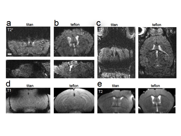
Fig. 1 Comparison of teflon and titanium ring: The maximum penetration depth of the MPLSM is about 700μm. On MRI, this section is outshined by artifacts of the titanium ring. New constructed teflon rings allow an artifact-free imaging.

Fig. 2 Longitudinal monitoring glioma growth in cranial window mice: The improved and artifact-free image quality with teflon rings allowed longitudinal imaging of tumor growth in the same animal.

Fig. 3 Study outline and NP imaging by T2*-w (80µm isotropic resolution): MRI showed the highest uptake of NP specifically in the tumor periphery, where macrophages/microglia mainly accumulate, and to a lesser degree in the tumor core.

Fig. 4 Correlated MPM in the same animal (NP in red; TAM in green): About 2h after intravenous NP injection 2-photon microscopy showed that most of the particles stayed intravascularly. Small amounts of NP also leaked into the brain parenchyma and got phagocytosed by resident microglia. 48 hours after NP injection, nanoparticles labeled the majority of tumor macrophages/microglia.