4362
Using iron oxides and perfluorocarbons with magnetic resonance imaging for simultaneous cell tracking of two populations in a model of stem cell rejectionOlivia C. Sehl1,2, Ashley V. Makela1,2, Amanda M. Hamilton1, and Paula J. Foster1,2
1Robarts Research Institute, London, ON, Canada, 2Medical Biophysics, The University of Western Ontario, London, ON, Canada
Synopsis
Mesenchymal stem cells (MSCs) are a promising cellular therapeutic. However, this therapy is limited by MSC apoptosis following administration, which can ultimately trigger immune rejection. In this study we explored the ability to label, detect, and quantify both MSCs and infiltrating macrophages using both iron and fluorine-19 (19F) based MRI cell tracking. With a dual-tuned surface coil and a 3D bSSFP sequence, we demonstrated that both cell populations can be monitored during the same imaging session. This is the first study to image macrophage infiltration in vivo using 19F on a 3T clinical MRI system.
Introduction
Mesenchymal stem cells (MSCs) have shown promising results as a cellular therapeutic1–5. MSCs play an important role in modulating inflammation within the local microenvironment through secretion of trophic factors2,6. Unfortunately, this therapeutic potential is often limited due to stem cell death. In the days following administration, many stem cells undergo apoptosis due to the stresses of administration and inadequate access to nutrients7. Cytokines released by the apoptotic stem cells attract macrophages to the implant site, triggering an immune response by the host. Ultimately, the resulting influx of immune cells is great enough to trigger stem cell rejection8. Fundamental questions with regard to the fate of cells after MSC transplantation remain unanswered. To implement successful cell-based therapies, methods must be developed to allow long-term monitoring of transplanted cells and infiltrating macrophages in vivo8,9. Cellular MRI has proven to be an effective technique for non-invasive and longitudinal tracking of cells1,5,9–13. To date, most cellular MRI has been performed with either iron oxides or perfluorocarbons (PFC)1. In this study we explored the ability to label, detect, and quantify two cell types (MSCs and macrophages) simultaneously using dual iron (Fe) and fluorine-19 (19F) based cell tracking in a model of stem cell rejection.Methods
Mouse mesenchymal stem cells (mMSCs) were labeled with 100µg Fe/mL ferumoxytol, an ultrasmall superparamagnetic iron oxide (USPIO), following protocol by Frank et al.14 (Figure 1). 1x10⁶ labeled (n=5 mice) or unlabeled (n=3 mice) mMSCs were implanted in the right hind limb muscle of C57Bl/6 mice. Immediately after, each mouse was administered 200μL 120 mg/mL PFC intravenously (iv) via the tail vein for in situ labeling of phagocytic macrophages. 24 hours later, both 1H and 19F images were acquired on a 3T clinical scanner using a 4.31cm dual-tuned surface coil (Figure 2) and a 3D balanced steady state free precession (bSSFP) sequence (day 1). Imaging was repeated on day 12 to investigate temporal changes in iron-associated signal voids and 19F signal. Regions of signal void due to mMSC were delineated manually and void volume calculated. The number of 19F spins was quantified by comparison of 19F signal in the region of interest to 19F signal in reference tubes. A two-tailed students t-test was completed to compare void volume at day 1 and 12, 19F signal between mice injected with unlabeled or iron labeled mMSCs, and also between day 1 and 12.Results
On day 1, regions of signal void were detected in 1H MRI of all mice which received iron-labeled mMSCs (Figure 3A). There were no signal voids found in 1H MRI of mice injected with unlabeled mMSCs (Figure 3C). Abundant 19F signal was localized to the right hind limb of mice at day 1 (Figure 3B,D) and 12 (Figure 4B,D), where injection of mMSCs occurred. On day 12, signal void was still present but the void volume had significantly decreased by 64% (Figure 4A&5A). There was also significantly more (25%) 19F signal on day 12 (Figure 5B). No differences in 19F signal were found between mice with iron labeled or unlabeled mMSCs (Figure 5C).Discussion
We have shown that it is possible to detect both implanted mMSCs and infiltrating macrophages in the same mouse during the same scanning session. The signal void created by iron positive MSCs declines over time, likely due to MSC death5,12,13. As a result, PFC-labeled macrophages accumulate near the MSCs within 24 hours, as an immune reaction. The 19F signal persists and rises over 12 days, indicating the increased infiltration of immune cells. Hitchens et al.11 previously demonstrated the proof of principle for dual iron and 19F cell imaging, showing that if iron and 19F agents are in different cells no signal quenching should be observed. This is the first study to utilize PFC to indicate inflammation associated with iron labeled stem cells and to track this over time in live mice. Compared to iron-based cell tracking, 19F MRI has lower sensitivity and consequently, preclinical 19F cell tracking has only been performed at relatively high magnetic field strengths (>3T)1,9–11. This is the first study to demonstrate the ability to image macrophage infiltration in vivo using 19F on a clinical MRI system. The bSSFP imaging sequence and surface RF coil (Figure 2) play a major role in our ability to detect and track 19F positive cells at 3T.Conclusion
1H and 19F MRI can be used in conjunction to noninvasively monitor the fate two cell populations in vivo. We propose that this cellular imaging technique could be used to detect the infiltration of macrophages at transplant sites to identify cell rejection.Acknowledgements
Thank you to the University of Western Ontario Graduate Scholarship (WGRS), the Ontario Graduate Scholarship (OGS), the Strategic Training Program in Cancer Research and Technology Transfer (CaRTT), and the Translational Breast Cancer Research Unit (TBCRU) for the financial support of this research.References
- Gaudet, J. M., Hamilton, A. M., Chen, Y., Fox, M. S., & Foster, P. J. (2017). Application of dual 19F and iron cellular MRI agents to track the infiltration of immune cells to the site of a rejected stem cell transplant. Magnetic resonance in medicine, 78(2), 713-720.2.
- Meirelles, S. L., & Nardi, N. B. (2009). Methodology, biology and clinical applications of mesenchymal stem cells. Frontiers in bioscience (Landmark edition), 14, 4281-4298.3.
- Chagastelles, P. C., Nardi, N. B., & Camassola, M. (2010). Biology and applications of mesenchymal stem cells. Science progress, 93(2), 113-127.4.
- Segers, V. F., & Lee, R. T. (2008). Stem-cell therapy for cardiac disease. Nature, 451(7181), 937.5.
- Makela, A. V., Murrell, D. H., Parkins, K. M., Kara, J., Gaudet, J. M., & Foster, P. J. (2016). Cellular imaging with MRI. Topics in Magnetic Resonance Imaging, 25(5), 177-186.6.
- Caplan, A. I., & Dennis, J. E. (2006). Mesenchymal stem cells as trophic mediators. Journal of cellular biochemistry, 98(5), 1076-1084.7.
- Reagan, M. R., & Kaplan, D. L. (2011). Concise review: Mesenchymal stem cell tumor‐homing: Detection methods in disease model systems. Stem cells, 29(6), 920-927.8.
- Wyburn, K. R., Jose, M. D., Wu, H., Atkins, R. C., & Chadban, S. J. (2005). The role of macrophages in allograft rejection. Transplantation, 80(12), 1641-1647.9.
- Hitchens, T. K., Ye, Q., Eytan, D. F., Janjic, J. M., Ahrens, E. T., & Ho, C. (2011). 19F MRI detection of acute allograft rejection with in vivo perfluorocarbon labeling of immune cells. Magnetic resonance in medicine, 65(4), 1144-1153.10.
- Gaudet, J. M., Ribot, E. J., Chen, Y., Gilbert, K. M., & Foster, P. J. (2015). Tracking the fate of stem cell implants with fluorine-19 MRI. PloS one, 10(3), e0118544.11.
- Hitchens, T. K., Liu, L., Foley, L. M., Simplaceanu, V., Ahrens, E. T., & Ho, C. (2015). Combining perfluorocarbon and superparamagnetic iron‐oxide cell labeling for improved and expanded applications of cellular MRI. Magnetic resonance in medicine, 73(1), 367-375.12.
- Gonzalez-Lara, L. E., Xu, X., Hofstetrova, K., Pniak, A., Chen, Y., McFadden, C. D., Martinez-Santiesteban, F.M., Rutt, B.K., Brown, A.,& Foster, P. J. (2011). The use of cellular magnetic resonance imaging to track the fate of iron-labeled multipotent stromal cells after direct transplantation in a mouse model of spinal cord injury. Molecular Imaging and Biology, 13(4), 702-711.13.
- Noad, J., Gonzalez‐Lara, L. E., Broughton, H. C., McFadden, C., Chen, Y., Hess, D. A., & Foster, P. J. (2013). MRI tracking of transplanted iron‐labeled mesenchymal stromal cells in an immune‐compromised mouse model of critical limb ischemia. NMR in Biomedicine, 26(4), 458-467.14.
- Thu, M. S., Bryant, L. H., Coppola, T., Jordan, E. K., Budde, M. D., Lewis, B. K., Chaudhry, A., Ren, J., Varma, R.N.S., Arbab, A.S., & Frank, J. A. (2012). Self-assembling nanocomplexes by combining ferumoxytol, heparin and protamine for cell tracking by magnetic resonance imaging. Nature medicine, 18(3), 463.
Figures
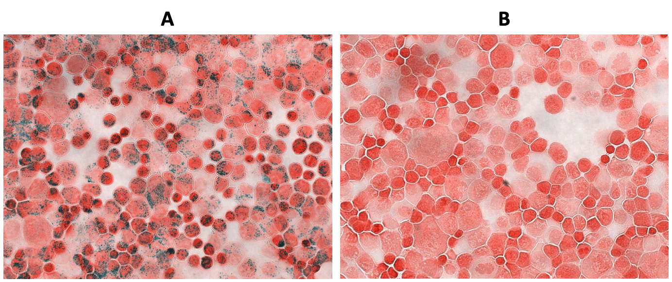
Figure 1. (A) Ferumoxytol labeled and (B) unlabeled mMSCs stained with Perl’s Prussian Blue (PPB), to assess iron labeling efficiency, and counterstained with nuclear fast red, to delineate cells. Ferumoxytol labeled mMSCs are PPB positive and demonstrate adequate labeling efficiency.
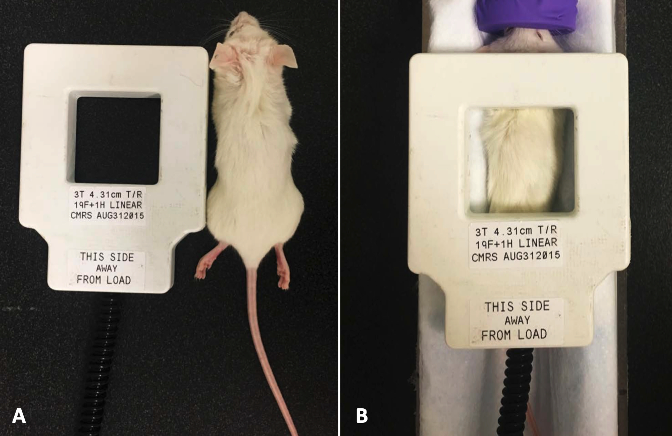
Figure 2. (A) 4.31cm 1H and 19F dual-tuned surface coil for use with 3T MRI, relative to mouse. (B) Surface coil is placed directly above the region of interest on the mouse during the imaging session.
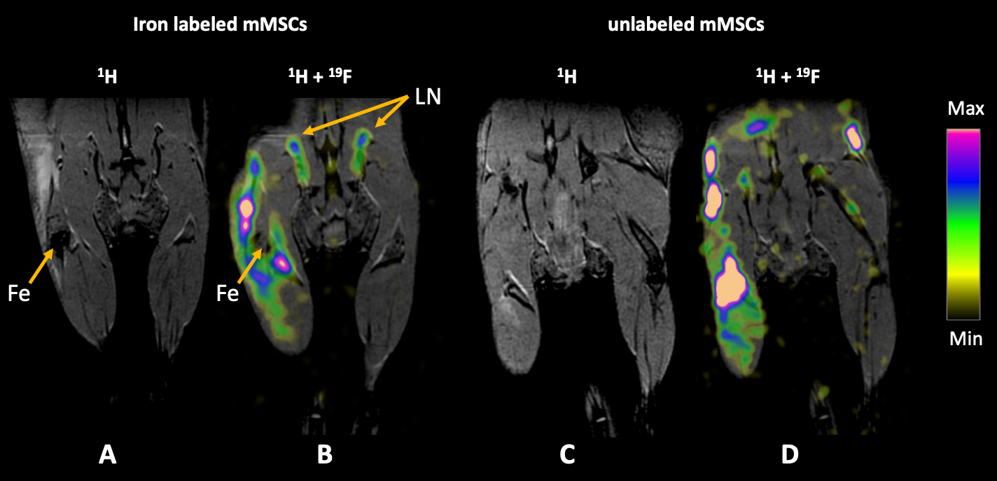
Figure 3. Coronal bSSFP images of mouse hind limbs, 1 day post mMSC and iv PFC injection. (A) 1H MRI showing signal void caused by iron labeled mMSCs, and (B) with 19F image overlay. The iron void remains visible and 19F signal surrounds mMSC implant site. (C) 1H MRI of mouse with unlabeled mMSCs. No iron signal void is present. (D) The same mouse with 19F image overlay. 19F signal surrounds mMSC implant site. 19F signal also presents in lymph nodes (LN) and reference tubes. The color bar illustrates the range of 19F spins. Maximum signal is peach colored.
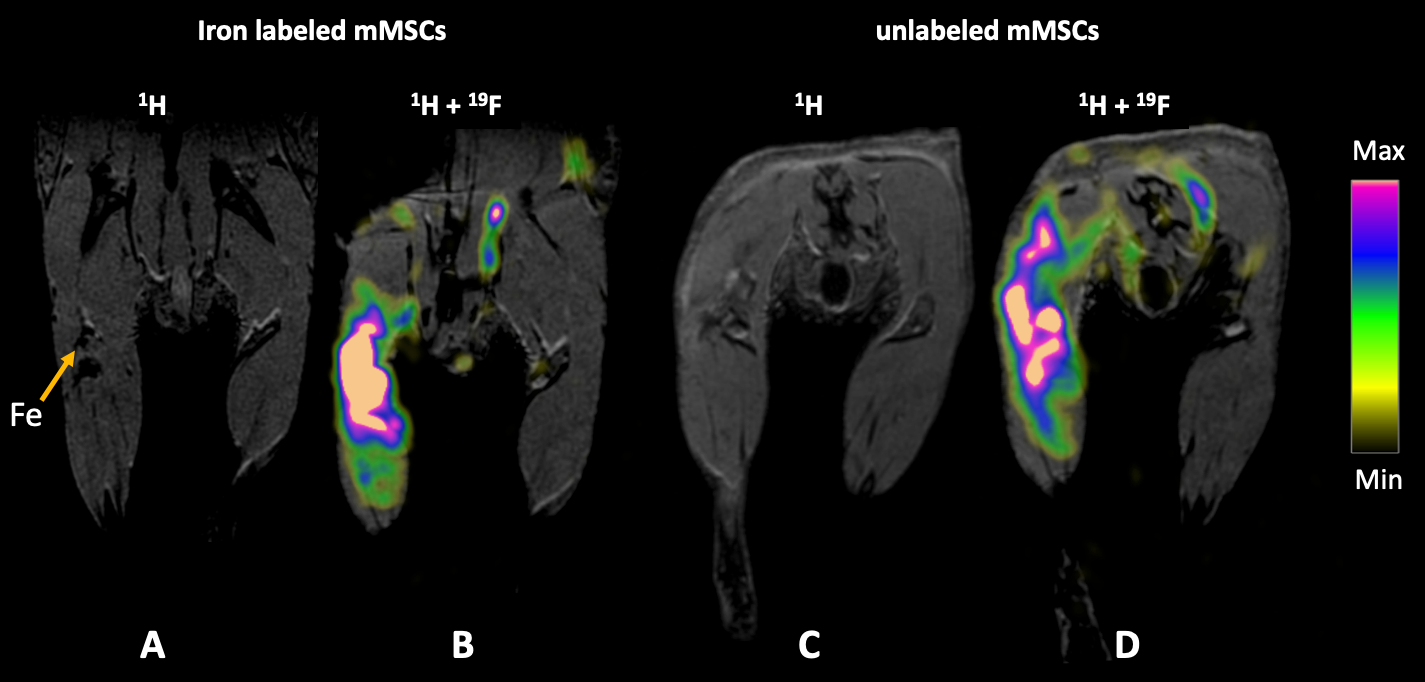
Figure 4. Coronal bSSFP images of mouse hind limbs, 12 days post mMSC and iv PFC injection. (A) 1H MRI showing signal void caused iron labeled mMSCs, and (B) with 19F image overlay. The iron void is not visible. 19F signal surrounds mMSC implantation site. (C) 1H MRI of mouse with unlabeled mMSCs. No iron signal void is present. (D) The same mouse with 19F image overlay. At day 12 it appears that more 19F signal has accumulated in right limb where mMSC injection occurred. The color bar illustrates the range of 19F spins. Maximum signal is peach colored.
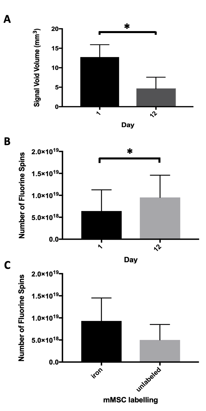
Figure 5. (A) The signal void volume created by implanted iron labeled mMSCs significantly decreased after 12 days, by 64%. (B) 19F signal quantified in the right hind limb at day 1 and 12. There is significantly more 19F signal measured on day 12, corresponding to a 25% increase. (C) There was no significant difference in 19F signal between mice with iron labeled or unlabeled implanted mMSCs.