4359
Simultaneous PET/MRI to detect brain tumour metabolic and functional heterogeneity1Department of Information Engineering, University of Padova, Padova, Italy, 2Padova Neuroscience Center, University of Padova, Padova, Italy, 3Department of Neuroscience, University of Padova, Padova, Italy, 4Department of Molecular Imaging, University of Padova, Padova, Italy
Synopsis
Demarcation for brain tumour neurosurgical resection is typically performed using Fluid Attenuated Inversion Recovery images hyperintensities. In this study, we investigated in glioma patients whether these hyperintensities represent structural alterations that are metabolically and functionally homogeneous using simultaneous measurements of quantitative 18-Fluoro-deoxyglucose PET and resting state functional magnetic resonance imaging. FLAIR hyperintense regions resulted to be heterogeneous both at metabolic and functional standpoint and only partially significantly altered when compared with contralateral homologous white matter. These results support the hypothesis that multi-parametric approaches might improve the outcome of brain surgery informing the resection procedure.
Introduction
Multimodal imaging offers the possibility of an accurate description of structural, metabolic and functional features of brain tumours1, exploiting simultaneous acquisition of both magnetic resonance imaging (MRI) and positron emission tomography (PET). In the surgical planning of tumour resection, current strategies employ the Fluid Attenuated Inversion Recovery (FLAIR) hyperintensities to delineate regions of brain alterations amenable to be removed. Although gross total resection is associated with better outcome and prolonged survival2, the benefit of a larger neurosurgical abscission needs to be balanced against the risk of significantly altering the patient’s quality of life by inflicting deficits in eloquent areas as well as essential brain functions1.The aim of this study was to investigate whether FLAIR hyperintensities represent structural alterations that are metabolically and functionally homogeneous, in order to provide a more comprehensive tool in the decision process that leads to the surgical planning. To this end we simultaneously measured quantitative 18-Fluoro-deoxyglucose ([18F]FDG) PET and resting state functional magnetic resonance imaging (rs-fMRI) in patients affected by brain tumours.Methods
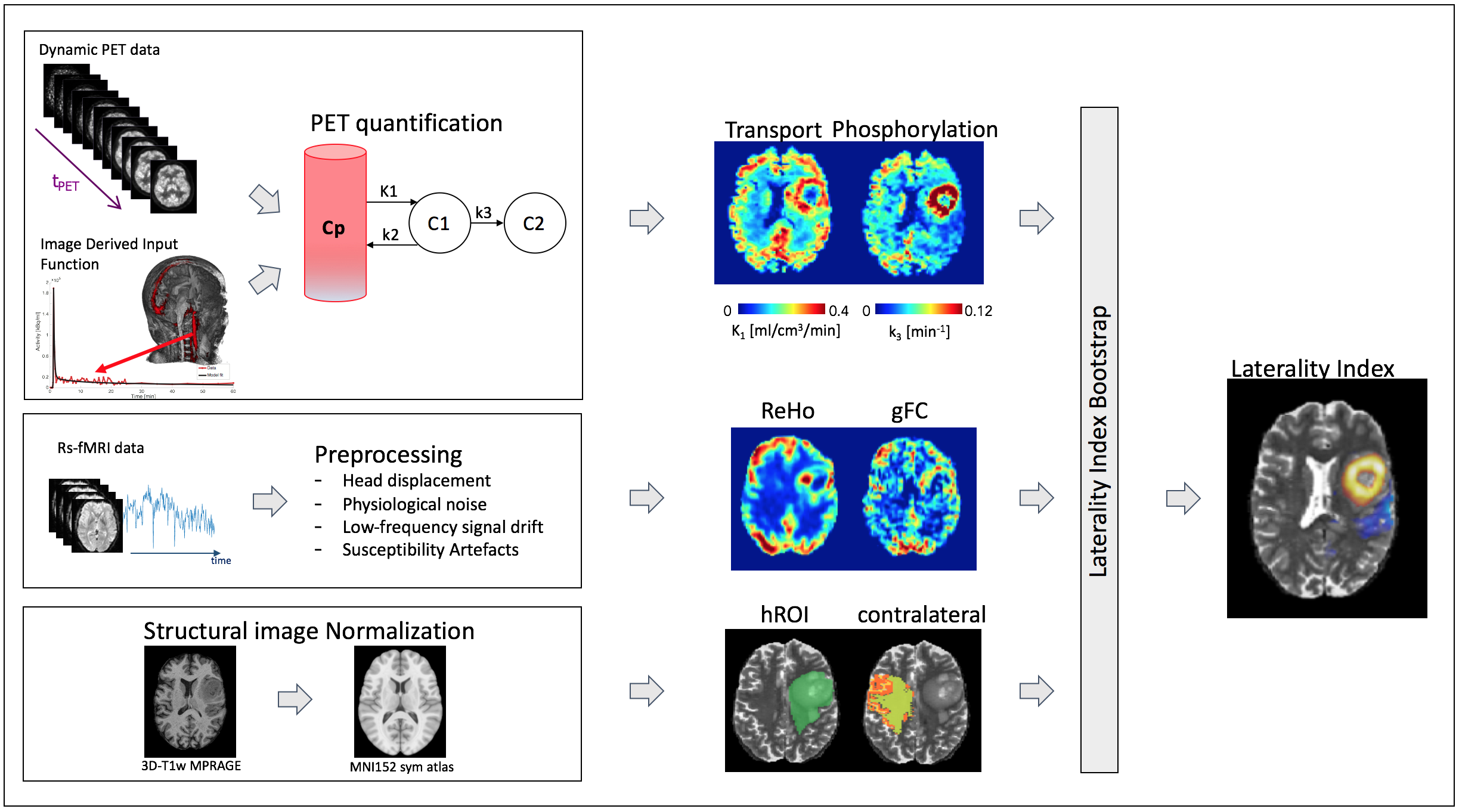
Twelve subjects (7 males, age 59±19y) with newly diagnosed glioma (8 high grade; 4 low grade)3 were scanned before surgery with a Siemens Biograph mMR PET-MR scanner. [18F]FDG were dynamically recorded for 60 minutes while acquiring MRI sequences used both to define the tumour morphology (3D-T2w, 3D-FLAIR, 3D-T1w-MPRAGE before and after contrast administration) and to assess the brain functional connectivity (rs-fMRI, 15 minutes, MBfactor 2, TR/TE 1260/30ms, voxel size 3x3x3mm3). Dynamic PET data were quantified using the standard 2-tissue 3-parameter compartmental model4 to separate the delivery of [18F]FDG from plasma to tissue (K1 [ml/cm3/min]) and intracellular phosphorylation (k3 [1/min]). From a physiological perspective, K1 represents the net influx transport from the plasma mediated by the blood brain barrier (BBB). As BBB is highly permeable to glucose, it could be employed as a proxy of brain tissue perfusion. Rs-fMRI data underwent a state-of-art pre-processing; voxel-wise maps of local and global connectivity were then obtained by means of respectively Regional Homogeneity (ReHo) and global functional connectivity (gFC) indexes. ReHo is a measure of the similarity or synchronization between the time activity of a given voxel and its nearest neighbours5, whereas gFC is a voxel-wise measure that is computed as the average of the zero-lag cross-correlation between the time activity of the given voxel and each of the other brain voxels. The 3D-FLAIR hyperintensities were manually segmented and mapped into the contralateral hemisphere using a nonlinear approach. The white matter of the contralateral region was automatically extracted and used as reference to calculate voxel-wise laterality index (LI) for each quantitative parameter.
$$LI = \frac{tumour - contralateral}{tumour + contralateral}$$
In order to identify voxels with a statistically significant positive or negative LI (SLI), a bootstrap approach was employed6.The percentage of positive or negative LI over the total number of voxels within the FLAIR segmentation was calculated (SLI%) to summarize regional heterogeneity. In addition, for each patient a voxel-wise correlation analysis was performed on LI to investigate the relationship between metabolic abnormalities and the local/global functional changes.
Results
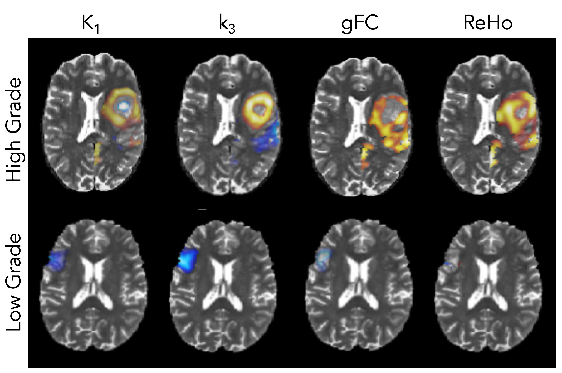
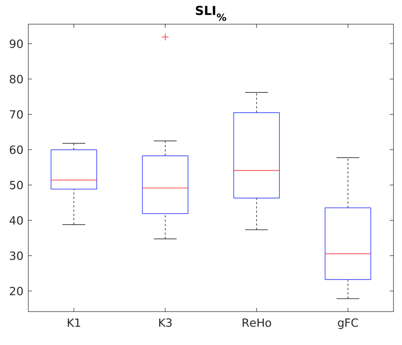
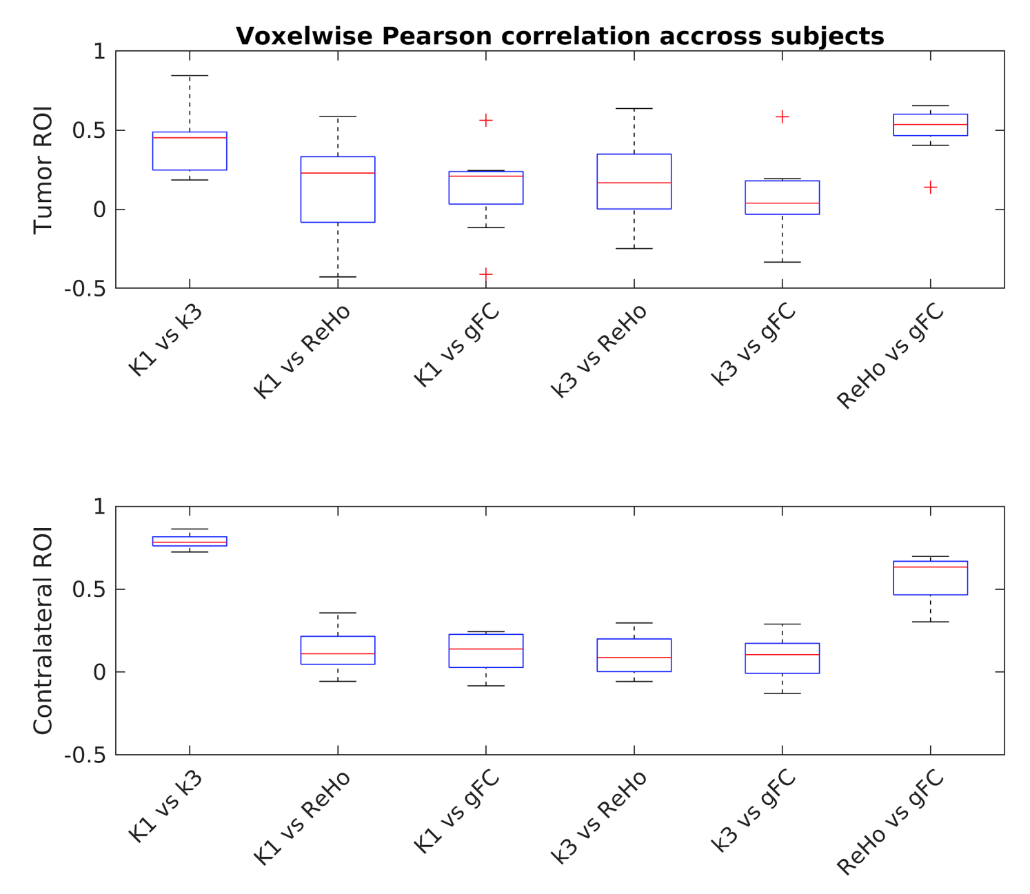
Regions of hyperintense FLAIR signal (hROI) are highly heterogeneous both metabolically and functionally. LI resulted to be negative in the larger part of low-grade gliomas. Concerning the high-grade gliomas, a more heterogeneous LI pattern was found with either a very high or low (necrotic) phosphorylation core surrounded by regions of either high or low [18F]FDG delivery (Figure2). Local functional connectivity shares similar patterns. About half of hROI voxels showed a metabolic alteration with SLI% for K1 of 52.6±7.8%, and SLI% for k3 of 54.3±18.2%. Analogous results were obtained for rs-fMRI ReHo with SLI% of 56.9±14.3%. Notably, a smaller altered area was detected by gFC with SLI% of 33.8±13.9% (Figure3). When compared with the contralateral correlation results (Figure4), the hROI’s results show changes in all the LI combination and higher variability. However, statistically significant Pearson correlation coefficients were found only between LI-K1 and LI-ReHo (ρ=0.40; p<0.05), and LI-k3 and LI-ReHo (ρ =0.53; p<0.05). No significant correlations were found between either both LI-K1, LI-k3 and LI-gFC.Discussion and Conclusions
FLAIR hyperintensities were heterogeneous both metabolically and functionally. Importantly, on average in each patient only part (about half) of the FLAIR hyperintense region showed statistically significant metabolic or functional connectivity alterations. Hence the FLAIR segmentations should be carefully evaluated by the surgeon and multi-parametric approaches might improve the outcome of brain surgery informing the resection procedure. Regarding functional connectivity contribute, local functional connectivity seems to be more informative than global as more related to metabolic features.Acknowledgements
We acknowledge that multiband EPI sequence was made available from University of Minnesota through the C2P Siemens sharing mechanism.References
1. Ghinda, D. C., Wu, J. S., Duncan, N. W., et al. How much is enough—Can resting state fMRI provide a demarcation for neurosurgical resection in glioma? Neuroscience and Biobehavioral Reviews, 2018;84:245-261.
2. Kreth F., Thon N., Simon M., et al. Gross total but not incomplete resection of glioblastoma prolongs survival in the era of radiochemotherapy, Annals of Oncology, 2018;24(12):3117-3123.
3. Louis D.N., Perry A. et al. The 2016 World Health Organization classification of tumors of the central nervous system: a summary. Acta neuropathologica 2016;131(6):803-820.
4. Phelps ME, Huang S-C, et al.. Tomographic measurement of local cerebral glucose metabolic rate in humans with [F-18]2-fluoro-2-deoxy-D-glucose: validation of method. Ann Neurol. 1979;6:371-388.
5. Zang, Y., Jiang, T., et al. Regional homogeneity approach to fMRI data analysis. Neuroimage 2004;22:394-400.
6. Wilke M. and Schmithorst V.J. A combined bootstrap/histogram analysis approach for computing a lateralization index from neuroimaging data. Neuroimage 2006;33(2):522-530.
Figures