4357
Investigation of halo-artifacts using improved scatter correction in 68-Ga-PSMA PET/MRI of the prostate1Highfield and Hybrid MR Imaging, University Hospital Essen, Essen, Germany, 2Department of Diagnostic and Interventional Radiology and Neuroradiology, University Hospital Essen, Essen, Germany, 3Siemens MR, Siemens Healthcare GmbH, Erlangen, Germany, 4Erwin L. Hahn Institute for Magnetic Resonance Imaging, Essen, Germany
Synopsis
A potential challenge of using radiotracer 68-Ga-PSMA for detection and staging of prostate cancer in PET/MR is a frequently observed halo-artifact around the urinary bladder caused by improper scatter correction (SC). Tumor manifestations in these regions might be non-detectable or show distorted SUVs. To evaluate the impact of SC on 68-Ga-PSMA PET/MR imaging, PET data sets of 100 patients were reconstructed twice using standard and improved SC. The improved SC significantly reduces the halo-artifact around the bladder. Measured SUVs in the halo margin on average increased by 325 %, and therefore, considerably affect quantitative assessment of prostate cancer in PET/MR.
Purpose:
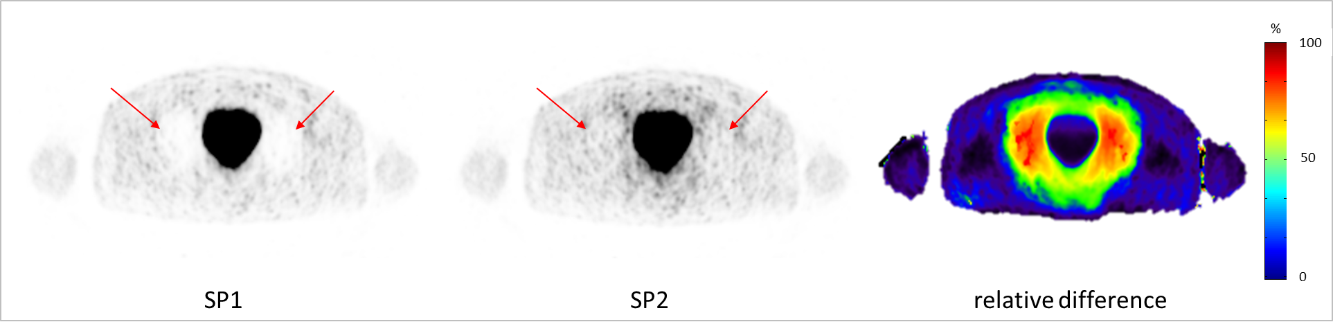
Since the introduction of [68-Ga] gallium-labelled prostate-specific membrane antigen (PSMA) ligand as positron emission tomography (PET) tracer, several studies show promising results for the detection of prostate cancer with PET/magnetic resonance imaging (MRI) [1-4]. A potential challenge when using 68-Ga-PSMA for detection and staging of prostate cancer is a frequently observed so-called “halo-artifact” or photopenic effect visible in PET data often caused by improper scatter correction (SC) [5-7]. The halo-artifact causes reduced signal intensity around the urinary bladder and at the levels of the kidneys in scatter-corrected PET images (Fig. 1). Therefore, tumor manifestations and PET active lesions in these regions might be non-detectable or show distorted standardized-uptake-values (SUVs), thus hampering PET quantification. The aim of this study is to investigate the impact of improved SC on 68-Ga-PSMA PET quantification in PET/MR hybrid imaging of the prostate in 100 patients.Methods:
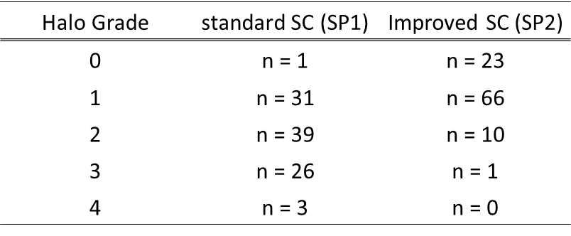
PET scatter correction typically employs a single Compton scatter simulation to compute a scatter sinogram, which is scaled to the emission data to account for multiple and possible external scatter from outside of the scanner’s field-of-view. Eliminating the sometimes problematic renormalizing (rescaling) of the PET emission image during the SC calculations for tracers like 68-Ga-PSMA can minimize the appearance of the halo-artifact and therefore, improve diagnostic image quality and PET quantification. To validate the impact of this “un-renormalized” SC on 68-Ga-PSMA PET/MR imaging, all 100 PET data sets were reconstructed twice: 1. standard SC (software version VE11 SP1) serving as the reference standard, and 2. improved un-renormalized SC (software version VE11 SP2). The presence and visibility of halo-artifacts in PET data was rated in each reconstruction (grade 0 = no halo-artifact, 1 = slight halo presence, 2 = moderate halo presence, 3 = strong halo presence, and 4 = severe halo). SUVs were measured in all detectable lesions, the bladder, the gluteus maximus and at the halo-artifact margin. Relative differences between standard and improved scatter correction were calculated.Results:
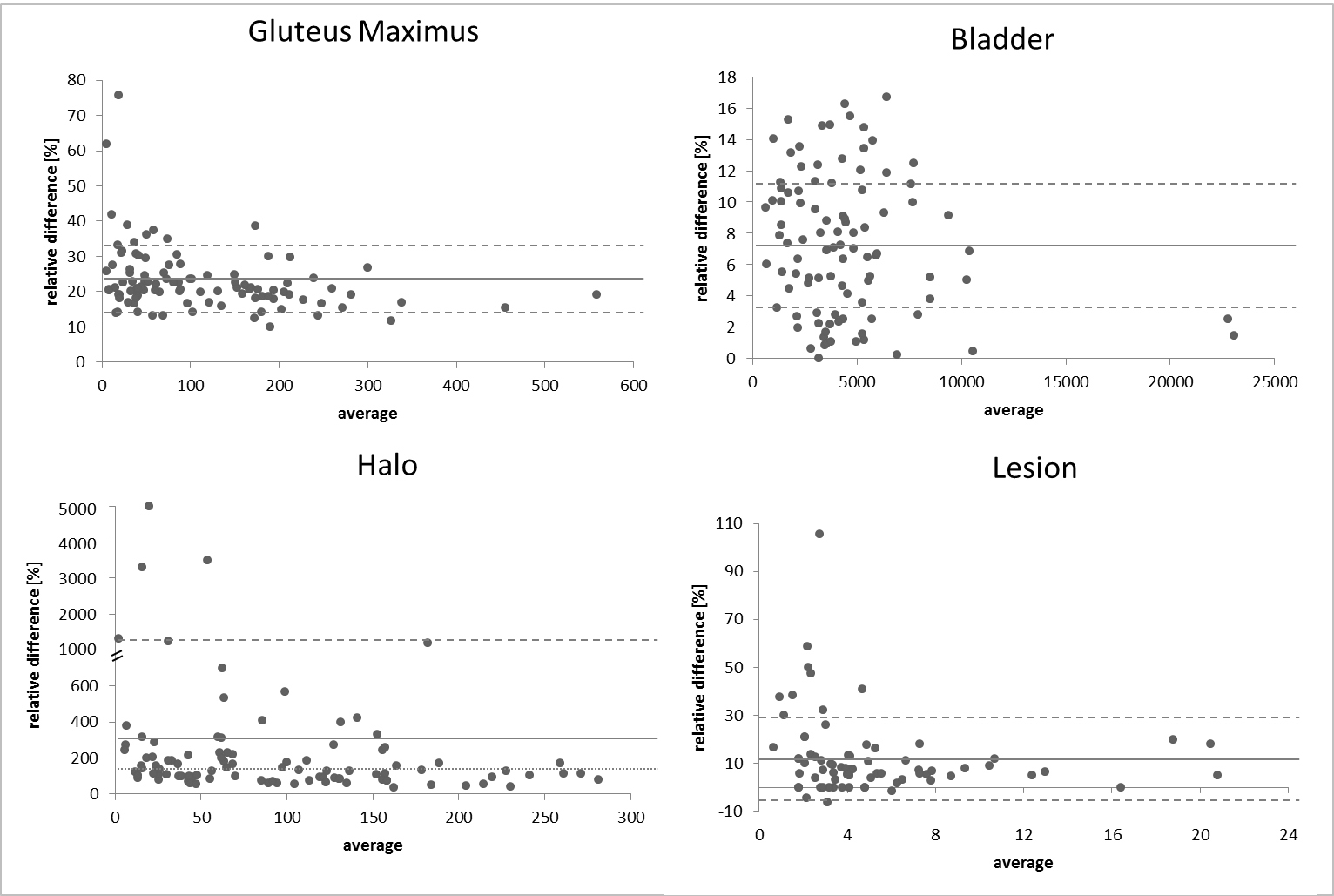
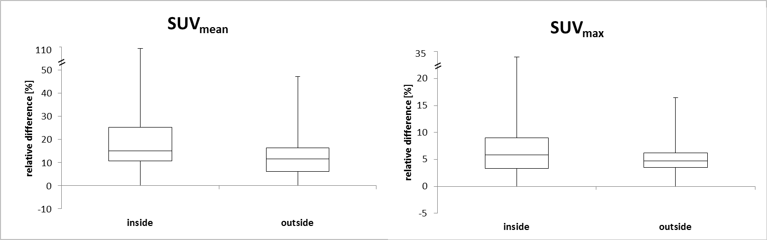
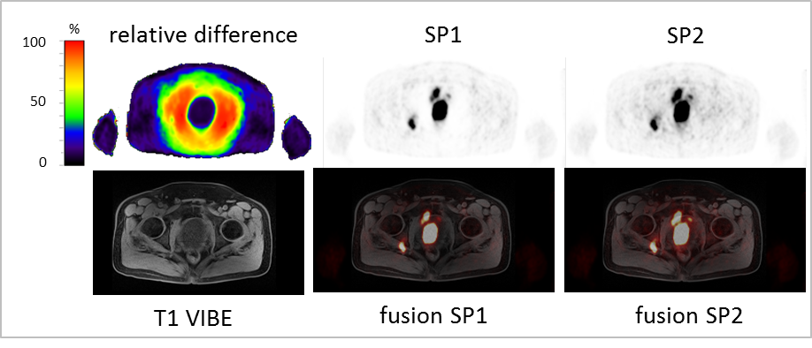
Table 1 shows the presence of halo-artifact rating in grades for 100 patients. With standard SC the average grade is 2 (moderate halo-artifact), whereas for improved SC the average grade is 0.9 (slight halo-artifact). With improved SC in 23 patients no halo-artifact was visible and no PET data set presented with a severe halo. In all 100 patients, the same number of congruent lesions (74) was detected for both PET data reconstructions. Thus, no lesion was completely missed-out due to halo-artifacts, independent of the scatter correction method used. Changes in PET signal-to-noise-ratio and image noise were statistically not significant. Bland-Altman plots (Fig. 2) show the relative difference in measured SUVmean in the gluteus maximus, the bladder, at the halo margin and in all detected lesions using standard and improved scatter correction. The average increase in SUVmean in the gluteus maximus using improved SC is 23.0 ± 9.2 % (mean ± SD) when compared to the standard SC (SP1). The average increase in the bladder using improved SC is 7.1 ± 4.5 %. The average increase in SUVmean in the halo-margin using SP2 is 325.4 ± 748.5 % when compared to SP1, the median is 127.3 %. The average increase in all 74 detected lesions using improved SC is 12.4 ± 16.8 %. All changes in SUVmean are statistically significant (p < 0.05). Fig. 3 shows relative differences in SUVmean and SUVmax between standard and improved scatter correction. Detected lesions were divided into lesions detected inside (38 lesions) or outside (36 lesions) the halo margin. The average increase using improved scatter correction in SUVmean for lesions inside the halo-margin is 17.5 % and outside the halo-margin 6.9 %, in SUVmax for lesions inside the halo margin is 7.4 % and outside 3.5 %. Fig. 4 shows a patient example with relative difference maps, PET data corrected with SP1 and SP1, MR data and fused images. Halo-artifact was reduced from severe halo presence to no halo-artifact using SP2. Relative differences up to 50 % in SUVmean in the lesions were calculated.Discussion and Conclusion:
For quantitative PET/MR hybrid imaging of prostate cancer using 68-Ga-PSMA a proper scatter correction is important to ensure best possible diagnostic quality and PET quantification. The improved SC significantly reduces the halo-artifact around the bladder. SUVs in the halo margin increase averaged around 325 %, and therefore, considerably affect the quantitative assessment of prostate cancer in PET/MR hybrid imaging.Acknowledgements
No acknowledgement found.References
[1] Eiber M, Nekolla SG, Maurer T, Weirich G, Wester HJ, Schwaiger M. (68)Ga-PSMA PET/MR with multimodality image analysis for primary prostate cancer. Abdom Imaging. 2015;40:1769-71.
[2] Afshar-Oromieh A, Haberkorn U, Schlemmer HP et al. Comparison of PET/CT and PET/MRI hybrid systems using a 68Ga-labelled PSMA ligand for the diagnosis of recurrent prostate cancer: initial experience. Eur J Nucl Med Mol Imaging. 2014;41:887-97.
[3] Freitag MT, Radtke JP, Hadaschik BA et al. Comparison of hybrid (68)Ga-PSMA PET/MRI and (68)Ga-PSMA PET/CT in the evaluation of lymph node and bone metastases of prostate cancer. Eur J Nucl Med Mol Imaging. 2016;43:70-83.
[4] Lütje S, Blex S, Gomez B et al. Optimization of Acquisition time of 68Ga-PSMA-Ligand PET/MRI in Patients with Local and Metastatic Prostate Cancer. PLoS One. 2016;11:e0164392.doi:10.1371.
[5] Heußer T, Mann P, Rank CM et al. Investigation of the halo-artifact in 68Ga-PSMA-11-PET/MRI. PLoS One. 2017;12:doi:10.1371.
[6] Afshar-Oromieh A, Wolf M, Haberkorn U et al. Effects of arm truncation on the appearance of the halo artifact in 68Ga-PSMA-11 (HBED-CC) PET/MRI. Eur J Nucl Med Mol Imaging. 2017;44:1636-46.
[7] Noto B, Büther F, Auf der Springe K et al. Impact of PET acquisition durations on image quality and lesion detectability in whole-body 68Ga-PSMA PET-MRI. EJNMMI Res. 2017;7:12.doi:10.1186/s13550-017-0261-8.
Figures