4353
Statistical Assessment of Nitroxide Radicals by T1-weighted Dynamic MRI in ex-vivo Porcine Aortic wall1Chair of Cellular and Molecular Imaging, Comprehensive Heart Failure Center (CHFC), University Hospital Wuerzburg, Germany, Wuerzburg, Germany, 2Chair of Tissue Engineering and Regenerative Medicine (TERM), University Hospital Wuerzburg, Germany, Chair of Tissue Engineering and Regenerative Medicine (TERM), University Hospital Wuerzburg, Germany, Wuerzburg, Germany, 3Translational Center ‘Regenerative Therapies’ (TLC-RT), Fraunhofer Institute for Silicate Research, Wuerzburg, Germany (ISC), Wuerzburg, Germany
Synopsis
Atherosclerosis genesis is attributed to a breakdown of homeostatic mechanisms undergoing increased oxidative stress followed by inflammation with extension to all aortic layers. Nitroxide radicals (NR) have been used as a redox-sensitive T1 contrast agent in NMR studies. Previous results have demonstrated assessment of exogenous induce redox state by using NR. In this study, we statistically assess the kinetics of NR-induced T1-weighted contrast in the ex-vivo porcine vascular wall. The results demonstrate persistence of the key values of NR induced T1-contrast kinetics and allow stepping further to quantitative assessment of the redox state within the vascular wall.
Introduction
Atherosclerosis genesis is attributed to a breakdown of homeostatic mechanisms undergoing increased oxidative stress and therefore local cell damage followed by inflammation with extension to all aortic layers. Nitroxide radicals (NR) are used as a MRI T1 contrast agent sensitive to the redox activity1,2. Previous results3,4 have demonstrated that by using NR, the variation of the redox state of the vascular tissue induced by exogenous redox factor (ascorbic acid) could be qualitatively assessed ex-vivo. However, further studies and the quantification of sensitivity of NR to endogenous ROS require comprehensive information on the kinetics of the nitroxide induced T1 contrast. The aim of this study was to statistically assess and analyze the kinetics of the NR-induced T1-weighted contrast in the ex-vivo porcine vascular wall.Materials and Methods
Materials and Methods: TEMPOL (4-Hydroxy-2,2,6,6-tetramethylpiperidine 1-oxyl´) (Merck KGa, Darmstadt, Germany) was dissolved at 1M in NaCl stock solution and stored at 4ºC. All subsequent dissolutions were performed using isotonic solution.
Segments of descending aorta (n=12) were provided from healthy German Landrace piglets of 18 to 55kg. The excised segments were immediately placed in isotonic 0.9% NaCl solution at room temperature. Aortic rings were carefully prepared by detaching blood clots, lymphatic nodes or loose connective tissue by simultaneous preservation of the adventitial layer. 5mm thick rings were manually sliced prior to the experiment.
MRI scans were performed at 1 to 7 hours after excision. TEMPOL-exposed aortic rings were incubated for 5min in 3ml of 10 and 30mmol solution at 37ºC. Unexposed rings were analogously incubated in 3ml saline solution at the same temperature. Prior to the scans, all samples were subsequently once washed in NaCl solution. Scans with a single sample were performed immediately after treatment for 30-40min.
All scans were performed at room temperature using a Bruker PharmaScan 7T scanner (Bruker BioSpin, Ettlingen, Germany) and a dual channel TX/RX 2.5/3cm int. /ext. diameter, 4cm length 1H-Cryoprobe. The kinetics of TEMPOL was assessed with a GRE pulse sequence with the following parameters: TR/TE=90/2.6ms, TA=46sec, FA=30/60/80°, matrix size=128x128, field-of-view=18-20x18-20mm, slice thickness=0.5mm.
The rate of TEMPOL induced T1 contrast variation was assessed both on the central left and the right sides of the aortic ring to keep a flip angle variation as small as possible. The ROI was manually set to separately assess each aortic layer. Additionally, the wall thickness was measured on both sides. The kinetics of the nitroxide radical induced T1 contrast was calculated by the ratio of the relative image contrast variation between the aortic layers as follows: adventitia/media(m) and intima/media(m) at the time points 0/480/960/1440sec (t1-t4) after the initial incubation as indicated: m(t1)-m(t2)/t2-t1.
Statistical analysis was performed by using ImageJ 1.41 (Bethesda, MD, USA) and GraphPad Prism 8 (La Jolla, CA, USA).
Results and Discussion
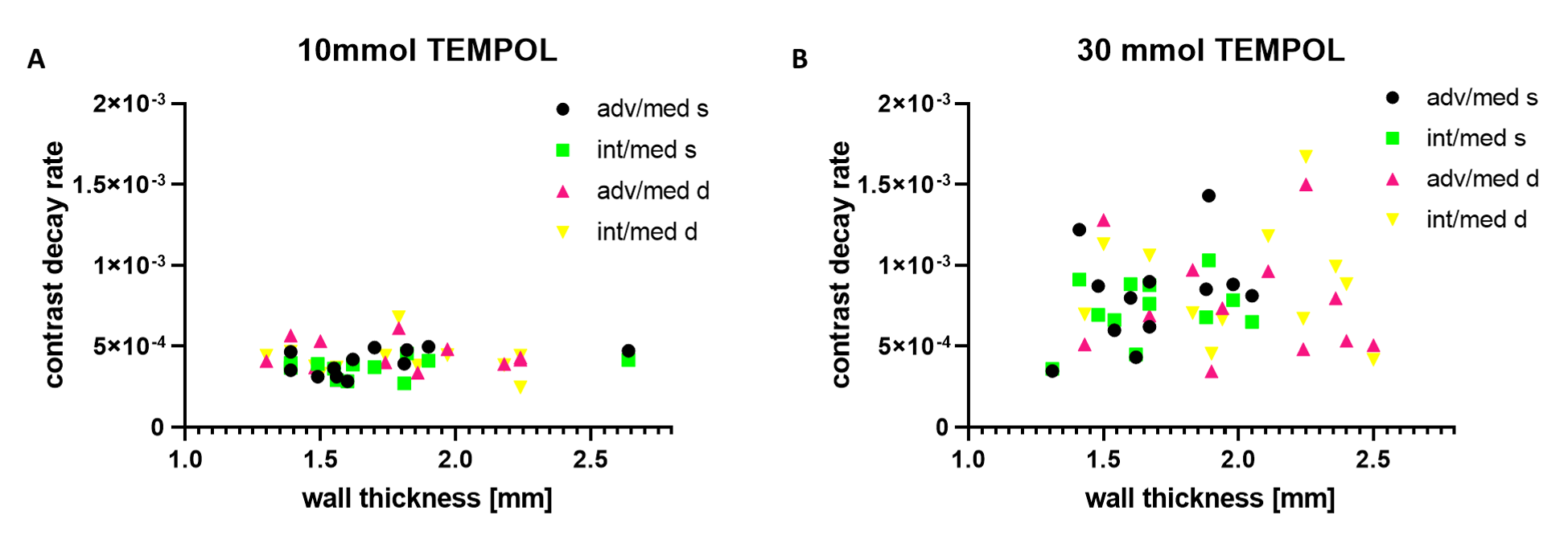
Fig.1 (A,B) shows two different aortic rings before and after the exposure to TEMPOL at the indicated four timepoints after treatment. Hyper intensity spots indicate that the homogeneity of the B1-field is affected by the restricted capacity of the surface elements of the cryoprobe leading to the contrast variation hotspots (C). (D) exemplarily indicates the manually assessed parameters as the coat-adjusted ROI as well as the wall thickness. Low nitroxides concentrations result in a slower distribution within the vascular wall by simultaneously strongly increased homogeneity of the T1 contrast variation rate over different samples (Fig.2). Fig.3 demonstrates significant diference of the contrast variation due to NR penetration from the adventitia and intima sides of the wall. The factor 3 higher rate of NR penetration from adventitia side could be expected considering essentially larger extracellular volume from the side of the coat. Fig.4 The kinetics of the TEMPOL induced contrast distribution shows only weak variation in the broad range of aortic wall thickness. The revealed outlying values result from the ROI placed in the B1-inhomogeniety hotspot, absent for the symmetrically positioned region.Conclusion
We have statistically assessed the kinetics of the stable nitroxids radical TEMPOL by T1-weighted dynamic MRI inside the ex-vivo porcine vascular wall. The results demonstrate persistence of the key values of NR induced T1 contrast kinetics and allows to step further to quantitative assessment of the redox state of the vascular wall tissue with stable nitroxide radicals.Acknowledgements
Organizational support by David Lohr and technical support by Christian Wittke, Steffen Baltes, Verena Burkard und Kerstin Körner is appreciated. Organs were provided under the approval numbers 55.2 2532-2-256, 55.2 2532-2-664 and 55.2.2-2532-2-681-12 approved by the local animal welfare committee according to the German Animal Welfare Act and the EU Directive 2010/63/EU. Financial support was obtained from the German Ministry of Education and Research (BMBF) under grant#01E1O1504.References
(1) Cui SX, Joy R, French BA, et al. Dynamic Nitroxide-Enhanced MRI Detects Oxidative Stress in the Hearts of Mice Subject to Angiotensin II Infusion. Abstract No. 3116, Proceedings of ISMRM, Honolulu, USA, 2017.
(2) Matsumoto K, Hyodo F, Matsumoto A, et al. High-resolution mapping of tumor redox status by magnetic resonance imaging using nitroxides as redox-sensitive contrast agents. Clin Cancer Res. 2006; 12(8):2455-62.
(3) Terekhov M, Pali M., Wittke C, et al.: Dynamic MRI of Nitroxide Radical for TEMPOL kinetics and Redox State Assessment in Porcine Aortic Wall. Abstract No. 7656, Proceedings of ISMRM, Paris, France, 2018.
(4) Pali M, Terekhov M, Wittke C, et al.: High-resolution and Semidynamic Vessel Wall Imaging Kinetics obtained From Stable Radical MRI in ex-vivo Porcine Aorta. Abstract No. 62012, ESC, Vienna, Austria, 2018.
Figures