4350
In vivo monitoring of Mesenchymal Stem Cells in a rat knee with Tissue Saturation SWIFT1CHOP, Philadelphia, PA, United States, 2Radiology, University of Minnesota, Minneapolis, MN, United States, 3UC Davis, Sacramento, CA, United States
Synopsis
Mesenchymal Stem Cells (MSCs) have potentials to be used for the treatment of bone diseases. We developed a new MRI method for detection of Fe-labeled MSCs. This protocol utilizes a resonance frequency shift induced by iron-oxide particles and allows imaging of grafted MSCs with a saturated signal from the host tissue. We compared retention of control and MSCs tagged with a high affinity to the bone tissue peptide. As compared with control, more tagged MSCs were detected at the injection area nine days after implantation. The new protocol allows
Introduction
In our previous work [1], we showed that the SWIFT sequence is capable of producing a hyperintense signal from iron labeled cells. However, in a knee joint, due to the similarity of signal intensities with surrounding bone tissues, detection of grafted cells is more challenging. Recently we introduced a new approach using the SWIFT sequence [2] to overcome this limitation. Iron oxide particles increase the resonance frequency of labeled cells and allowed us to implement a chemical selective RF pulse to saturate host tissue and improve the detectability of grafted cells. The goal of this study was to demonstrate the benefits of the new technique for the detection of grafted cells used in treating bone disorders. We quantitatively compared the detection of a control and LLP2A-Ale-tagged MSCs in the knee joint of rats.Methods
Cell culture: Mouse MSCs were maintained on uncoated flasks in media with DMEM and 10% FBS and labeled with 200 μg/ml of Feraheme overnight. After labeling cells with iron oxide particles, MSCs were incubated with 4 μg of a synthetic high-affinity and specificity peptidomimetic ligand (LLP2A-Ale) that increases the homing and osteogenic differentiation of MSCs on the surface of trabecular and cortical bone [3,4]. The 20 μl of 4x10⁶MSCs were injected into rat knee joints. Three groups of animals were examined: 1) Control rats injected with iron free and untagged MSCs (n=3); 2) Iron labeled but untagged MSCs (n=5); and 3) Iron labeled and LLP2A-Ale-tagged MSCs (n=5). Imaging: 3D MB-SWIFT images [5] were acquired at 9.4 T with 4 phase shifted hard pulses of 5.2 μs length (with 2.6 μs dead time), TR = 1.9 ms, excitation/acquisition bandwidths 192/384 kHz, number of views = 96000. A frequency-selective RF pulse (Gaussian, 7 ms, 90-degree flip angle) tuned to the water proton resonance was implemented to saturate signal from host tissue. Image analysis: AKT-snap software was used for measurements of volume donor cells from collected imagesResults
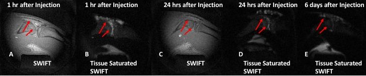
Figure 1 exhibits images of rat knee injected with iron labeled MSCs acquired with (A) regular MB-SWIFT, and (B) tissue-saturated version of MB-SWIFT, one hour after implantation of labeled MSCs. A hyperintense signal from grafted cells was detected on both images. Images on panels (C) and (D) depict the same animals but 24 hrs after injection. An identification of grafted cells on regular MB-SWIFT images was challenging while the tissue saturated images showed cells very clearly. The detection of the cells six days after implantation was possible only on tissue saturated images (panel E).
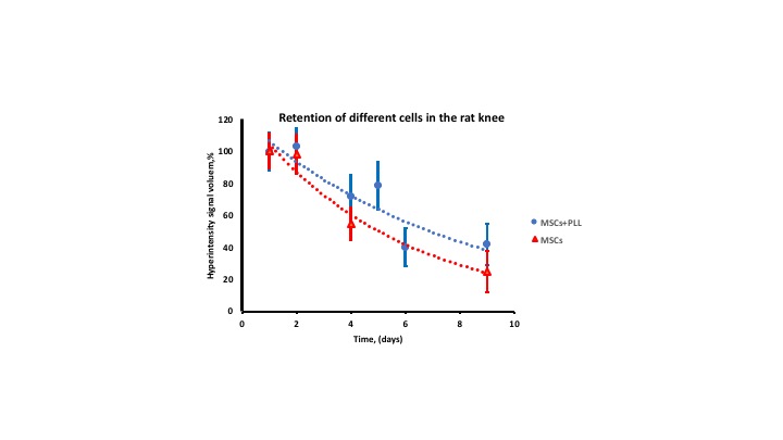
We compared the retention of the control and LLP2A-Ale-tagged cells after implantation into a rat knee. The volume of the hyperintense signal from the donor cells was measured on tissue-saturated MB-SWIFT images. The LLP2A-Ale-tagged cells have higher tendency to be retained in the injected area relative to control MSCs (Figure 2).
Discussion and conclusions
Development of new methods for the detection of the grafted cells is an essential area of imaging research. We modified the original the MB-SWIFT pulse sequence and implemented a tissue saturation scheme. This technique permits monitoring of grafted cells with minimum background signal from the host tissue. Results of our experiments indicate that the new method allows the in vivo detection of quantifiable signal from iron labeled MSCs for nine days which was not possible with conventional MRI.
Development of new target reagents for more effective delivery of therapeutic cells to an injured area is a crucial area of cell therapy research. Histological study showed that LLP2A-Ale-peptide improved delivery of MSCs to the bone tissues. Results of our experiments showed that control and LLP2A-Ale-tagged cells both were detected for 9 days after injections. More LLP2A-Ale-tagged than control MSCs were found at the endpoint of our study. Our results may suggest that 1) studies for an extended period are needed to improve a statistical difference between control and LLP2A-Ale-tagged cells, and 2) direct injection of the iron labeled MSCs into a knee joint is not optimal to detect the benefit of tagging MSCs with LLP2A-Ale-peptide. The new imaging protocol allows to detect grafted cells in a knee joint and opens new preclinical opportunities to investigate new cells reagents for bone disorders.
Acknowledgements
Acknowledgment. This study was supported by NIH grants R21A06850, P41 EB015894 and WM KECK Foundation. We also would like to thank Tony Huynh and Jaskanwaljeet Kaur for the help with MRI acquisition and animal preparations.References
1. Magnitsky S, Zhang J, Idiyatullin D, Mohan G, Garwood M, Lane NE, Majumdar S.”Imaging of Grafted Mesenchymal Stem Cells in Bone Tissue”. MRM, 2017 Nov;78(5):1900-1910
2. Magnitsky S, Pickup S, Garwood M, Idiyatullin D, “Imaging of a high concentration of iron labeled cells with positive contrast in a rat knee” MRM, 2018, 10
3. Guan M, Yao W, Liu R, et al. Directing mesenchymal stem cells to bone to augment bone formation and increase bone mass. Nature medicine 2012;18:456-62.
4. Yao W, Guan M, Jia J, et al. Reversing Bone Loss by Directing Mesenchymal Stem Cells to the Bone. Stem Cells2013.
5. Idiyatullin D, Corum CA, Garwood M. Multi-Band-SWIFT. J Magn Reson2015;251:19-25.
Figures