4344
Approaching the suppression of arm truncation artifact on 18F-FDG-PET/MR attenuation correction map: a feasibility study on MR image synthesis from cycle-GAN1Zhongshan Hospital, Shanghai, China, 2United-Imaging, Shanghai, China
Synopsis
Arm truncation artifact in PET/MR system was introduced by 1) smaller MR FOV to cover subject’s arm and 2) distorted gradient encoding at the boundary of FOV. PET imaging normally has larger FOV and non-distorted property. In this study, a cycle-GAN model trained with paired inputs of A (PET + pseudo-truncated-MR images) and B (pseudo-none-truncated-MR images) was proposed to synthesize none-truncated-MR images from PET and real-truncated-MR images then used for the generation of attenuation correction map. PET/MR whole body images from 10 patients were used for this feasibility evaluation. The cycle-GAN model was able to synthesize none-truncated-MR images for patients either with partial-arm covered or with distortion artifact in arm region.
Introduction
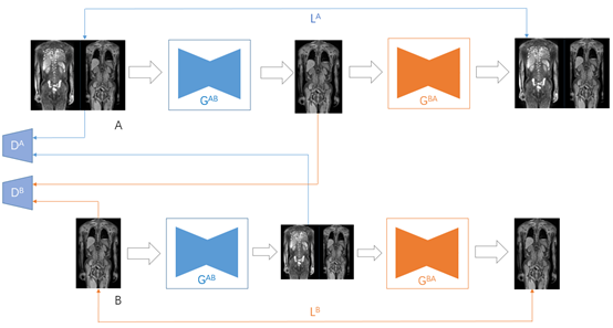
Arm truncation artifact was the common artifact in PET/MR, and may lead to misidentification of smaller lesions or quantification error [1]. Conventionally the contour extracted from 18-F-FDG-PET images was combined with arm-truncated-MR water / fat images for 5-compartment attenuation correction (AC) map generation. However, the PET contour ignored the tissue context, and may lead to incorrect estimation of AC map. The cycle generative adversarial networks (cycle-GAN) trained with paired or un-paired A-B datasets was able to synthesize B from A [2]. In this study, we modified generator part of the cycle-GAN with fast convergence to map domain A (PET + truncated MR water and fat images) and B (none-truncated MR water and fat images) for PET/MR AC map generation.Methods:
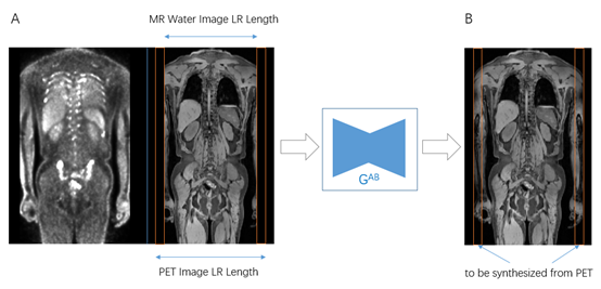
10 patients’ whole body PET/MR images with partial-arm covered (acquired at uPMR-790, United-Imaging Healthcare, Shanghai, China) were respectively included into this study. 8 patients’ images were used for training and 2 patients’ images were for test purpose. The input shape to the cycle-GAN A was 256x256x3 representing PET + MR water + MR fat images (matrix size: 256x256), and the input shape to cycle-GAN B was 256x256x2 designed for MR water + MR fat images (Figure 1). An open dataset for style transferring (horse vs. zebra, 1067 images) [3] was processed to accommodate the inputs and used as the base for transfer-learning. In the training phase, the preparation of inputs to A and B includes 1) cropping both A and B in the left-right (LR) direction to 80%, 2) cropping MR images (water and fat images) in A by 80% in order to have B as reference (Figure 2). In the testing phase, the generator for A-to-B were used to take the original PET + MR water + MR fat images as input. 20 coronal, transverse continuous slices of corresponding contrast (PET, MR water,MR fat) images were selected from each patient’s PET/MR whole body scan. Flip, mirror and scale operations were performed to augment image sets, and total 480 slices were used for training, where 8 coronal slices were used for testing. All the images were normalized and scaled to [0, 1] before the input to network. Tissue compartment consistency was evaluated by comparing the consistency between synthesized tissue compartment with common knowledge (fat, muscle, bone), where consistency in one compartment distribution contributes to score by adding 1 point. The shape consistency was scored visually in range of 0 – 3, with 1 point representing roughly 30% consistency.Results:
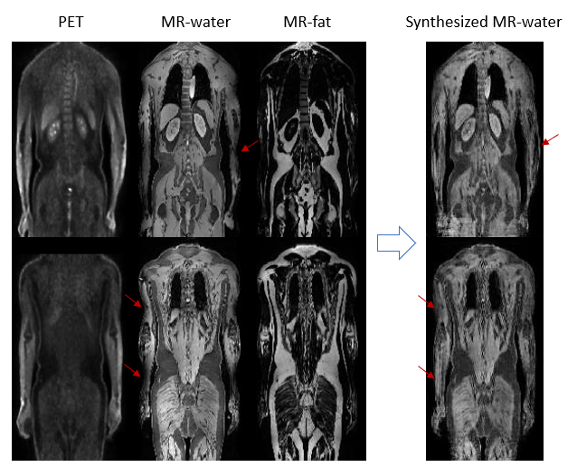
The synthesized MR water images were used for performance evaluation. The mean absolute errors of normalized images were 8.9, 9.6 for train, test image sets respectively. The shape tissue compartment and shape consistency were visually evaluated in 10 slices for each case. The shape consistency score was 2.5, where tissue compartment consistency score was 2.1 (Figure 3).Discussion:
In this study, we successfully adapted cycle-GAN to compensate the truncated MR images from PET and MR images, and achieved a well preserved shape with reasonable tissue compartment consistency in the truncated region. This synthesized MR images can be further used for better attenuation correction map generation as compared to the PET contour based correction. The 18F-FDG-PET and MR images have the similar structure, for example, in the truncated arm region, the bone, muscle, skin boundary, fat share the similar shape pattern both in PET and MRI, and it forms the image base for the link between PET and MRI contrast. By using created pseudo-truncated-MR images, we established the links between PET and MRI contrast in the truncated region by exploring the linkage in body region (none truncated region). A further exploration on attenuation correction map generation might be feasible by style(contrast)-transferring PET and MR images to CT images using unpaired PET+MR and CT images and cycle-GAN.Conclusion:
We demonstrated the feasibility of synthesis none-arm-truncated MR water and fat images from PET, truncated MR water and fat images. It provides new opportunity for PET/MR attenuation correction.Acknowledgements
No acknowledgement found.References
1. Lindemann ME, Oehmigen M, Blumhagen JO, et al. MR-based truncation and attenuation correction in integrated PET/MR hybrid imaging using HUGE with continuous table motion. Med Phys. 2017 Sep. 44(9):4559-4572.
2. Jun-Yan Zhu, Taesung Park, Phillip Isola, and Alexei A. Efros. Unpaired Image-to-Image Translation using Cycle-Consistent Adversarial Networks, IEEE International Conference on Computer Vision (ICCV), 2017.
3. https://people.eecs.berkeley.edu/~taesung_park/CycleGAN/datasets/ horse2zebra.zip
Figures